

Detection assays employing magnetic nanoparticles
a technology of magnetic nanoparticles and detection assays, applied in the direction of material testing goods, measurement devices, instruments, etc., can solve the problems of consuming a lot of time, consuming a lot of labor, and long western blotting workflows
- Summary
- Abstract
- Description
- Claims
- Application Information
AI Technical Summary
Benefits of technology
Problems solved by technology
Method used
Image

Examples
example 1
Dot Blot Analysis Using 100 Nm Fluorescent MNP-Capture Agent Complexes
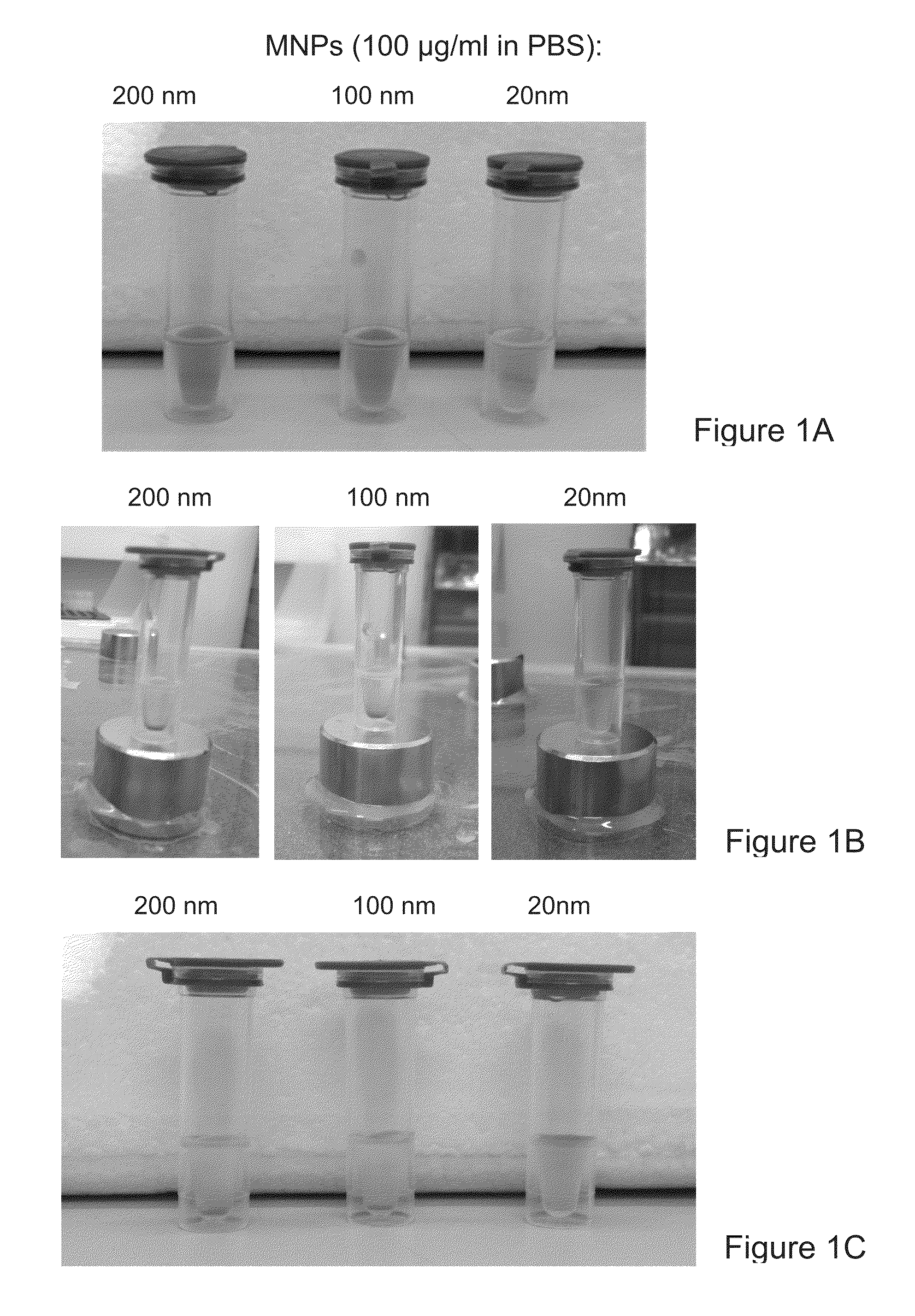
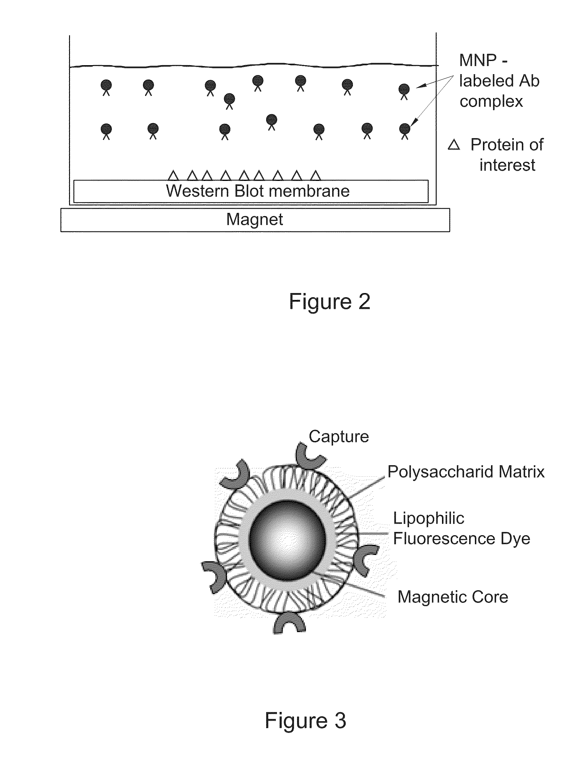
[0234]Fluorescent magnetic nanoparticles (MNPs) of 100 nm size (hydrodynamic diameter) were purchased from Chemicell. These MNPs contain a shell of lipophilic fluorescence dye which was formed around a magnetic core and coated with a polysaccharide matrix. Capturing agents were embedded within the polysaccharide matrix (FIG. 3). MNPs with three capture agents—Protein G, anti-mouse IgG and streptavidin—were tested. All three MNP types had fluorescence with excitation pick at 547 nm and emission pick at 580 nm.
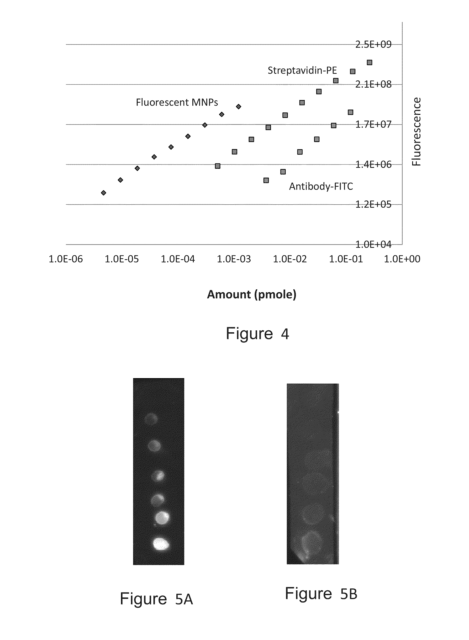
[0235]Preliminary experiments were done to evaluate the fluorescence level of these MNPs. Small drops (2 μl) of solutions containing MNPs at a series of concentrations were spotted over a 0.45 μm nitrocellulose (NC) membrane. Similar spotting was done with standard capturing agents that were labeled with the commonly used fluorophores fluorescein (as FITC) and phycoerythrin (PE). Fluorescent detection was done...
example 2
Characterization of Smaller (≦30 nm) MNP-Antibody Complexes
[0239]To overcome the disadvantages of the system described in Example 1, magnetic nanoparticle-antibody complexes comprising small sized iron oxide nanoparticles 30 nm) were purchased from Ocean Nanotech. These iron oxide (IO) nanoparticles are coated with linear amphiphilic polymer carrying end carboxylic groups, which can be used to immobilize antibodies or other proteins (FIG. 6).
[0240]IO magnetic nanoparticles associated with horseradish peroxidase (HRP)-labeled anti-rabbit IgG secondary antibody (Ocean Nanotech) were used in the following experiment. Three nanoparticle sizes were used: 10 nm, 20 nm, and 30 nm. Enzyme (HRP) activity of the nanoparticles was tested using a colorimetric assay in solution, with comparison to the free labeled antibody (anti-rabbit IgG-HRP from Jackson). This analysis was done by placing a concentration series of MNP or antibody solution in wells of non-binding 96 well plate (50 μl solution ...
example 3
Dot Blot Analysis with Small MNPs Carrying a Secondary Antibody
[0241]Dot Blot Assay with Rabbit IgG / Anti Rabbit IgG Antibody
[0242]Rabbit IgG solution (2 μl) was spotted on two 0.45 μm NC membrane at a series of 16 concentrations (×1.5 dilution series), yielding spotted amounts from 5 μg down to 11 ng. One membrane was reacted with the 10 nm IO magnetic nanoparticles described in Example 2 (0.43 nM). The second, reference membrane was reacted with free anti-rabbit HRP under similar conditions (e.g. the same molar concentration). Color signal was developed and detected using 4-CN substrate and ChemiDoc MP imager.
[0243]The results are shown in FIG. 8. It can be seen that the results with the 10 nm MNPs are comparable to the results with the free anti-rabbit HRP. For example, the lowest amount of 11 ng Rabbit IgG could be similarly detected by the two methods.
Dot Blot Assay with Transferrin / Anti-Transferrin Antibody
[0244]Human Transferrin (hT) was spotted on NC membrane, reacted with an...
PUM
 Abstract
Description
Claims
Application Information