
Electron microscopic observation method for observing biological sample in shape as it is, and composition for evaporation suppression under vacuum, scanning electron microscope, and transmission electron microscope used in the method

a technology of electron microscopic observation and biological sample, which is applied in the direction of material analysis using wave/particle radiation, instruments, biomass after-treatment, etc., can solve the problems of difficult to observe a biological/living body sample in the living state and difficult to acquire an image in the living state, and achieve high magnification

Active Publication Date: 2014-08-14
JAPAN SCI & TECH CORP
View PDF0 Cites 9 Cited by
  • Summary
  • Abstract
  • Description
  • Claims
  • Application Information

AI Technical Summary

Benefits of technology

The invention allows for observing a sample at a high magnification without damaging the sample. It also allows for observing a living biological sample as it is alive, with the ultrastructure of the sample in a moving state using an electron microscope.

Problems solved by technology

For this reason, the surface shape thereof is significantly deformed in some cases, and thus it is difficult to observe a biological / living body sample in the living state as it is using an electron microscope.
For that reason, it is difficult to acquire an image in the living state as it is at a high magnification in the observation of a biological / living body sample, as well.

Method used


Image

  • Electron microscopic observation method for observing biological sample in shape as it is, and composition for evaporation suppression under vacuum, scanning electron microscope, and transmission electron microscope used in the method
  • Electron microscopic observation method for observing biological sample in shape as it is, and composition for evaporation suppression under vacuum, scanning electron microscope, and transmission electron microscope used in the method
  • Electron microscopic observation method for observing biological sample in shape as it is, and composition for evaporation suppression under vacuum, scanning electron microscope, and transmission electron microscope used in the method

Examples


example 1

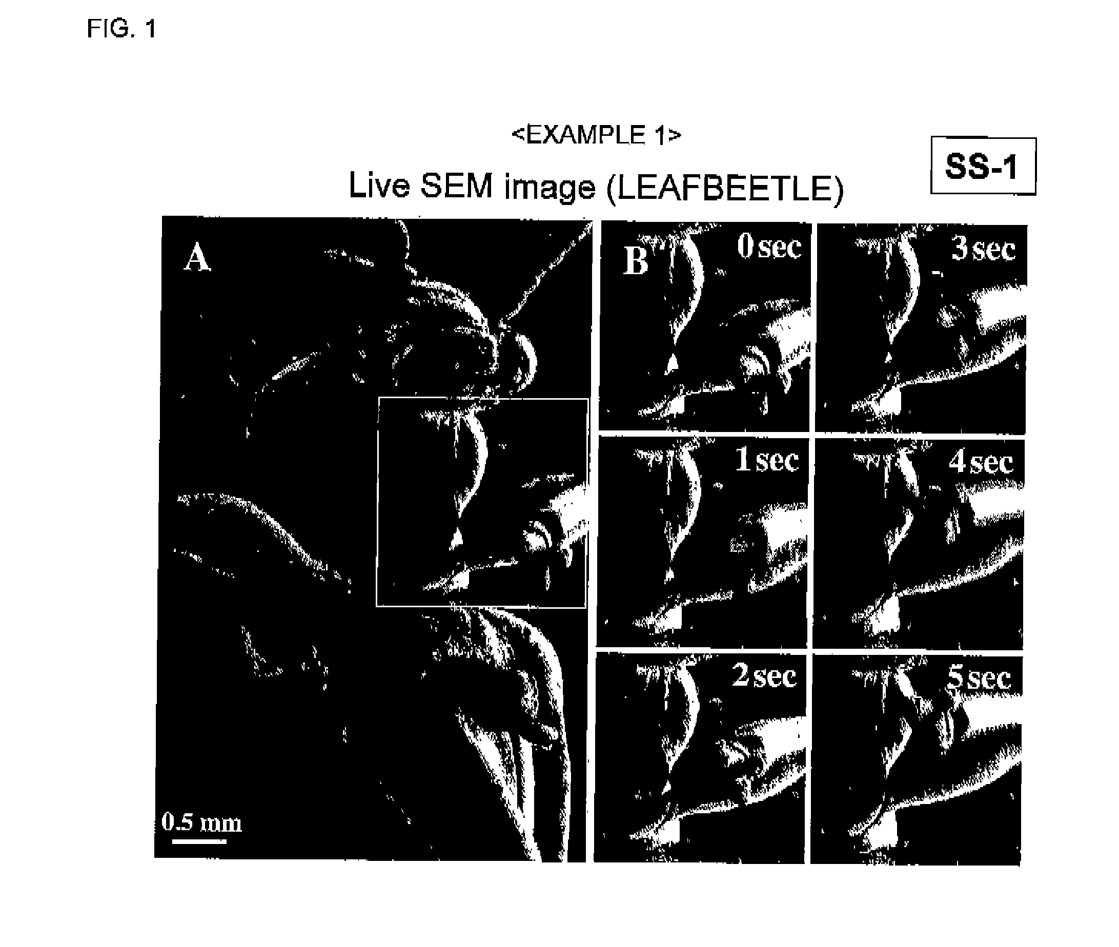
[0216]A composition for evaporation suppression was prepared using a 0,1% aqueous solution of sodium laurylbenzenesulfonate as an amphiphilic compound and 0.01 wt % of ethylenediamine nickel complex as a metal compound.

[0217]A living leaf beetle was immersed in the composition for evaporation suppression for 1 minute, taken out therefrom, and the excess was wiped off, thereby covering the body surface of the leaf beetle with a thin film.

[0218]Thereafter, the leaf beetle covered with a thin film was introduced in the sample chamber of a SEM, and videotaped (FIG. 1). The situation that the leaf beetle was moving was observed even after the initiation of electron beam irradiation.

[0219]Moreover, the leaf beetle was alive even after taken out from the sample chamber after SEM observation.

example 2

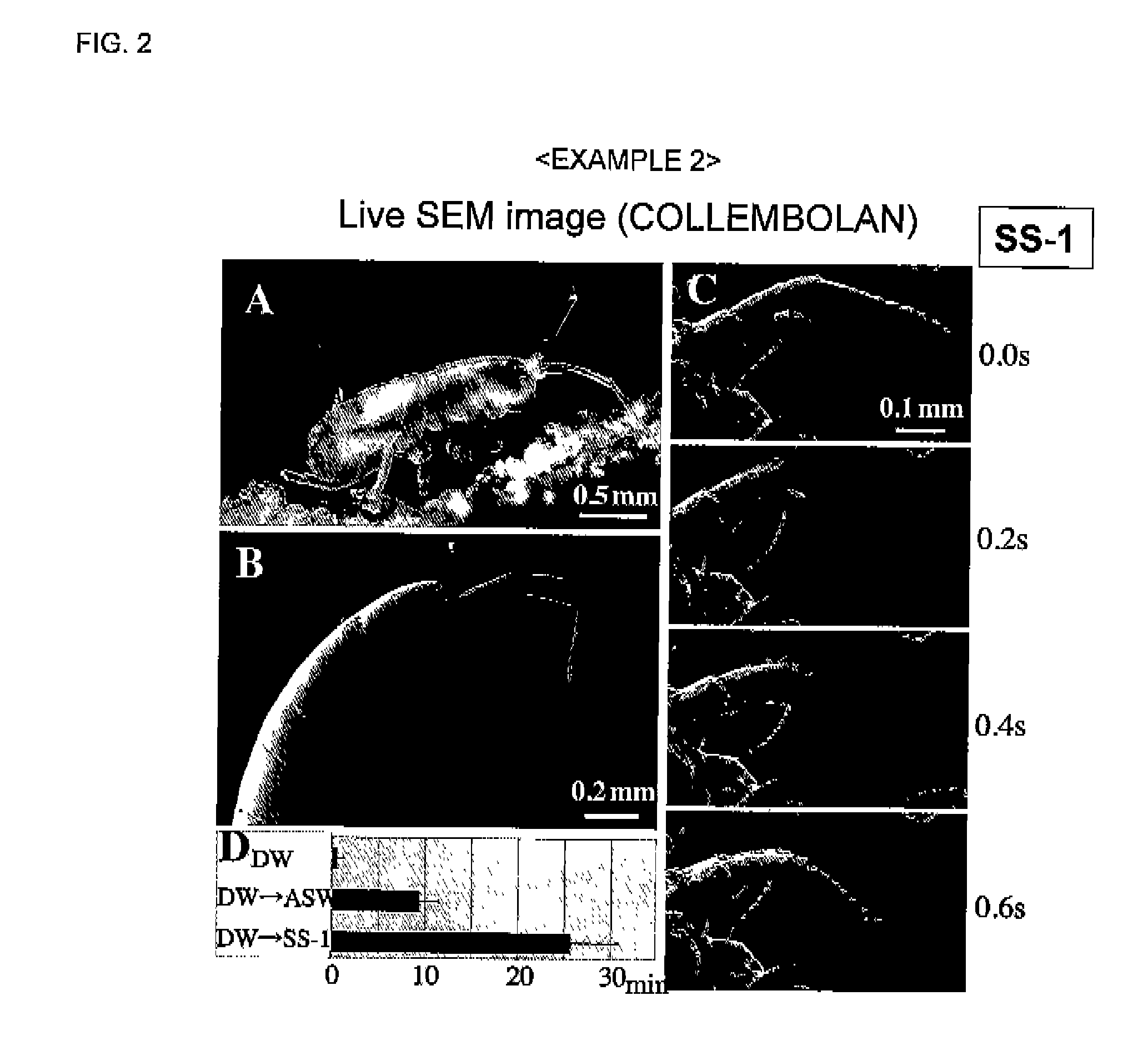
[0220]A composition for evaporation suppression was prepared using a 0.1% aqueous solution of sodium laurylbenzenesulfonate as an amphiphilic compound and 0.01 wt % of ethylenediamine nickel complex as a metal compound.

[0221]A living collembolan was immersed in the composition for evaporation suppression for 1 minute, taken out therefrom, and the excess was wiped off, thereby covering the body surface of the collembolan with a thin film.

[0222]Thereafter, the collembolan covered with a thin film was introduced in the sample chamber of a SEM, and videotaped (FIG. 2). The situation that the collembolan was moving was observed even after the initiation of electron beam irradiation.

[0223]Moreover, the collembolan was alive even after taken out from the sample chamber after SEM observation.

example 3

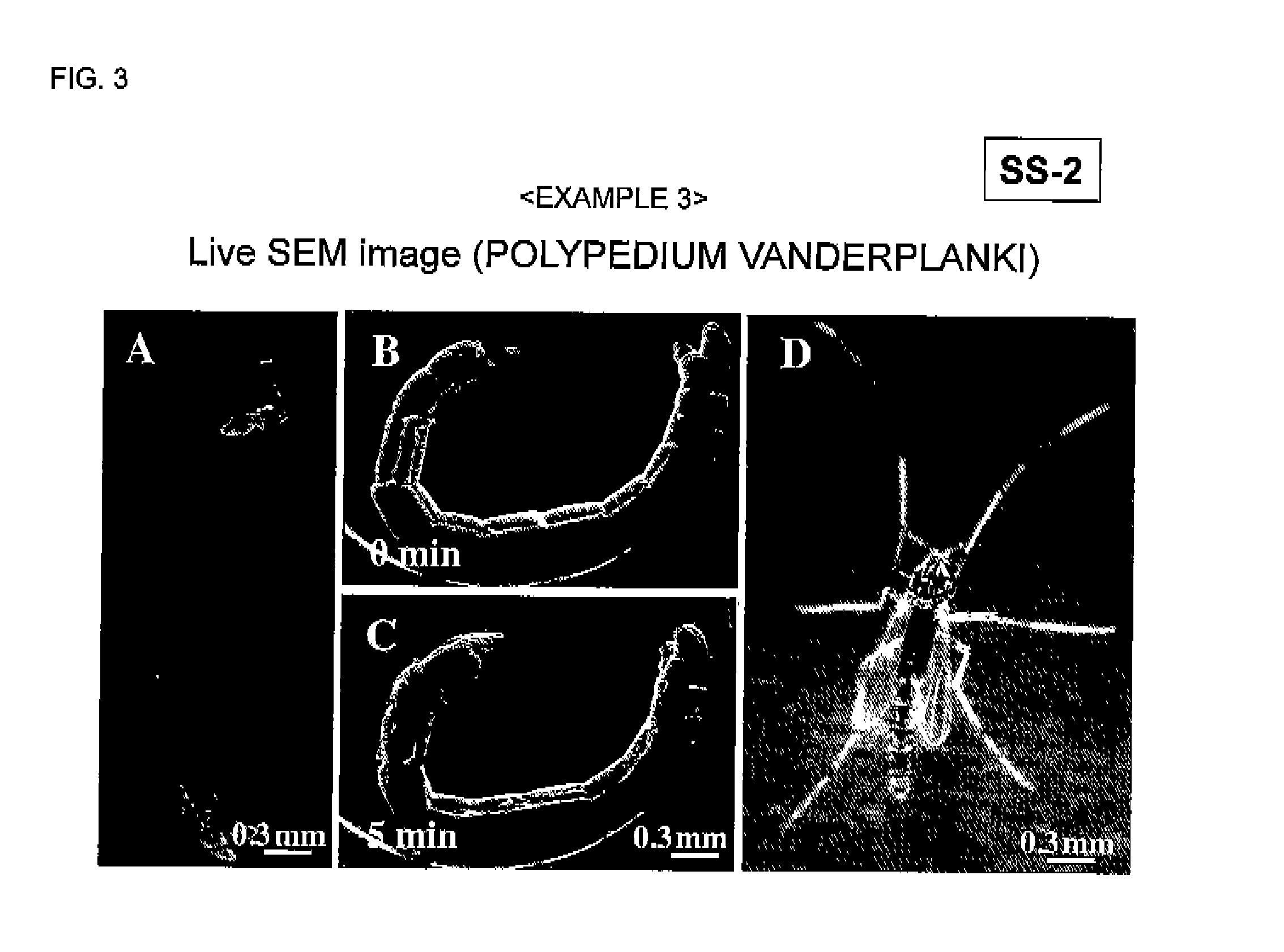
[0224]A composition for evaporation suppression was prepared using a 10% aqueous solution of Tween 20 as an amphiphilic compound, and 1% (w / v) of trehalose and 0.1% (w / v) of pullulan as saccharides.

[0225]A living larva of Chironomus yoshimatsui was immersed in the composition for evaporation suppression for 1 minute, taken out therefrom, and the excess was wiped off, thereby covering the body surface of the larva of Chironomus yoshimatsui with a thin film.

[0226]Thereafter, the larva of Chironomus yoshimatsui covered with a thin film was introduced in the sample chamber of a SEM, and videotaped (FIG. 3). The situation that the larva of Chironomus yoshimatsui was moving was observed even after the initiation of electron beam irradiation.

[0227]Moreover, the larva of Chironomus yoshimatsui was alive even after taken out from the sample chamber after SEM observation, and was metamorphosed into an imago.


PUM

PropertyMeasurementUnit
viscosityaaaaaaaaaa
thicknessaaaaaaaaaa
pressureaaaaaaaaaa

Abstract

Provided is an observation method by an electron microscope, in which a biological sample can be observed as it is alive and a situation that the biological sample is moving can be observed using an electron microscope, and a composition for evaporation suppression under vacuum, a scanning electron microscope, and a transmission electron microscope used in the method.The sample observation method by an electron microscope according to the invention includes applying a composition for evaporation suppression containing at least one kind selected from an amphiphilic compound, oils and fats, and an ionic liquid to the surface of a sample to form a thin film, and covering the sample with the thin film, and displaying an electron microscopic image of the sample, which is covered with the thin film and accommodated in a sample chamber under vacuum, on a display device.

Description

TECHNICAL FIELD[0001]The present invention relates to an electron microscopic observation method for observing a biological sample in the shape as it is in a scanning electron microscope, a transmission electron microscope, or the like, and a composition for evaporation suppression under vacuum, a scanning electron microscope, and a transmission electron microscope used in the method.BACKGROUND ART[0002]A sample is put under vacuum in order to perform sample observation using a scanning electron microscope and a transmission electron microscope, and thus the sample is desired to be resistant to vacuum and to be imparted with electrical conductivity required to acquire an image thereof.[0003]The preparation of a sample for scanning electron microscope is performed as follows. The sample is dehydrated by vacuum drying in advance, and then an electrically conducting material (platinum, carbon, gold, palladium, osmium, and the like) is coated on the surface of the sample by a means of v...

Claims


Application Information

Patent Timeline
Patent Type & Authority Applications(United States)
IPC IPC(8): G01N1/44G01N23/225
CPCG01N23/225G01N23/04H01J37/20G01N2223/612G01N1/2853G01N1/44G01N23/2252H01J2237/2002H01J2237/2004H01J37/26
Inventor HARIYAMA, TAKAHIKOTAKAKU, YASUHARUSUZUKIMURANAKA, YOSHINORIOHTA, ISAOSHIMOMURA, MASATSUGUISHII, DAISUKE
Owner JAPAN SCI & TECH CORP
Learn More