

Detection, isolation and analysis of rare cells in biological fluids
a technology of biological fluids and rare cells, applied in the field of immunological methods and kits for detection, capture and isolation of rare cells from biological fluids, can solve the problems of high invasiveness of procedures, prone to significant risk of fetal loss (up to 1%) and/or maternal complications
- Summary
- Abstract
- Description
- Claims
- Application Information
AI Technical Summary
Benefits of technology
Problems solved by technology
Method used
Image

Examples
examples
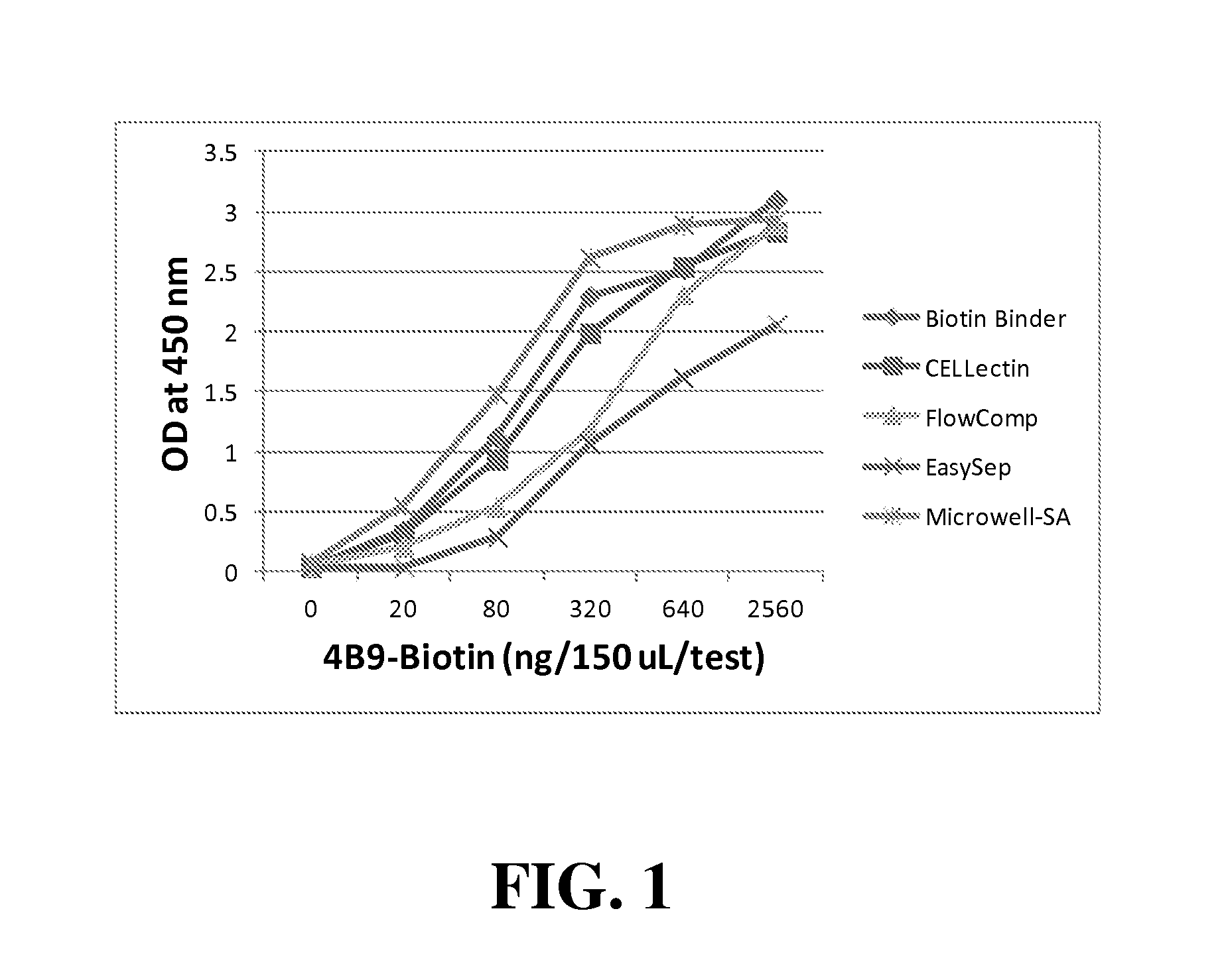
Cell-Free 4B9 ELISA
[0065]The 4B9 ELISA of one embodiment of the present invention involves direct or indirect coating of 4B9 antibody onto solid-supports, detecting bound 4B9 using goat ant-mouse IgM (Fab)2 labelled with the enzyme horseradish peroxidase (HRPO), and colorimetric quantification of the reaction using HRPO substrate Tetramethylbenzidine (TMB). Whereas in direct coating, 4B9 was detected by incubation with the detection anti-mouse antibody-HRPO conjugate (0.025 ug / mL of assay buffer; 10 mM NaPO4, pH 7.4, containing 8.8 g NaC1, 0.5 g BSA, 0.5 mL Tween-20, and 2.5 mL proclin / L), the indirect coating involved pre-incubation of unlabeled or biotinylated 4B9 (10 ug / mL) with second antibody or streptavidin coated support, respectively.
[0066]In general, comparative evaluation of microtitration wells, magnetic particles in test tubes, and glass slides in 16-well partitioned assemblies (Grace Bio labs) were performed under nearly identical conditions of antibody volume (50 uL / re...
experiment # 1
Experiment #1
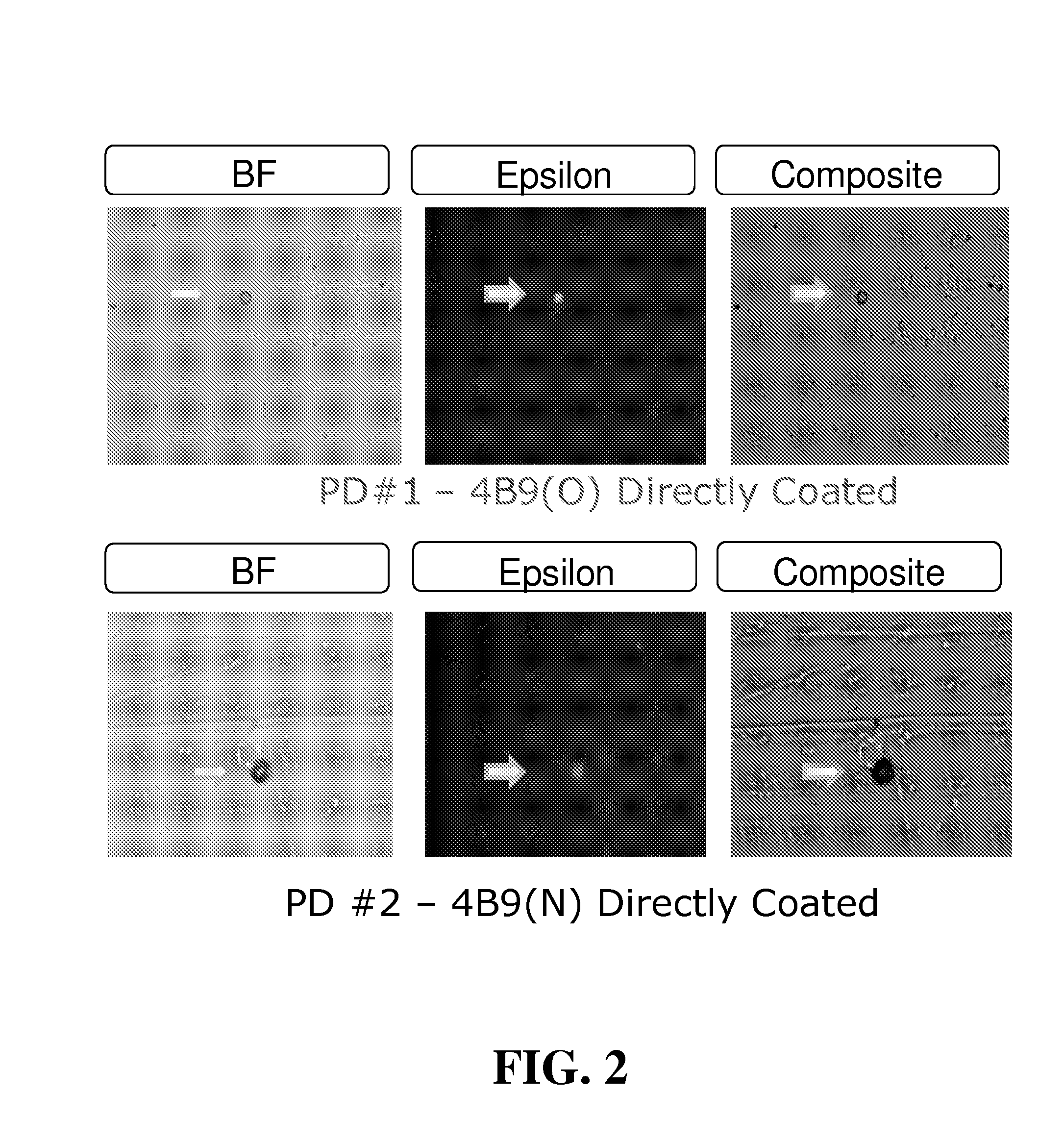
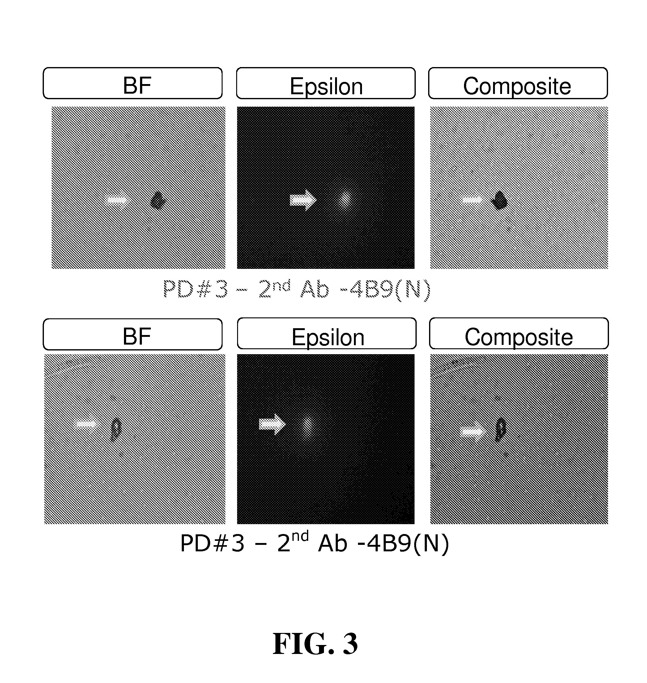
[0104]Maternal blood from four first trimester pregnancies (30 mL total volume) were washed, resuspended to original volume, and pooled. Equal volumes of pooled blood (10 mL) were added to each of three Petri dishes coated with 4B9 antibody old lot (PD#1; 4B9-O), 4B9 antibody new lot (PD#2; 4B9-N), or anti-mouse IgM coupled with 4B9-O antibody (PD#3). After 60 minutes incubation with gentle mixing on a flat orbital shaker, cells were washed 5× with PBS and stained for detection of fetal epsilon globulin. From this first trimester pooled blood, the total number of isolated fetal cells stained positively for epsilon globulin in PD#1, PD#2, and PD#3, were 909 (91 / mL of blood), 1192 (119 / mL of blood), and 580 (58 / mL of blood), respectively (See Table 3).
TABLE 3ISOLATION OF FETAL NRBC FROM POOLEDFIRST TRIMESTER MATERNAL BLOODHbNo. ofSolidCapturePooledBloodPretreatment / detectionFixed &CellsSupportAbBloodVol / TestRBC lysisantibodyPermeabilizedIsolatedPD#14B9(O)1st10 mLnoAMCAyes...
experiment # 2
Experiment #2
[0107]In the second experiment, five different glass microscope slides from three different manufacturers were first coated directly as well as indirectly with 4B9 and analyzed comparatively for their binding capacity with Cell-Free 4B9 ELISA. Glass slides demonstrating higher binding capacity were coated with biotinylated 4B9 via streptavidin coating (Slide #1), or unlabeled 4B9 via second antibody (Slide #2). Blood from another first trimester pregnancy (8 ml) was washed, resuspended to original volume, and incubated with slide #1 and slide #2 as above. The isolated cells were subsequently stained for fetal epsilon globulin and nuclei with TO-PRO. Microscope glass slides were coated with streptavidin or 2nd antibody followed by incubation with biotinylated 4B9 antibody (SA; Slide#1) or untouched 4B9 antibody (Slide#2). Peripheral blood from a single first trimester pregnancies (about 8 mL) was washed and used in equal volumes. Isolated cells were stained for epsilon h...
PUM
| Property | Measurement | Unit |
|---|---|---|
| pH | aaaaa | aaaaa |
| volume | aaaaa | aaaaa |
| volume | aaaaa | aaaaa |
Abstract
Description
Claims
Application Information