Method and device for monitoring retinopathy
a technology of retinopathy and monitoring devices, applied in the field of methods and devices for monitoring retinopathy, can solve problems such as pupil restriction
- Summary
- Abstract
- Description
- Claims
- Application Information
AI Technical Summary
Benefits of technology
Problems solved by technology
Method used
Image
Examples
example 1
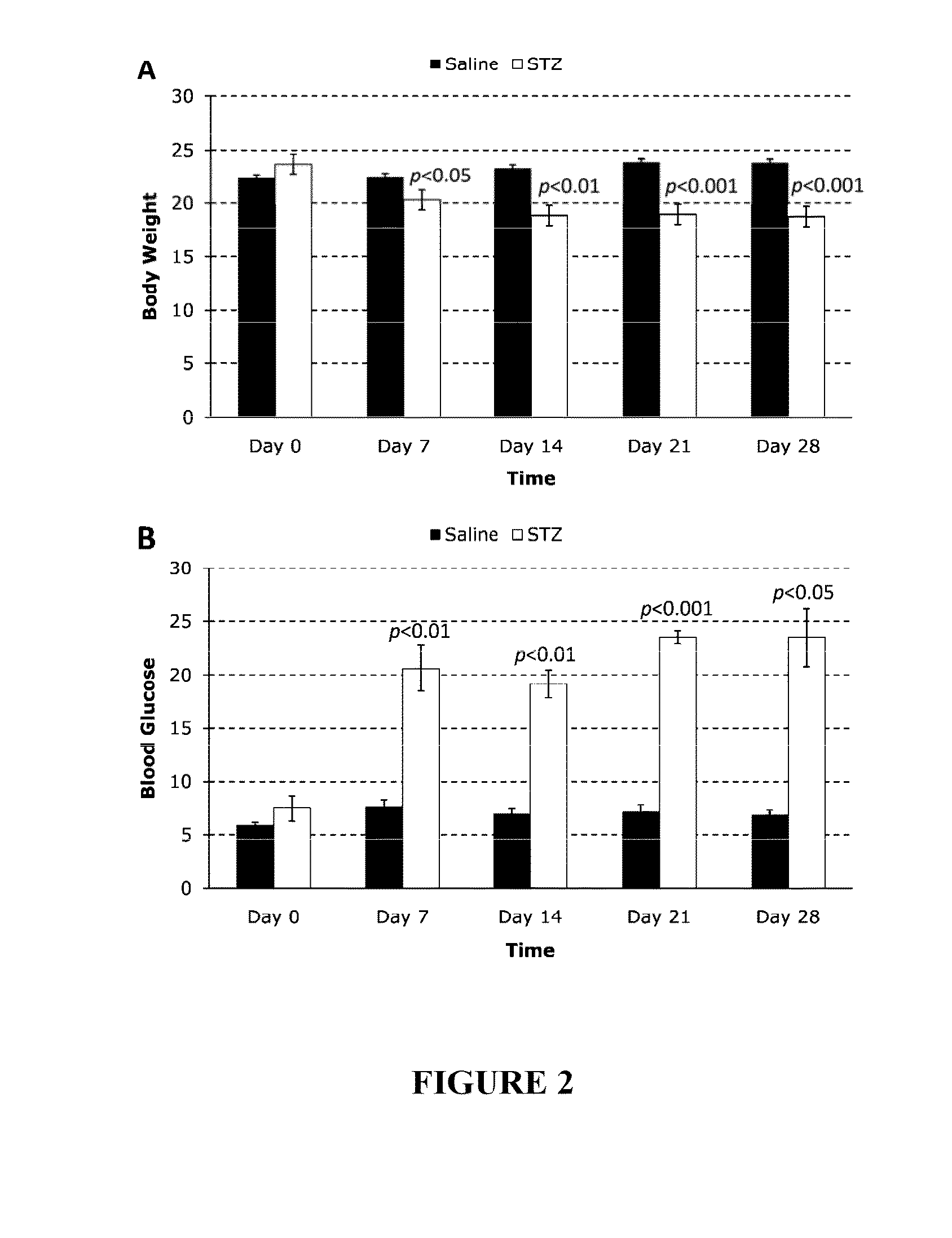
[0155]The present study was designed to develop a novel method for quantifying PLR based on AF intensity (AFI) emitted due to confocal retinal blue light excitation (cRBLE) and to study longitudinal PLR alteration in a type 1 diabetic mouse model. Diabetes was triggered via a single intraperitoneal injection of streptozotocin (STZ) into wild type C57BL / 6J mice. Anaesthethized mice were subjected to cRBLE weekly over a period of four weeks. At each time point, PLR was quantified by introducing the concept of the ‘area under the curve’ (AUC) of the intensity profile of retinal AF measured at 5 second (s) intervals over a period of 275 s. The mice develop diabetes as early as three days after STZ induction. The blood glucose levels peaked at approximately 23 mmol / L and the body weight decreased by approximately 20% after one month post-treatment. A progressive decrease in diabetic AUC occurred during this period but control AUC remained relatively unchanged. PLR was initiated despite s...
example 2
[0184]In this study, the method involves the use of (i) a scanning laser ophthalmoscope (cSLO) with a reflectance IR and a 488 nm blue laser in dual-mode operation and (ii) an image processing software for the automated quantification of pupil size. A total of 9 subjects volunteered for this study (3: negative control, 1: diabetic but no DR, 1: early DR and 4: moderate DR). Confocal scanning laser ophthalmoscopy was used to induce and to record PLR. The experiments were conducted in the morning where the left eye, and subsequently right eye, was subjected to 488 nm blue light excitation for 3 minutes, during which time changes in pupil size were monitored.
[0185]Research Design and Methods
[0186]Patient Recruitment.
[0187]Patients were recruited at SNEC Diabetic Retinopathy Services (DRS). A total of 9 subjects volunteered for this pilot study (3: negative control, 1: diabetic but no DR, 1: early DR and 4: moderate DR).
[0188]PLR Acquisition.
[0189]PLR measurements were taken in the morn...
example 3
[0232]62 diabetic patients were screened using the dual mode method.
[0233]Methods.
[0234]Patient eyes were subjected to infrared light over a total 3 minute period. Pupil IR image was captured every 1 second over the 3 minutes. Simultaneously, for the 3 minute period, the eyes were subjected to blue light, which was repeatedly turned on for 5 seconds, off for 5 seconds. The pupil region was segmented as described in Example 2 for each IR data frame captured and the pupil size (in pixels) was calculated. For each light ON / OFF cycle, the ratio of pupil maxima / pupil minima was computed, where pupil maxima denotes the largest pupil size during light OFF and pupil minima denotes smallest pupil size during light ON. The average ratio was computed for data combined from all ON / OFF cycles within the 3 minute period.
[0235]FIG. 22.
[0236]Bar chart depicting the average ratio of pupil maxima to pupil minima; from left to right: NO DR (patients with diabetes but no diabetic retinopathy), MILD DR ...
PUM
 Abstract
Description
Claims
Application Information
 - R&D
- Intellectual Property
- Life Sciences
- Materials
- Tech Scout
- Unparalleled Data Quality
- Higher Quality Content
- 60% Fewer Hallucinations
Browse by: Latest US Patents, China's latest patents, Technical Efficacy Thesaurus, Application Domain, Technology Topic, Popular Technical Reports.
© 2025 PatSnap. All rights reserved.Legal|Privacy policy|Modern Slavery Act Transparency Statement|Sitemap|About US| Contact US: help@patsnap.com


