

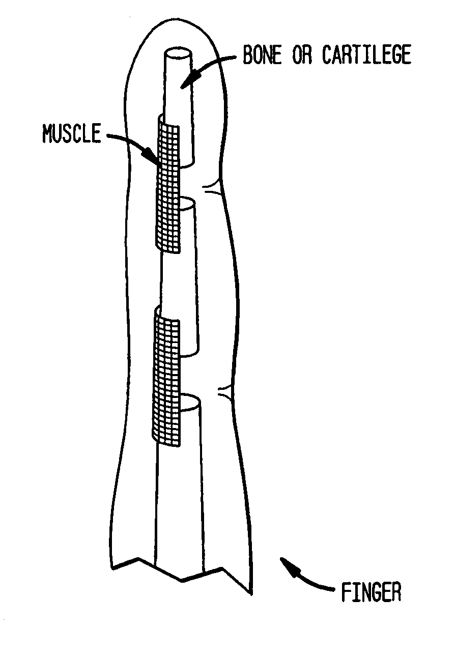
Production of tissue engineered digits and limbs
a technology of artificial tissue and limbs, which is applied in the field of artificial tissue constructs, can solve the problems of injuring connective and interstitial tissues, affecting finger amputations, and damage to bones, and achieves the effects of enhancing tissue maturation and function, enhancing vascularisation of muscle components, and increasing biological demands of engineering large tissues
- Summary
- Abstract
- Description
- Claims
- Application Information
AI Technical Summary
Benefits of technology
Problems solved by technology
Method used
Image

Examples
example 1
Methods and Materials
(i) Scaffold Preparation
[0165]Electrospun nanofiber scaffolds have been developed using a solution of collagen type I, elastin, and poly(D,L-lactide-co-glycolide) (PLGA, mol. ratio 50:50, Mw 110,000) (Boeringer-Ingelheim, Germany). Collagen type I from calf skin (Elastin Products Company, Owensville, Mo.), elastin from ligamentum nuchae (bovine neck ligament), (Elastin Products Company, Owensville, Mo.), and PLGA are mixed at a relative concentration by weight of 45% collagen, 40% PLGA, and 15% elastin. The solutes are dissolved in 1,1,1,3,3,3-hexafluoro-2-propanol (99+%) (Sigma Chemical Company, St. Louis, Mo.) at a total solution concentration of 15 w / v % (150 mg / mL). High molecular weight PLGA, previously used for electrospinning tissue scaffolds is added to the solution to increase mechanical strength of the scaffold and increase viscosity and spinning characteristics of the solution.
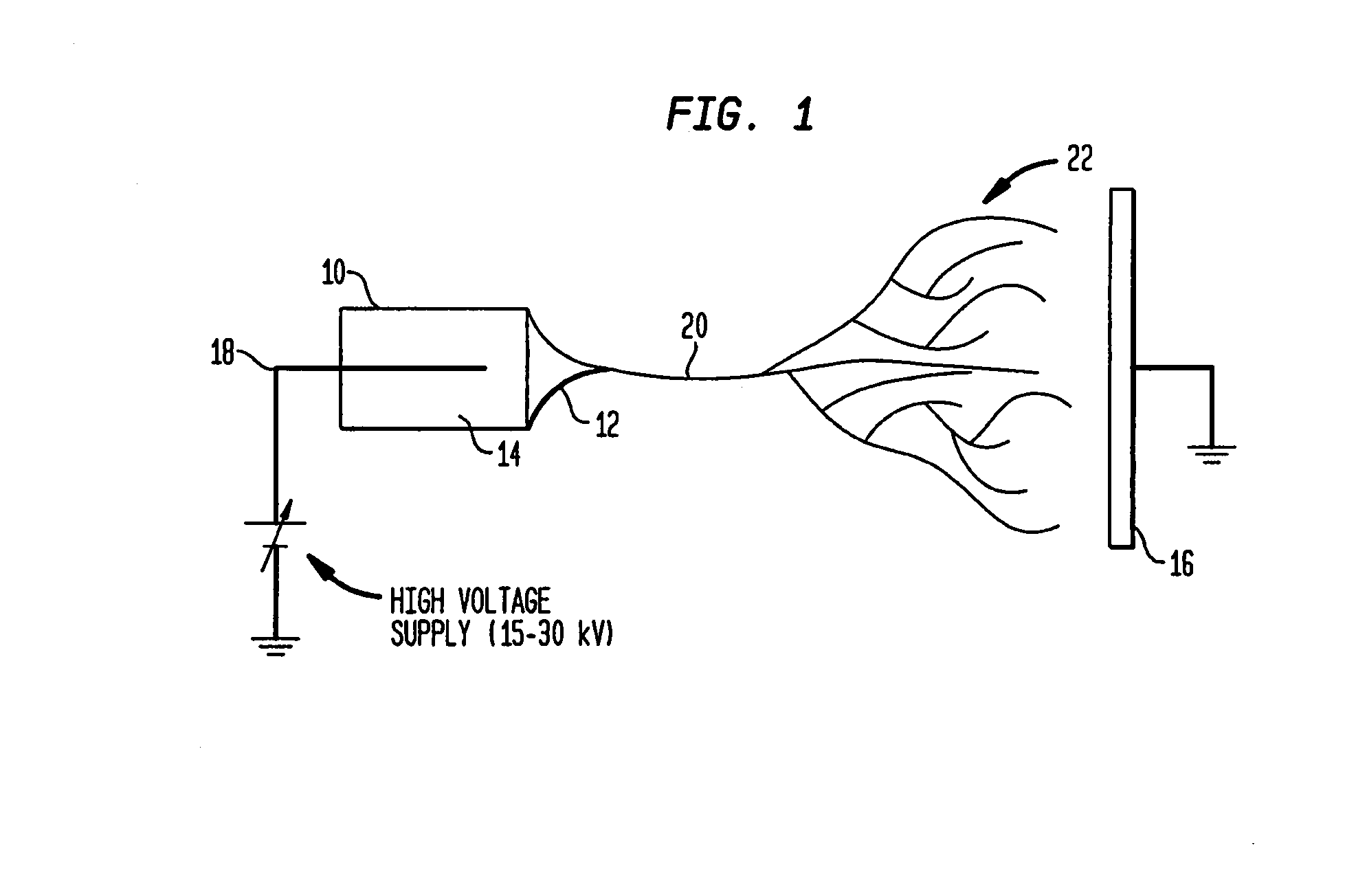
[0166]Physically, the electrospinning method requires a high voltage power ...
example 2
Preparation of Decellularized Organs
[0184]The following method describes a process for removing the entire cellular content of an organ or tissue without destroying the complex three-dimensional infra-structure of the organ or tissue. An organ, e.g. a liver, was surgically removed from a C7 black mouse using standard techniques for tissue removal. The liver was placed in a flask containing a suitable volume of distilled water to cover the isolated liver. A magnetic stir plate and magnetic stirrer were used to rotate the isolated liver in the distilled water at a suitable speed for 24-48 hours at 4° C. This process removes the cellular debris and cell membrane surrounding the isolated liver.
[0185]After this first removal step, the distilled water was replaced with a 0.05% ammonium hydroxide solution containing 0.5% Triton X-100. The liver was rotated in this solution for 72 hours at 4° C. using a magnetic stir plate and magnetic stirrer. This alkaline solution solubilized the nuclear...
example 3
Electrospun Matrices
[0188]An electrospun matrix was formed using the methods outlined in Example 1. A solution of collagen type I, elastin, and PLGA, were used. The collagen type I, elastin, and PLGA were mixed at a relative concentration by weight of 45% collagen, 40% PLGA, and 15% elastin.
[0189]The resulting fibrous scaffold had a length of 12 cm with a thickness of 1 mm. A 2 cm representative sample is depicted in FIGS. 3A-3C. This demonstrates the feasibility of spinning Type I Collagen and elastin into fibers from nanometer to micrometer diameter using concentrations from 3% to 8% by weight in solution. These results also show that by adding PLGA (Mw 110,000) to the mixture, solutions with higher viscosity and improved spinning characteristics could attained. By increasing the solution concentration to 15%, thicker, stronger scaffolds were able to be built while maintaining the collagen and elastin components.
[0190]Collagen type I stained positively on the decellularized scaffo...
PUM
 Abstract
Description
Claims
Application Information