

Systems and methods for frameless image-guided biopsy and therapeutic intervention
a frameless, biopsy technology, applied in the field of frameless image-guided biopsy and/or therapeutic intervention, can solve the problems of over-invasiveness, headache, and complicated traditional systems and methods for collecting a biopsy sample, and achieve the effects of avoiding the associated risk of skull damage, avoiding complications, and enhancing patient comfor
- Summary
- Abstract
- Description
- Claims
- Application Information
AI Technical Summary
Benefits of technology
Problems solved by technology
Method used
Image

Examples
Embodiment Construction
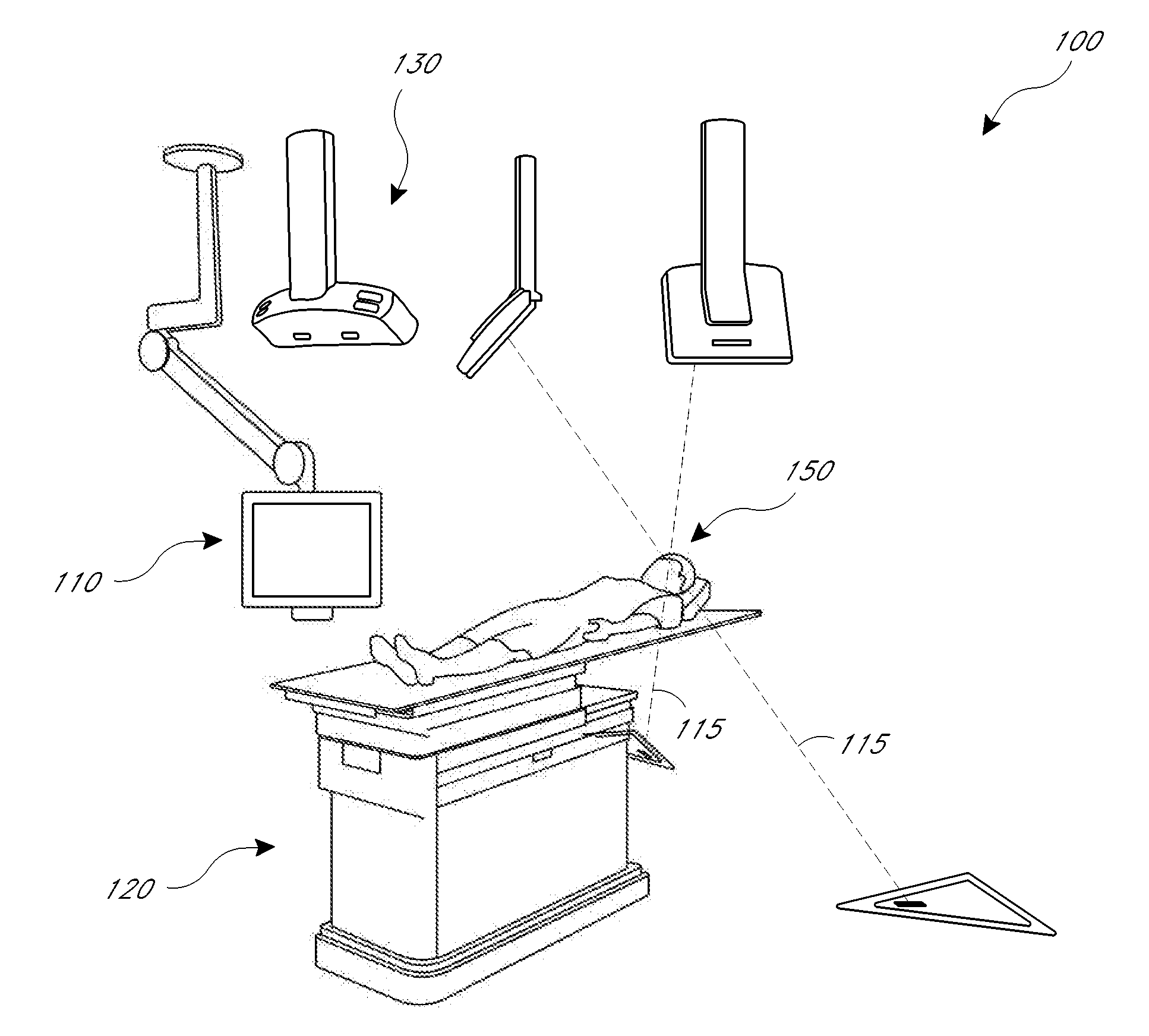
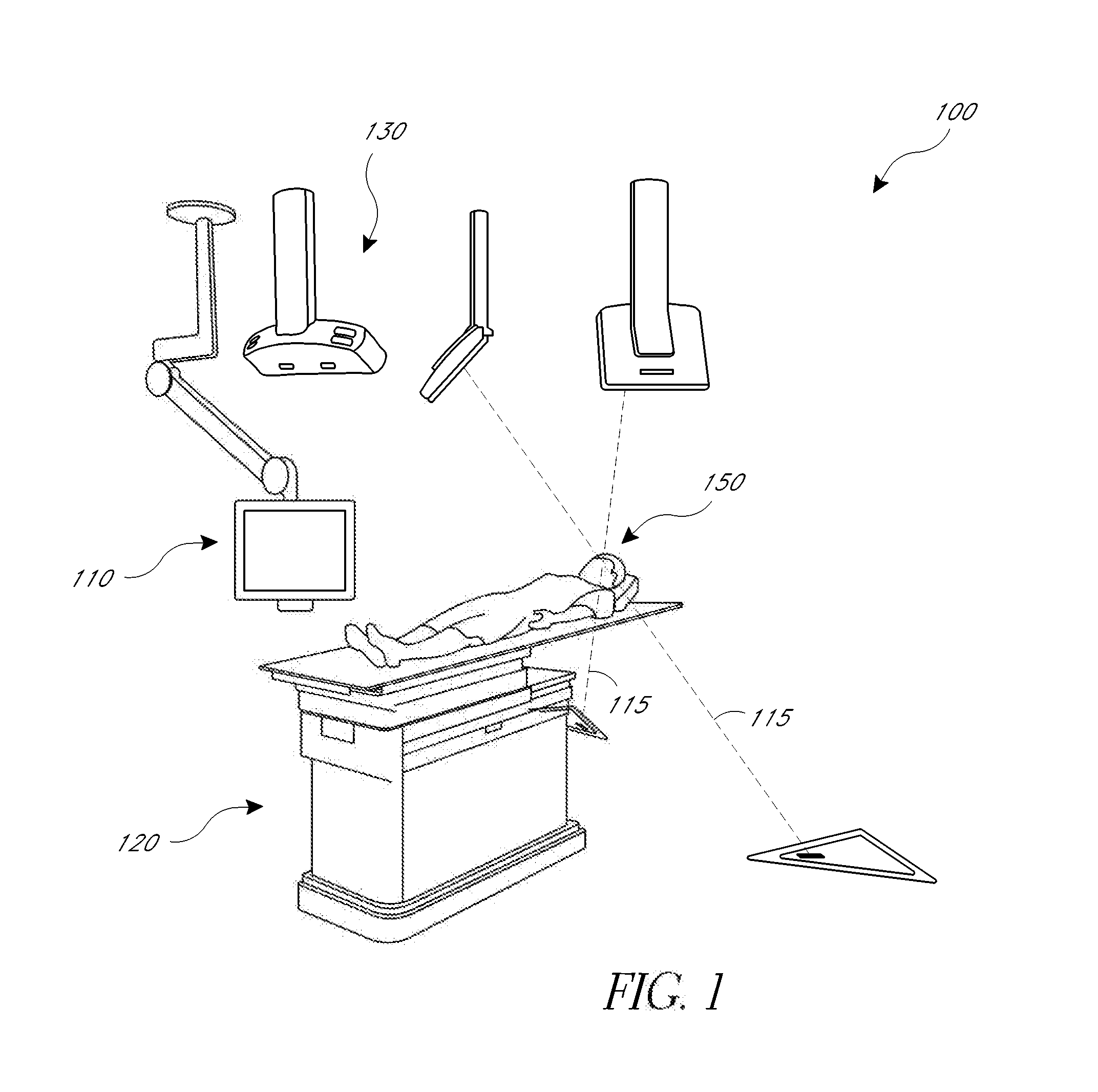
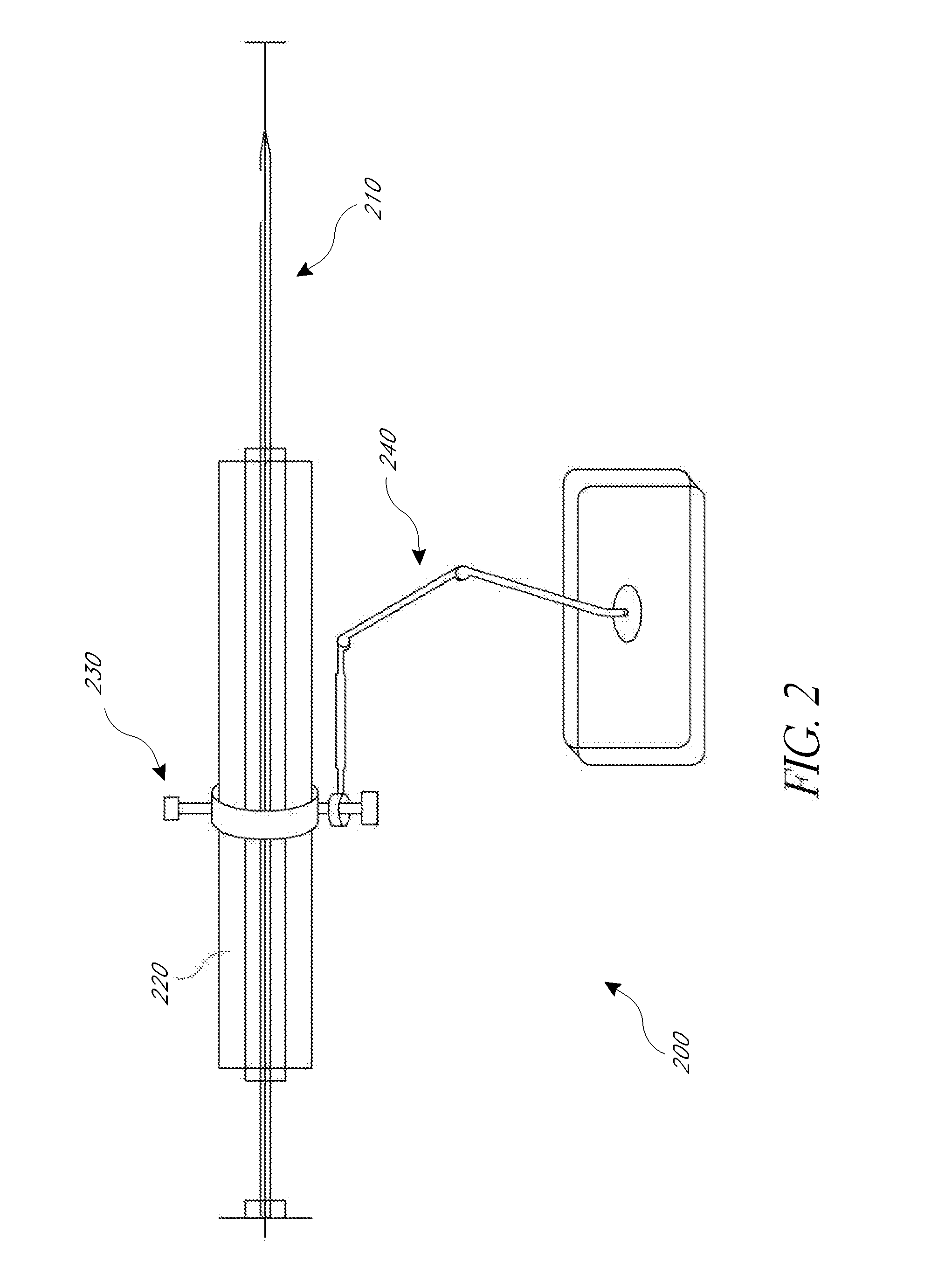
[0010]The present application provides for improved biopsy retrieval, drug delivery, and other treatments. According to one embodiment of the present application, a frameless image-guided biopsy system utilizes stereoscopic imaging (CT and / or MRI imaging can be used in some embodiments), a 6-Dimentional (6D) Robotic couch system along with infrared cameras, an optical distance indicator, laser guidance for treatment, and a needle positioning device. The depth of the target at any point is preferably determined using the optical distance indicator (ODI) installed on an isocentric C-arm. The accuracy of needle placement to the target can preferably be achieved to within 1 mm. Some treatments may include a technique for frameless stereotactic radiosurgery, as described herein.
[0011]According to one aspect of the application a method of performing a frameless image-guided biopsy uses stereoscopic imaging, a six-dimensional robotic couch system along with infrared cameras, a laser guidan...
PUM
 Abstract
Description
Claims
Application Information