Device for determining metastasis of cancer to sentinel lymph node

a technology for lymph nodes and metastases, which is applied in the field of devices for determining the presence or absence of cancer cell metastasis in sentinel lymph nodes, can solve the problems of low accuracy of determination, insufficient basis, and decreased qol of patients receiving axillary lymph node dissection, etc., and achieves low invasiveness and high patient comfort.

Inactive Publication Date: 2015-02-05
SBI PHARMA CO LTD
View PDF6 Cites 7 Cited by
  • Summary
  • Abstract
  • Description
  • Claims
  • Application Information

AI Technical Summary

Benefits of technology

[0017]According to the device of the present invention, the presence or absence of cancer cell metastasis can be confirmed in a sentinel lymph node around which fat is thickly accumulated without performing biopsy or extirpation; PDT can be performed in that state, if necessary; and a sign

Problems solved by technology

However, the dissection of the lymph node cannot completely prevent recurrence when metastasis to other organs has already occurred, and for early breast cancer, steps to remove the lymph node actually free of metastasis are performed and have often resulted in decreased QOL of a patient receiving the axillary lymph node dissection, such as the patient's trouble with a secondary disease, for example, lymphatic edema.
Metastasis can be determined in a case where extra-nodal invasion, the same pleomorphic calcification and the like in the node as those in the main lesion are observed; however, the accuracy of the determination is known to be not high for the present because the frequency of the case is extremely low and the sensitivity thereof is

Method used


Image

  • Device for determining metastasis of cancer to sentinel lymph node
  • Device for determining metastasis of cancer to sentinel lymph node
  • Device for determining metastasis of cancer to sentinel lymph node

Examples


first embodiment

Device I (1)

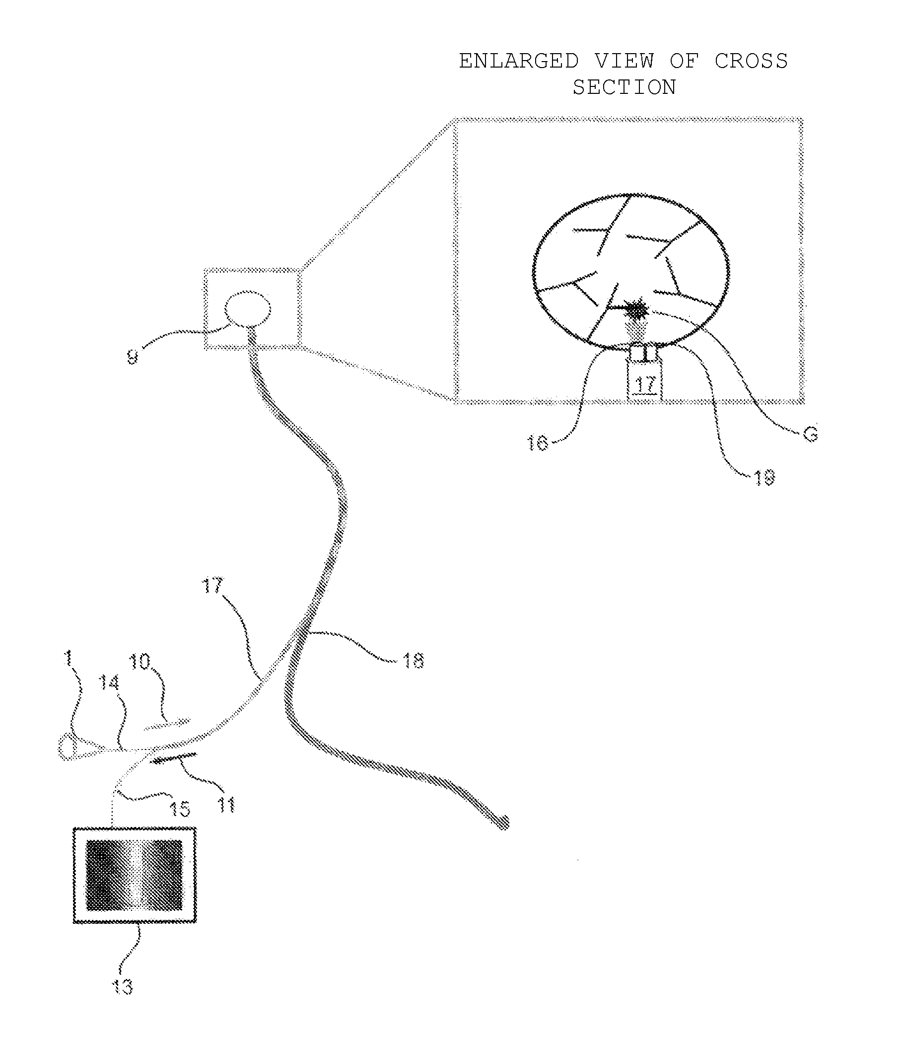
[0068]As shown in FIGS. 1 and 2, a hollow tube 17 into which a small-diameter optical fiber for launching light 14 and a small-diameter fiber for receiving light 15 are inserted passes through an afferent lymph vessel 18, and the small-diameter optical fiber for launching light 14 can guide excitation light 10 output from a light source section 1 to a launching end 16 to launch the excitation light from the launching end 16 to a sentinel lymph node 9. Reflected light, autofluorescence, and fluorescence from a photosensitizing substance 11 from cancer metastasis G in the sentinel lymph node 9 generated by irradiation with the excitation light are received by a light-receiving end 19 of the small-diameter optical fiber for receiving light 15 and enter the measuring section through a spectroscope 13 on which an excitation light cut filter is mounted.

second embodiment

Device II

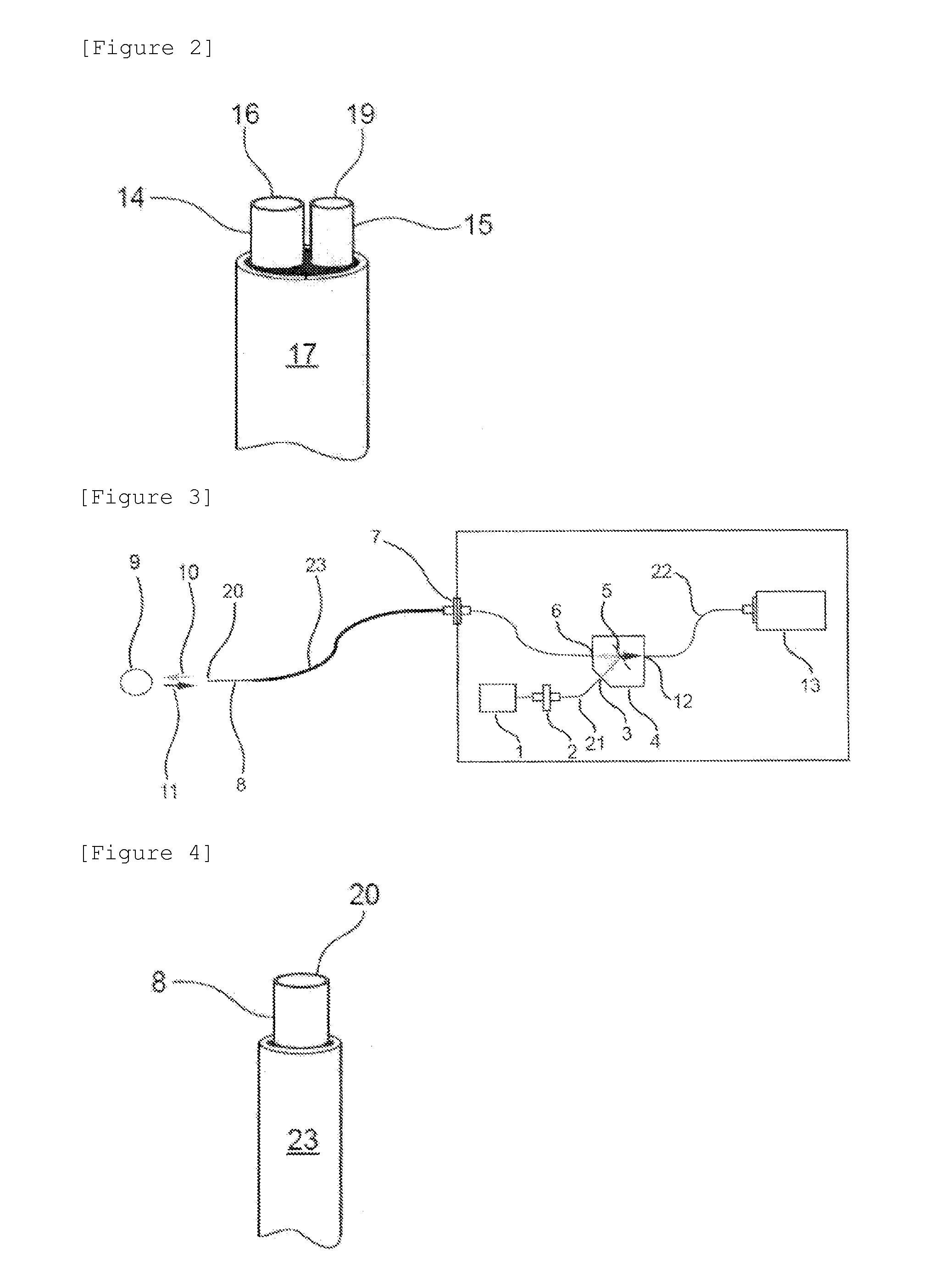
[0069]As shown in FIGS. 3 and 4, the excitation light output from the light source section 1 is optically connected to the light-output end of an optical fiber for outputting light 21 through a connection adaptor (1) 2. When the excitation light arrives at a light mixing / separating 3-port module 4 through a reflection port 3, the excitation light is reflected by a band reflection / transmission mirror 5; the reflected excitation light is guided to a small-diameter optical fiber for launching-cum-receiving light 8 inserted into a hollow narrow tube 23 passing through the lymph vessel through a common port 6 and a connection adaptor (2) 7; and the excitation light 10 is launched from a light-launching-cum-receiving end 20 towards the sentinel lymph node 9. The light 11 generated at the sentinel lymph node is received by the light-launching-cum-receiving end 20 and guided to the light mixing / separating 3-port module 4; the excitation light is reflected by the band reflection / tra...


PUM


Abstract

Provided is a device capable of determining whether or not cancer has metastasized to a sentinel lymph node, such as the axillary lymph node, thickly covered with fat, safely and in a simple manner without confirmation by biopsy or extirpation. Fluorescence generated in a sentinel lymph node upon the excitation of protoporphyrin (a photosensitizing substance) derived from 5-ALA administered to a subject can be detected by passing a hollow tube, into which a small-diameter optical fiber for launching light and the small-diameter optical fiber for receiving light are inserted, through the lymph vessel to identify the presence or absence of the metastasis of cancer cells even before the surgery of cancer or during prognosis.

Description

TECHNICAL FIELD[0001]The present invention relates to a device for determining the presence or absence of cancer cell metastasis in a sentinel lymph node, and more specifically to a device for detecting / identifying cancer metastasis to a sentinel lymph node by detecting fluorescence occurring when a photosensitizing substance, such as protoporphyrin IX (hereinafter also referred to as “PpIX”), accumulated in the sentinel lymph node of a subject is excited.[0002]Cancer (malignant tumor) is the biggest cause of death in Japan, and is said to be experienced by 1 of 2 Japanese people. Cancer is also highly ranked as the cause of death in other developed countries. Thus, it is an earnest desire for people around the world, including Japanese people, to develop an effective therapy for cancer. Reportedly, breast cancer is currently top of all cancers experienced by females and has an incidence of 1 in 23 females.[0003]Examples of currently performed therapeutic methods for cancer can incl...

Claims


Application Information

Patent Timeline
IPC IPC(8): A61B5/00
CPCA61B5/418A61B5/0084A61B5/0071A61B1/00165A61B1/043A61B10/0041F04C2270/041G01N2021/6484
Inventor INOUE, KATSUSHITANAKA, TOHRUISHII, TAKUYANAKAJIMA, MOTOWO
Owner SBI PHARMA CO LTD
Why Eureka
Try Eureka