
Method and kit for detection of Anti-beta amyloid antibodies

Inactive Publication Date: 2015-04-02
UNIV DEGLI STUDI DI MILANO BICOCCA
View PDF1 Cites 10 Cited by
  • Summary
  • Abstract
  • Description
  • Claims
  • Application Information

AI Technical Summary

Benefits of technology

The patent describes a method for detecting the concentration of antibodies in a sample using magnetic micro beads. By concentrating on the micro beads, the sensitivity of the detection process is increased. This allows for the quantitative detection of low levels of antibodies in cerebrospinal fluid, which can help to monitor the effectiveness of immunotherapy treatments for Alzheimer's disease and prevent the onset of symptoms. The method can also be used as a diagnostic tool to detect the presence of antibodies in patients who may be eligible for treatment. The invention provides a more sensitive and specific tool for diagnostic, prognostic, and teragnostic purposes compared to individual biomarkers.

Problems solved by technology

Due to the impossibility of monitoring antibody production in the patient by this strategy, besides the fact that the beta amyloid protein is ubiquitously distributed in the body and highly dishomogeneous from one patient to another in terms of deposition and circulating levels, these clinical trials were discontinued in phase 1 or 2, due to occurrence of episodes of acute meningoencephalitis.
In fact despite the greater safety of these therapeutic strategies some collateral risks do persist, among which the most relevant one is the development of vasogenic edema (VE).
However, these techniques are very costly, not yet fully validated and acknowledged in clinical routine, require highly qualified staff and specific instruments, are not indicative of a real biological response and therefore are used only in exceptional cases and accompanied by a chemical-physical investigation.
Moreover, it has to be pointed out that these techniques show nothing with regard to the concentration of antibodies spontaneously present in the CSF (autoantibodies), or to how much they have increased following the treatment or, again, to their connection with cerebro-vascular impairment.
To date, however, net evidences indicating the exact distribution of the plasma antibody titre in AD subjects compared to healthy population remain few and hazy.
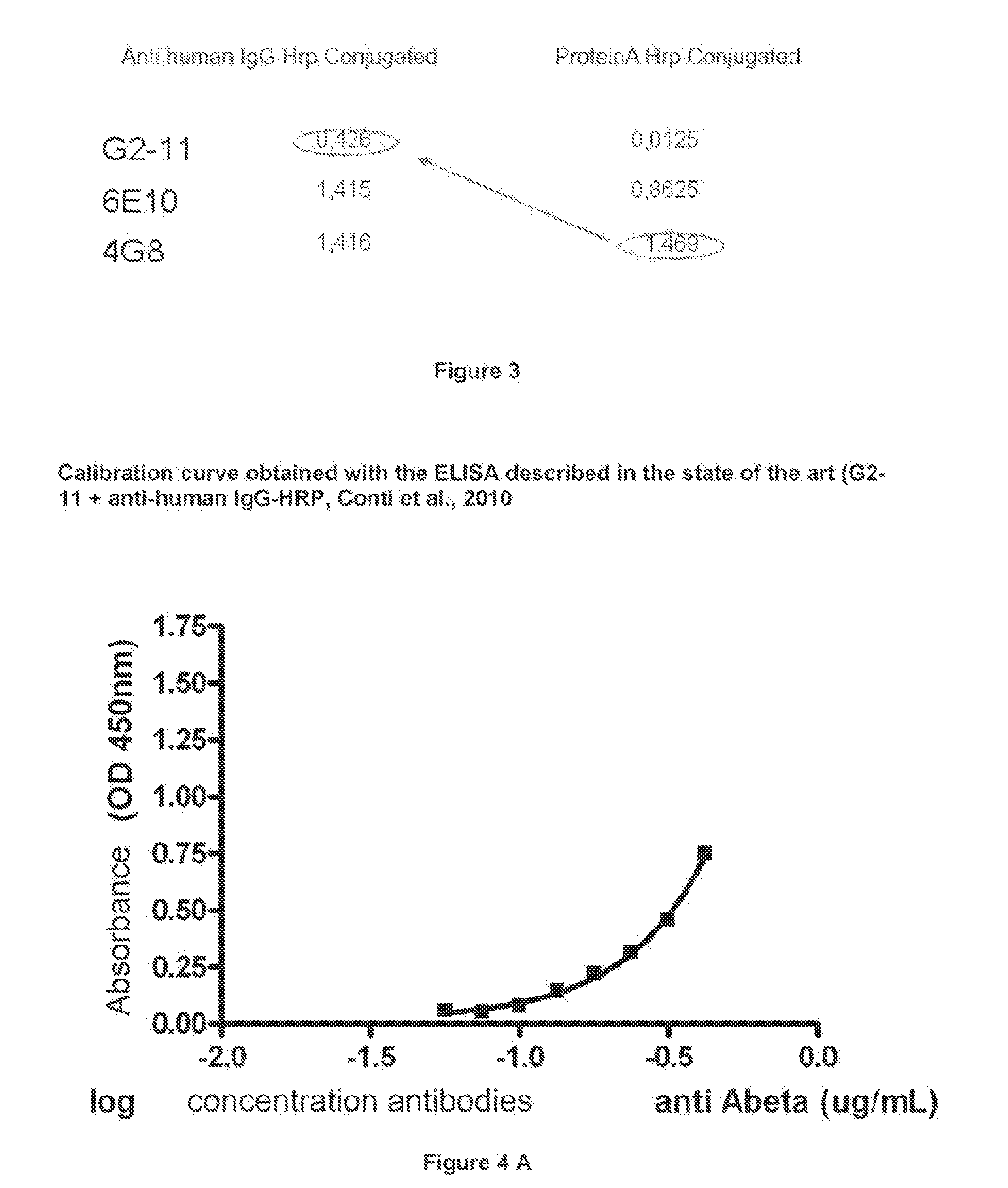
In fact, despite in plasma the antibody titre is sufficiently high to be dosed relatively straightforwardly, the method described by Du Y et al is nearly useless for dosage in the CSF, in which autoantibodies are present at an extremely low concentration, and therefore non-dosable with sufficient sensitivity and reliability by the ELISA methodology used by Du Y et al.
As for the dosage of antibodies anti-beta amyloid protein in CSF samples, the main problem remains therefore the very low concentration of autoantibodies present, values so .low that it is demonstrable that all are encompassed between zero, i.e. blank, and the first point of the calibration curve standard, i.e. too easily mistaken for background noise.
However, due to the technical-experimental difficulty in dosing autoantibodies, present in a very low concentration at CSF level, it is not yet known exactly whether AD subjects actually exhibit a decreased antibody titre prior to therapy and, above all, it is not known how many therapeutic antibodies actually succeed at passing the blood-brain barrier and therefore at having a therapeutic effect in the cerebral district, or, again, which be their concentration trend during therapeutic administration, or even more importantly, which be their concentration during the appearance of the radiological abnormalities (ARIA).
Hence, the problem of providing a method and a kit for dosing with a high sensitivity the concentration of antibodies anti-beta amyloid protein present in CSF samples is highly felt.

Method used


Image

  • Method and kit for detection of Anti-beta amyloid antibodies
  • Method and kit for detection of Anti-beta amyloid antibodies
  • Method and kit for detection of Anti-beta amyloid antibodies

Examples


example 1

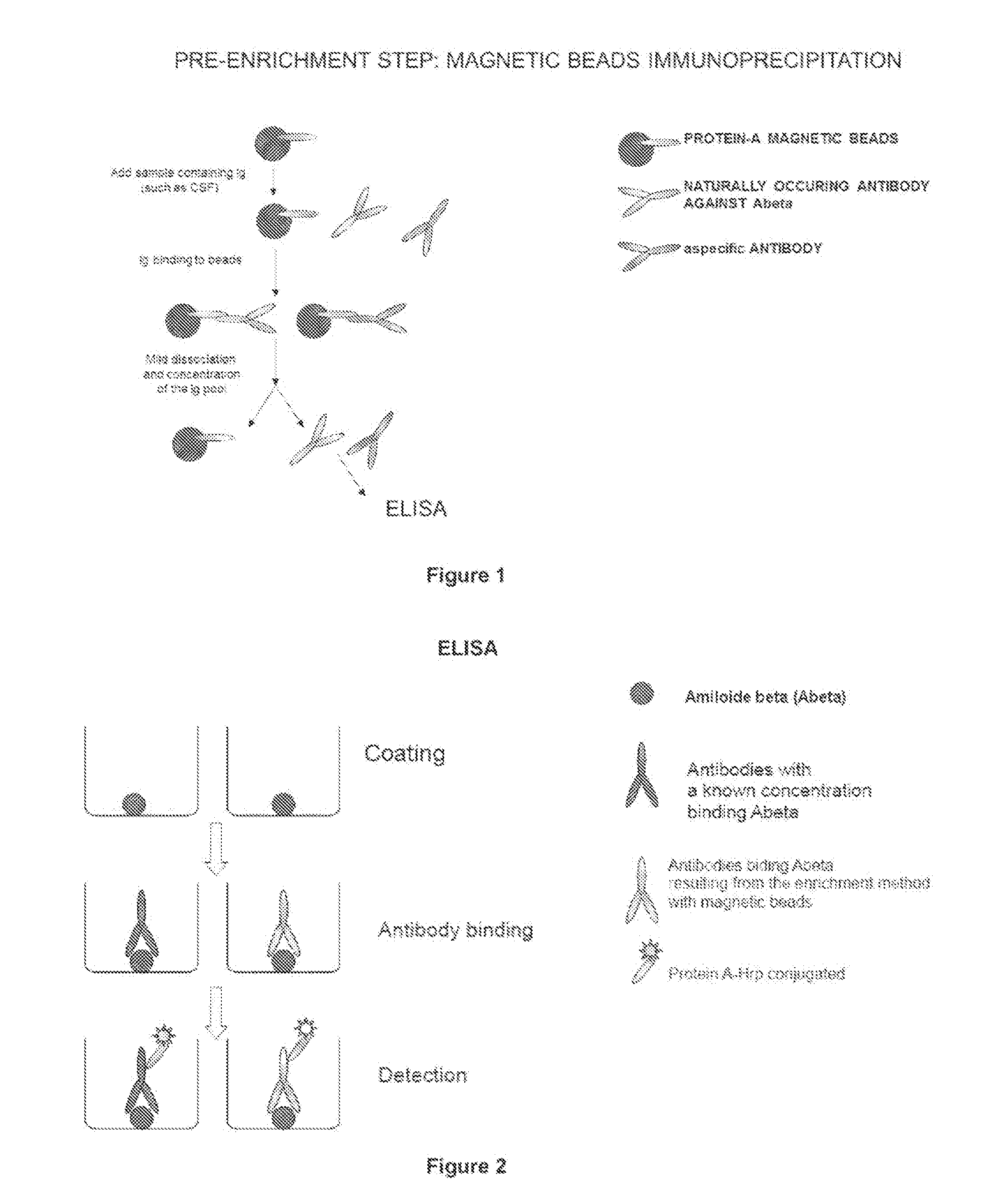
Procedure for Concentrating with Magnetic Micro Beads

[0156]1) Wash twice with PBS. pH 7.4, +0.02% Tween, 75 μl of magnetic micro beads conjugated to protein A (Invitrogen Dynal AS Dynabeads® protein G) for each sample to be analysed;

[0157]2) add to the washed magnetic micro beads a sample volume comprised between 200 and 500 μl of CSF from a patient and incubate under stirring for 40 min. at room temperature;

[0158]3) place the magnetic micro beads in the magnet for 2 min.; optionally store supernatant in another test tube (it will act as control for process efficiency);

[0159]4) remove from magnet and wash with 500 μl of citrate-phosphate buffer, pH 5+0.02% Tween, repeat twice;

[0160]5) then proceed with the elution phase, by adding 30 μl of 0.1 M citrate at pH 3.1 to the magnetic micro beads;

[0161]6) stir for 2 min;

[0162]7) place the tube in the magnet for 1 min and immediately transfer supernatant in a clean test tube (Eppeodorf polypropylene low affinity tube) containing 40 μl of 1...

example 2

Coating the Microplate with Abeta42 Protein

[0165]1) dissolve Abeta42 protein in 1M Tris Base buffer, pH 9, to a concentration of 1 μg / μl;

[0166]2) prepare a coating solution of Abeta in 50 mM NaHCO3, pH 9.6, to a concentration of 0.01 μg / μl;

[0167]3) sonicate for 1 min on ice, wait 1 min, sonicate again for 1 min;

[0168]4) add 100 μl of a coating solution per ELISA plate well (end concentration: 1 μg / μl);

[0169]5) incubate overnight

example 3

Immunoenzymatic Dosage

[0170]1) on the day following the coating performed as described in example 2, carry out 3 washings with PBS+0.05% Tween (PBST), 200 μl / well;

[0171]2) perform blocking to exclude a specific binding, by using a solution comprised of 5% serum and 1%BSA in PBST, 1.5 h at room temperature under oscillation;

[0172]3) repeat, the washing step described at 1);

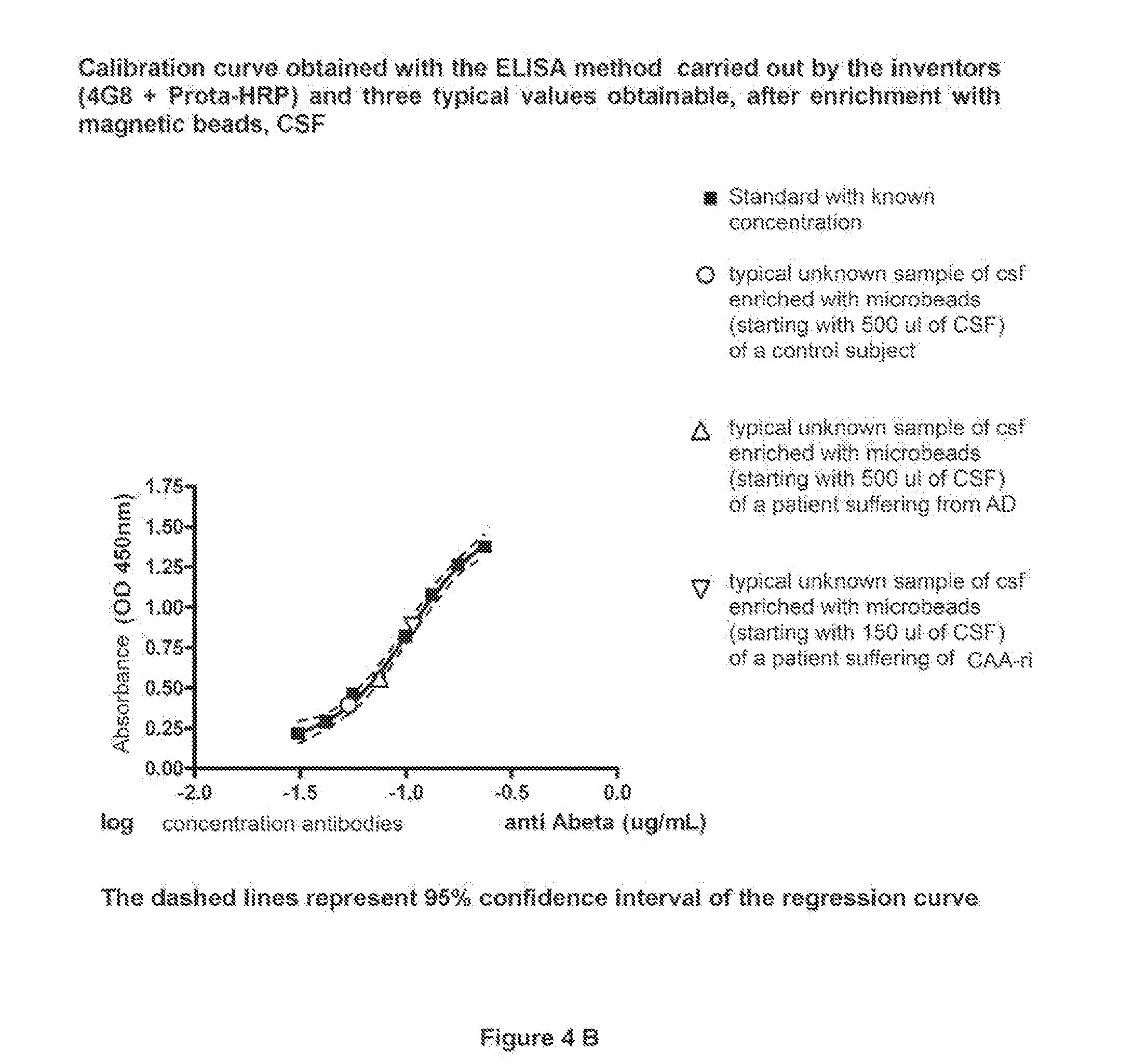
[0173]4) add 100 μl of the solution containing the primary antibody 4G8 (directed toward the amino acid portion 17-24 of the Abeta protein), to construct the standard curve. Standards are carded out by serial dilutions of the primary antibody. The range suggested for the standard curve for antibodies dosage in the CSF is of from 0.24 to 0.03 μg / ml. In walls to be used as blank, add only 100 μl of PBS. Moreover, a further blank for eluates is provided, comprised of 60 μl of 0.1 M citrate, pH 3.1, plus 40 μl of 1M Tris Base, pH 9.

[0174]5) add 100 μl of the sample of CSF as concentrated in Example 1 into the wells. Le...


PUM

PropertyMeasurementUnit
Densityaaaaaaaaaa
Densityaaaaaaaaaa
Densityaaaaaaaaaa

Abstract

An in vitro method and a kit for the quantification of antibodies anti-beta amyloid protein in a sample of cerebrospinal fluid comprising the following steps a) concentrating the quantity of said antibodies of said sample with magnetic micro beads coated with macromolecules capable of binding said antibodies b) analysing the concentrated sample obtained in step a) by immunoenzyme assay or by radioimmunoassay; c) analysing a sample comprising antibodies anti-beta amyloid protein having a known titre with the same assay used in step b) and elaborating the related calibration curve, wherein said antibody having a known titre is a murine antibody belonging to the IgG2a or IgG2b class or a human or humanized antibody belonging to the IgG2b or IgGi class.

Description

[0001]The present description relates to an in vitro method and a kit for the determination of antibodies anti-beta amyloid protein and of endothelial damage (by dosage of biomarker Tissue Factor Pathway Inhibitor, (TFPI) in samples of cerebrospinal fluid.STATE OF THE PRIOR ART[0002]Cerebral and cerebro-vascular accumulates of a 4-kDa peptide termed beta-Amiloyd (Abeta) or beta amyloid protein, is widely accepted as the key neuropathological event in Alzheimer disease (AD). AD main features are in fact amyloid plaques, mainly formed by Abate aggregates (Abeta40 and Abeta42), and Abeta deposition at vessel level, also referred to as Cerebral Amyloid Angiopathy, deriving by proteolysis of the amyloid precursor protein (APP) by beta- and gamma-secretase.[0003]Various mechanisms seem to be accountable for the formation of these accumulations, among which a malfunctioning of toxic aggregate clearance systems or a malfunctioning of cell defence systems at cerebral level. Therapeutic appro...

Claims


Application Information

Patent Timeline
IPC IPC(8): G01N33/564G01N33/68
CPCG01N33/564G01N33/6896G01N2446/00G01N2800/2821G01N2333/4704C07K16/18C07K2317/34G01N2333/4709G01N2800/2871G01N2800/52G01N33/54326
Inventor PIAZZA, FABRIZIOFERRARESE, CARLO
Owner UNIV DEGLI STUDI DI MILANO BICOCCA
Learn More