Preparing biological samples for analysis

a biological sample and analysis method technology, applied in the field of biological sample preparation, can solve the problems of many analyses missing the presence, difficult to detect and identify, and difficult to impede sample degradation, so as to facilitate effective heating, shorten the time needed, and uniform and rapid heating

Inactive Publication Date: 2015-07-16
DENATOR
View PDF0 Cites 0 Cited by
  • Summary
  • Abstract
  • Description
  • Claims
  • Application Information

AI Technical Summary

Benefits of technology

[0016]The present invention comprises a method for preparing a biological sample for analysis. The biological sample comprises at least one protein or polypeptide having an amino acid sequence, from an organism. The method comprises causing a volume of the sample to adopt a shape wherein the shape permits uniform and rapid heating, thereby forming a shaped sample; and heating the shaped sample so that the secondary structure of the macromolecule is disrupted, but the primary structure is not. In an embodiment, the biological sample is given a shape that facilitates an effective heating in terms of the heating being uniform and fast. This helps to shorten the time needed to reach a disrupted secondary structure. By blocking certain biological processes driven by proteins, degradation of other constituents of the sample is avoided. Because the time between taking the biological sample and performing a biological analysis has a large impact on the level of degradation, even after a short time, e.g., after as little as 1-3 minutes, it is important that heating takes place immediately after taking the sample. By heating the tissue proteins that function as proteases, their secondary and tertiary structure, and thereby their function, is lost.

Problems solved by technology

Sample degradation, however, is both hard to impede, and hard to detect.
The result is that many analyses miss the presence of species that have degraded long before the analysis is carried out; correspondingly, such analyses may in fact identify degradation products of critical components in place of the original components.
Despite their ubiquity, proteins are extremely sensitive to their environments and thus are not always easy to detect and to identify because they can degrade very quickly.
Once an organism dies, or once a sample of tissue is extracted from a-living organism, the regulatory balance of the organism or in the sample is lost and key proteins start to break down.
For example, some proteins whose natural role is to digest other proteins (a “proteolytic” function), and whose natural levels are kept in check while an organism is alive, may go out of control after death.
However, the study of tissue samples from patients or model organisms usually exposes the samples to a certain period of oxygen and nutrient depletion before homogenization and protease inactivation occurs.
Use of aldehyde solutions is problematic because it doesn't arrest natural degradation of proteins (though it is somewhat effective at maintaining large-scale structure of tissues).
Microwave irradiation is problematic because it is generally non-uniform, that is, some parts of the sample reach a temperature that is high enough to cause sample breakdown.
Furthermore, microwave irradiation has formerly been applied to living (non-human) subjects as part of a sacrificial protocol and thus has yet to be established as a tool for analyzing samples that have been extracted from subjects, both human and non-human.

Method used


Image

  • Preparing biological samples for analysis
  • Preparing biological samples for analysis
  • Preparing biological samples for analysis

Examples


example 1

Analysis of Mouse Brain Samples

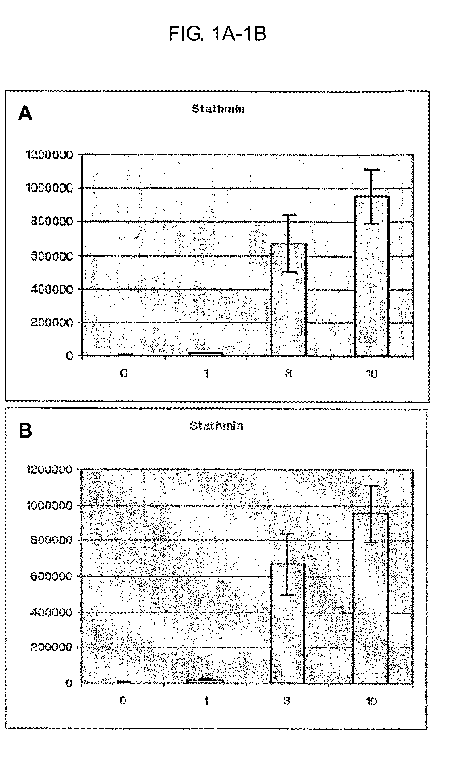
[0090]Twelve mice (C57 / BL6) were euthanized by cervical dislocation. The twelve mice were divided into three groups and each mouse brain was kept at room temperature (22° C.) for 1, 3, or 10 minutes. At their respective time point, the mouse brains were irradiated in a small animal microwave for 1.4 seconds at 4.5-5 kW. A fourth group was euthanized by focused microwave irradiation (the control group). The striatum, hypothalamus and cortex were thereafter rapidly dissected out of all of the mice after microwave radiation and stored at −80° C.

[0091]An additional group of four mice were also euthanized by cervical dislocation and the mouse heads were immediately cooled in liquid nitrogen. The striatum was rapidly dissected out on dry ice and frozen at −80° C. The frozen samples were shaped into thin slices. Half of the group of samples were then immediately heated to near 100° C. using a contact heating device. The other half of the samples were thawed a...


PUM

PropertyMeasurementUnit
timeaaaaaaaaaa
thickaaaaaaaaaa
threshold distanceaaaaaaaaaa

Abstract

Methods and devices for preparing a biological sample for analysis are described. The biological sample from an organism has at least macromolecule having a primary structure that naturally degrades after the sample is removed from the organism. The method includes causing the biological sample to adopt a shape to permit rapid and uniform heating. The shaped sample is then rapidly and uniformly heated, thereby altering a secondary structure of the macromolecule while preserving its primary structure.

Description

CROSS-REFERENCE TO RELATED APPLICATIONS[0001]This is a continuation of U.S. National Phase application Ser. No. 11 / 996,545 filed 27 Aug. 2006; which claims the benefit of priority to International Application No. PCT / SE2006 / 000979 filed 27 Aug. 2006; which is a continuation-in-part of U.S. application Ser. No. 11 / 212,454, filed 26 Aug. 2005; each of which is incorporated herein by reference in its entirety.STATEMENT REGARDING FEDERALLY SPONSORED RESEARCH OR DEVELOPMENT[0002]Not Applicable.INCORPORATION-BY-REFERENCE OF MATERIAL SUBMITTED IN COMPUTER READABLE FORMAT (CRF)[0003]Not Applicable.FIELD OF THE INVENTION[0004]The present invention relates to methods and apparatus for preparing a biological sample for analysis. The invention more particularly relates to methods and apparatus for heating a biological sample soon after it is extracted so that the primary structures of proteins in it are not degraded.BACKGROUND OF THE INVENTION[0005]It is key to any analysis of a biological samp...

Claims


Application Information

Patent Timeline
Patent Type & Authority Applications(United States)
IPC IPC(8): G01N1/44G01N33/68G01N1/28
CPCG01N1/44G01N33/68G01N1/286B01L7/00B01L2300/0832B01L2300/1805B01L2300/1838B01L2300/1866B01L2300/1883Y10T436/143333Y10T436/25B01J2219/00051C07K14/435
Inventor SKOLD, KARLSVENSSON, MARCUSPALMERS, GORANANDREN, PER
Owner DENATOR
Why Eureka
Try Eureka