Methods and systems for detecting biological components

a biological component and detection method technology, applied in the field of biological component detection methods and systems, can solve the problems of difficult detection of ctcs, hampered efforts to count ctcs, and complicated or prevent accurate identification

Active Publication Date: 2015-08-20
RGT UNIV OF CALIFORNIA
View PDF3 Cites 53 Cited by
  • Summary
  • Abstract
  • Description
  • Claims
  • Application Information

AI Technical Summary

Problems solved by technology

The number and diversity of such components in a biological sample often complicates or prevents the accurate identification and / or quantification of specific components of interest within the sample, which would enable the diagnosis or monitoring of a condition in the subject, such as cancer.
Efforts to count CTCs have been hampered by the fact that CTCs are extremely difficult to detect: they are exceptionally rare, and may be difficult to distinguish from healthy cells.
However, such approaches have limitations in sensitivity and / or specificity, leading to many healthy cells being mischaracterized as cancerous, and many cancer cells being missed in the analysis.
Further, microdroplets may or may not be stabilized by surfactants and / or particles.

Method used


Image

  • Methods and systems for detecting biological components
  • Methods and systems for detecting biological components
  • Methods and systems for detecting biological components

Examples


example 1

Microfluidic System for Performing Single-Cell PCR Reactions

[0454]Device Manufacturing:

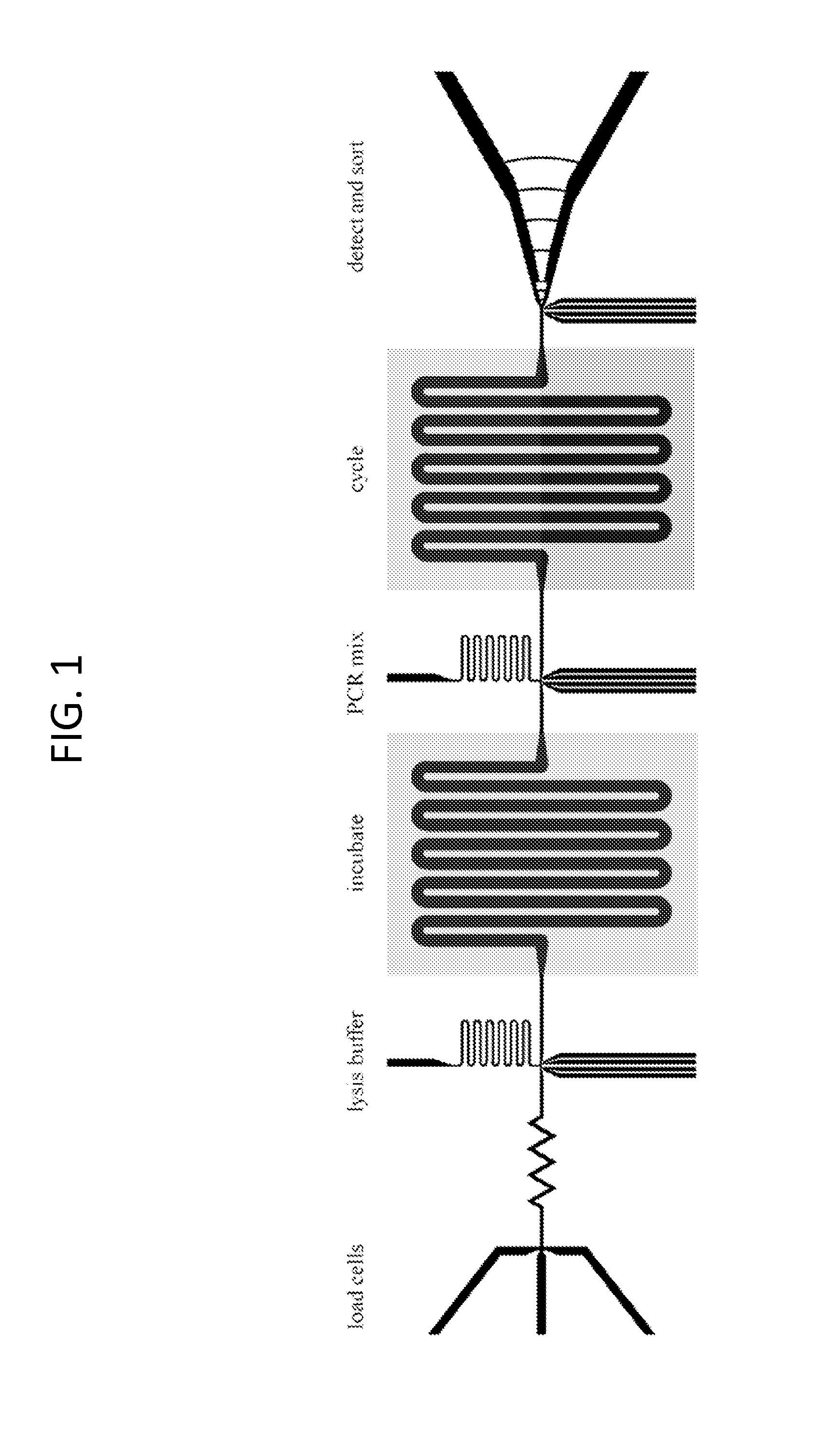
[0455]The chips were made using the same photolithographic processes in polydimethylsiloxane as the other devices described above. A general schematic of the chips is shown in FIG. 1. The general approach carried out by such chips is depicted in FIG. 6.

[0456]Sample Preparation:

[0457]5-25 mL whole blood samples were extracted from a subject via syringe. Nucleated cells were separated using on-chip pinched-flow fractionation, as generally described in Lab on a Chip, 2005, 5, 778-784; the disclosure of which is incorporated herein by reference. Nucleated cells were collected for subsequent analysis.

[0458]PCR Reactions:

[0459]The assay requires the execution of an RT-PCR reaction in drops containing concentrated cell lysates; however, cell lysates inhibit RT-PCR (FIG. 7). To overcome this inhibition, a protocol has been developed that utilizes proteinase K to digest inhibitory proteins in cell lysates....

example 2

Quantitative Multiplexed Assay

[0462]To screen more than one gene simultaneously, a multiplexed qPCR reaction may be utilized. Reactions were initially performed in bulk with PCR tubes to optimize reaction conditions. Using these methods, successful multiplexing was achieved during digital droplet RT-PCR for three TaqMan® probes, EpCAM, CD44 and CD45. An example of this multiplexing is shown in FIG. 11, where EpCAM and CD44 probes were multiplexed in drops containing both target transcripts. All PCR primer sets were designed to span large introns, making these larger genomic PCR products highly unlikely in multiplex reactions. Additionally, all TaqMan® probes are designed to hybridize to exon-exon junctions. The current probe sets do not recognize gDNA.

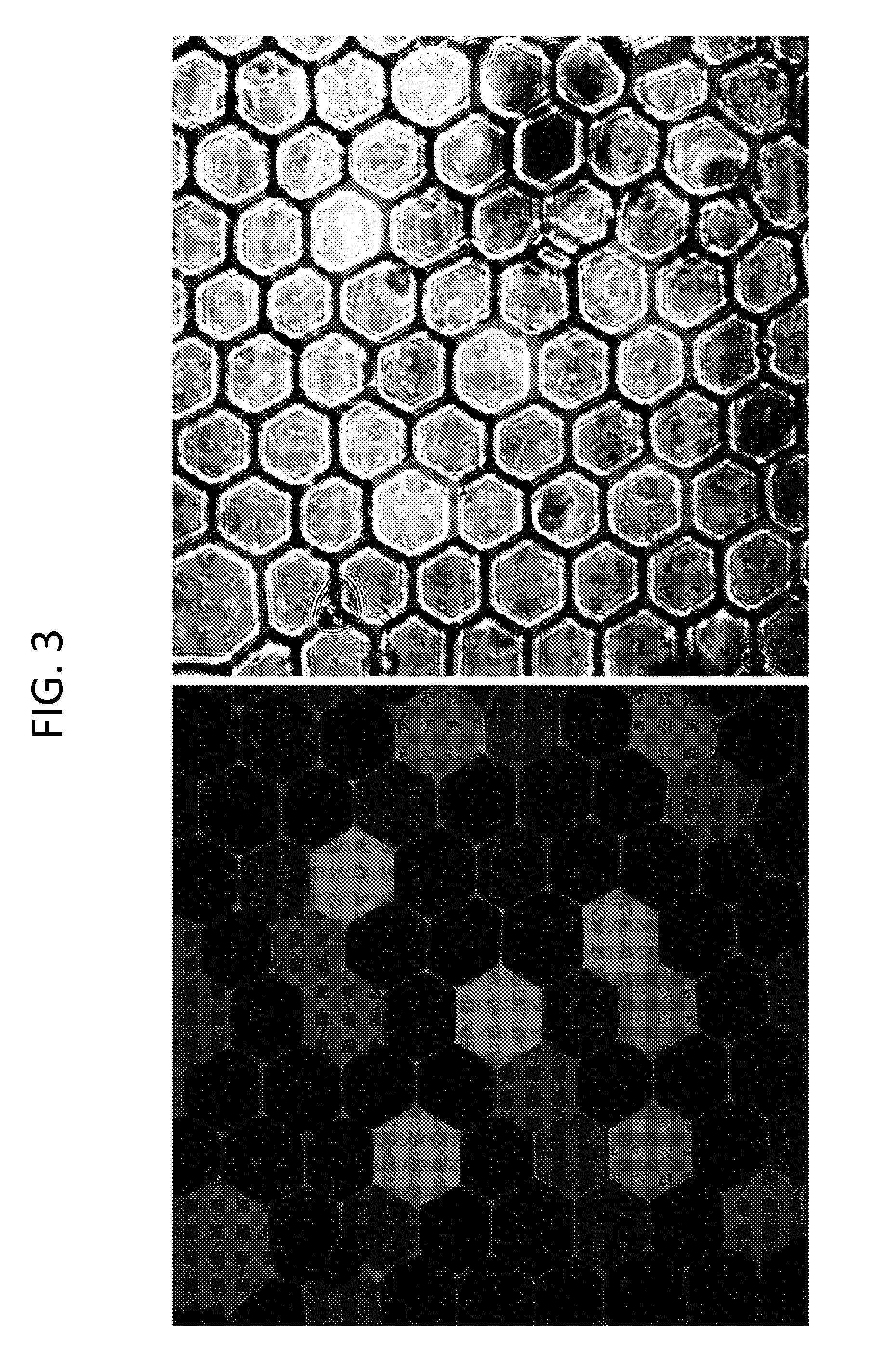
[0463]Single-Cell qPCR with Megadroplet Arrays:

[0464]To perform qPCR analysis on single cells, the drops are imaged as they are thermal cycled. This requires that the drops be held at fixed positions during thermal cycling so they can ...

example 3

Electrode-Free Picoinjection of Drops of Microfluidic Drops

[0467]Microfluidic devices were fabricated in poly(dimethylsiloxane) (PDMS) using soft photolithographic techniques. The devices had channel heights of 30 μm, optimal for the picoinjection of water-in-oil droplets that are 50 μm in diameter. The device design is similar to those described previously by Abate, et al. Proc. Natl. Acad. Sci. U.S.A., 2010, 107, 19163; the disclosure of which is incorporated herein by reference. An important difference, however, is that the channels for the metal solder electrodes are removed. Further, a “Faraday Mote”—an empty channel filled with a conducting aqueous solution—is implemented that runs between the injection site and the droplet spacer, as shown in FIG. 15, Panel B. The mote electrically isolates re-injected drops upstream of the picoinjection site from electric fields emanating from the picoinjector, preventing unintended merging. The emulsion that was picoinjected consists of mon...


PUM

PropertyMeasurementUnit
lengthaaaaaaaaaa
widthaaaaaaaaaa
heightaaaaaaaaaa

Abstract

Methods for the detection of components from biological samples are provided. In certain aspects, the methods may be used to detect and / or quantify specific components in a biological sample, such as tumor cells (e.g., circulating tumor cells). Systems and devices for practicing the subject methods are also provided.

Description

CROSS-REFERENCE TO RELATED APPLICATIONS[0001]This application claims priority to U.S. Provisional Application No. 61 / 682,707, filed Aug. 13, 2012; and to U.S. Provisional Application No. 61 / 784,754, filed Mar. 14, 2013; which applications are incorporated by reference herein in their entireties and for all purposes.INTRODUCTION[0002]Biological samples from a subject often contain a large number of different components. For example, a sample of a subject's blood may contain free floating DNA and RNA, circulating cells, and many other components. The number and diversity of such components in a biological sample often complicates or prevents the accurate identification and / or quantification of specific components of interest within the sample, which would enable the diagnosis or monitoring of a condition in the subject, such as cancer.[0003]For instance, circulating tumor cells (CTCs) are cells shed from tumors that enter into a subject's blood stream. Once in the blood, these cells c...

Claims


Application Information

Patent Timeline
Patent Type & Authority Applications(United States)
IPC IPC(8): C12Q1/68
CPCC12Q1/6886C12Q2600/118C12Q2600/16C12N15/1096C12P19/34C12Q1/686F04B13/00C12Q1/6806B01L3/502784B01L7/52B01L2200/0652B01L2300/0816B01L2300/0864B01L2300/0867B01L2300/0883B01L2300/1822B01L2400/0415B01L2400/0487B01F23/41B01F25/4335B01F33/3011B01F33/3031C12Q2563/159C12Q2537/143C12Q2565/629C12Q2600/158C12Q1/6844
Inventor ABATE, ADAM R.EASTBUM, DENNIS JAYSCIAMBI, ADAM R.
Owner RGT UNIV OF CALIFORNIA
Why Eureka
Try Eureka