Mechanically rotating intravascular ultrasound probe

a technology of ultrasound probe and rotor, which is applied in the field of medical devices, can solve the problems of increasing 1-2 mm2 ultrasonic dead band easily occurring around the catheter, and low image resolution, and achieves accurate control of the rotational speed of the rotor, and reduce the volume of the probe

Active Publication Date: 2015-10-22
SHENZHEN UNIV
View PDF4 Cites 11 Cited by
  • Summary
  • Abstract
  • Description
  • Claims
  • Application Information

AI Technical Summary

Benefits of technology

[0020]Compared with the prior art, in the mechanically rotating intravascular ultrasound probe provided by the present invention, a narrow-neck micro motor is designed to be located in the catheter, which may achieve an outside diameter of the catheter between 1.5 mm and 2 mm, further reduce the volume of the probe, and solve the problem of rotation distortion of an image when the catheter passes through a lesion with high-grade stenosis or a curved blood vessel section. Additionally, by adjusting an angle of the ultrasonic transducer, the present invention achieves forward scanning imaging and side scanning imaging for a blood vessel wall. The design of the present invention is ingenious in that the ionic liquid is used as an electrical liquid brush to achieve electrical connection of the ultrasonic transducer during rotation. As the ionic liquid has a characteristic of acoustic impedance close to that of a biological tissue of a human body, it not only acts as the acoustic coupling agent, but also achieves a function of electrical conduction, thus enabling a top electrode of the ultrasonic transducer during rotation to be effectively grounded, and obtaining an electrical shielding effect. The rotor of the ultrasound probe drives the high-resolution high-frequency ultrasonic transducer to implement forward-looking conical scanning imaging for the blood vessel wall, and also utilizes the rotational speed sensor to measure the rotational position and the rotational speed of the rotor in real time, thus, in combination with a closed-loop control system, achieving accurate control over the rotational speed of the rotor as well as scanning synchronization with an imaging system.

Problems solved by technology

However, there are disadvantages such as a lower image resolution and a 1-2 mm2 ultrasonic dead band easily occurring around the catheter.
Although using more array elements may improve the imaging resolution, in the meantime, it will increase the volume of the probe, thus severely affecting application thereof as the intravascular probe.
The type of mechanical sector probe, compared with the electronically scanned array probe, is advantageous in a higher imaging resolution, but the greatest disadvantage thereof is, when the catheter passes through a lesion with high-grade stenosis or a blood vessel section being curved, a friction may occur between a main shaft of the probe which is conducting rotary scanning and an inner cavity of the catheter to a great extent, thus obstructing free rotation of the catheter and causing rotation distortion to the image.
In addition, existing commercialized IVUS catheters may only help the doctors see an image of the blood vessel wall on a side of the ultrasound catheter, but fail to present an image of the blood vessel in front end thereof, such that their use is much restricted in Chronic Total Occlusion (CTO) lesions.

Method used


Image

  • Mechanically rotating intravascular ultrasound probe
  • Mechanically rotating intravascular ultrasound probe

Examples


Embodiment Construction

[0025]The present invention will be further illustrated in detail below with reference to the accompanying drawings and specific embodiments. The following embodiments are only described for explaining the present invention, but the present invention is not limited to the following embodiments.

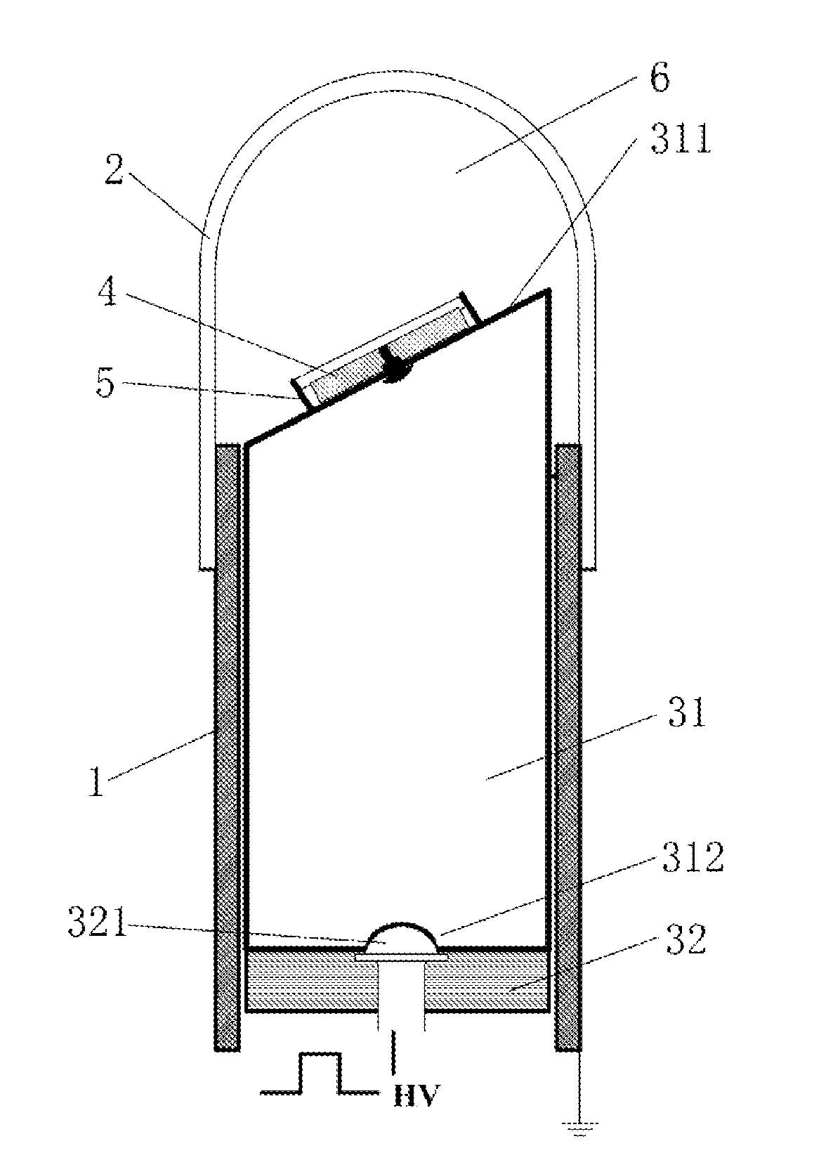
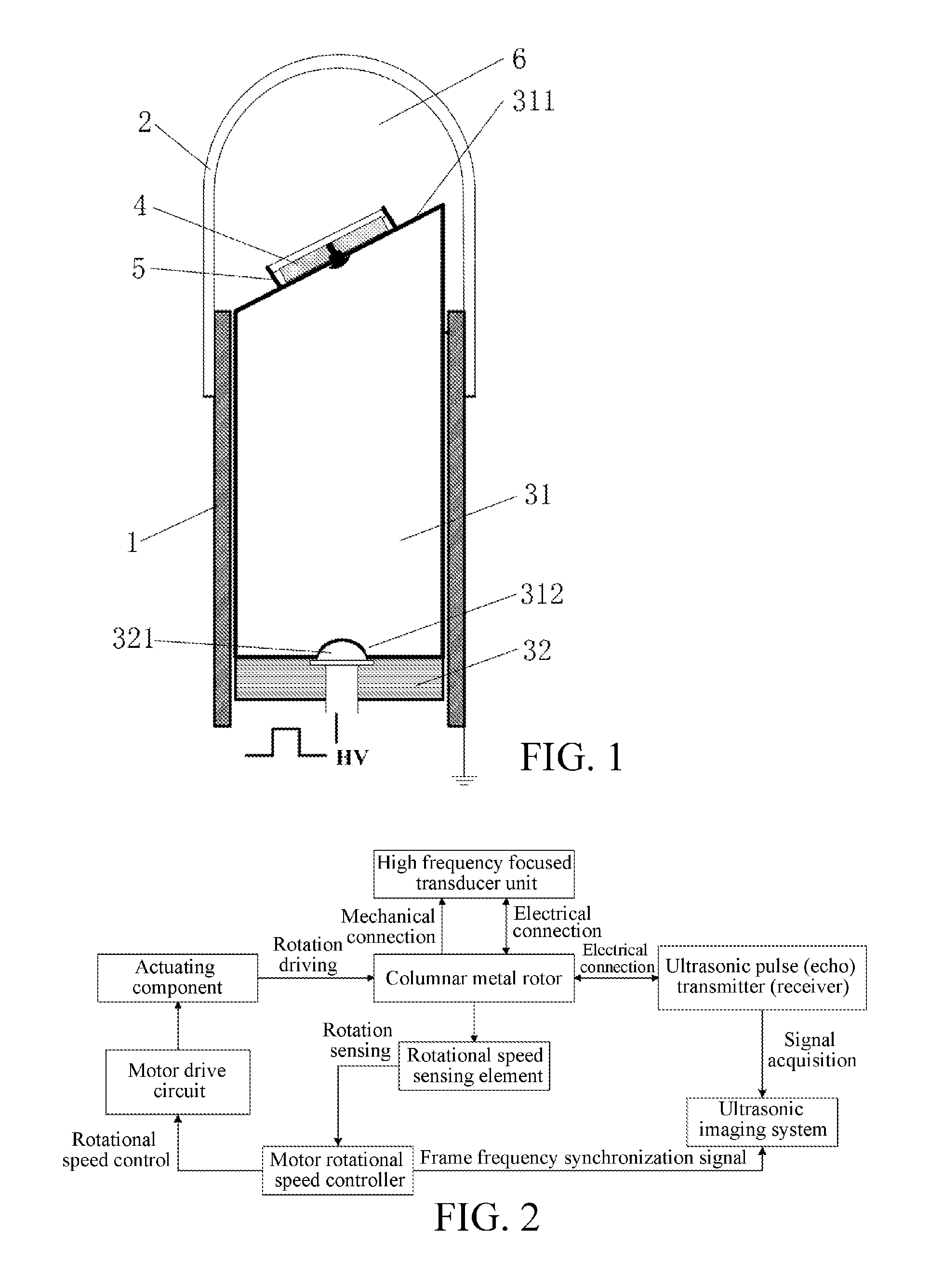
[0026]FIG. 1 is a structural diagram of a mechanically rotating intravascular ultrasound probe according to one embodiment of the present invention. In this embodiment, the intravascular ultrasound probe comprises a catheter 1, an acoustic window 2, a micro motor 3 and a high-frequency ultrasonic transducer 4, where the catheter 1 is a magnetic metal tube with a diameter between 1.5 mm and 2 mm, and an exterior wall thereof is coated with a biologically compatible material; the acoustic window 2, which has a spherical tip, and allows ultrasonic waves to pass through, is installed in an front end of the catheter 1 to enclose the front end of the catheter 1; the micro motor 3 is installed in a c...


PUM


Abstract

A mechanically rotating intravascular ultrasound probe relates to the technical field of medical devices, and aims to provide a forward-looking mechanically rotating intravascular ultrasound probe having a small volume, a high image resolution and good imaging stability. The intravascular ultrasound probe includes a catheter, an ultrasonic transducer disposed at a front end of a cavity of the catheter and a driving apparatus that drives the ultrasonic transducer to rotate mechanically. The driving apparatus is a micro motor disposed in the cavity of the catheter, including a rotor and a stator. The ultrasonic transducer is installed on top of the rotor and electrically connected to the rotor, and the rotor is also electrically connected to the stator; the catheter is a magnetic metal tube, and a front end thereof is enclosed by an acoustic window which has a spherical tip and allows ultrasonic waves of the ultrasonic transducer to pass through; the acoustic window is filled with an ionic liquid having a function of a ultrasonic coupling agent. The ultrasound probe solves a problem of rotation torsion of an image when the catheter passes through a lesion with high-grade stenosis or a curved blood vessel section, and achieves forward scanning imaging and side scanning imaging for a blood vessel wall.

Description

BACKGROUND[0001]1. Technical Field[0002]The present invention relates to the technical field of medical devices, and in particular to the technical field of intravascular ultrasound imaging devices used for interventional diagnosis and treatment.[0003]2. Related Art[0004]Intravascular ultrasound tomography (IVUS) is a novel diagnostic method combining a non-invasive ultrasonic diagnosis technology and a minimally invasive technology of interventional catheterization. As IVUS may accurately present a complex 3D anatomical structure of a blood vessel wall in real time, in addition to evaluating stenosis of lumen, it may further detect vulnerability of an atherosclerotic plaque and load of the plaque. Therefore, in the percutaneous coronary intervention treatment, IVUS has gradually replaced coronary angiography (CAG) that is originally deemed as a “gold standard” for diagnosis and treatment of a coronary heart disease, and becomes a novel diagnostic method widely applied clinically. T...

Claims


Application Information

Patent Timeline
Patent Type & Authority Applications(United States)
IPC IPC(8): A61B8/00A61B8/12
CPCA61B8/4461A61B8/12A61B8/4494A61B8/4281A61B8/445A61B8/56G01S15/894
Inventor PENG, JUECHEN, SIPINGWANG, TIANFU
Owner SHENZHEN UNIV
Why Eureka
Try Eureka