Plumeria acuminata extracts and methods of use
- Summary
- Abstract
- Description
- Claims
- Application Information
AI Technical Summary
Benefits of technology
Problems solved by technology
Method used
Image
Examples
example 1
Preparation of Candidate Substance
[0121]Extraction Protocol
[0122]Plumeria acuminata flower components were gathered and then chopped. The chopped flower components were then ground and a 50% ethanol solution was added to the chopped flower components. The solution was filtered and the filtrate vacuum evaporated to yield a concentrated extract of approximately 600 ml, which was then diluted with pure water. The diluted extract was vacuum evaporated, then again diluted with pure water. The diluted extract was left to stand at 4 C for 12 hours, then centrifuged and filtered. 3× extraction with hexane followed this filtration step, with the hexane layer and aqueous layers subsequently being separated. Charcoal was added to the aqueous layer, and the charcoal / aqueous layer mixture was then centrifuged and filtered. The filtered extract was then extracted 3× with water-saturated n-butanol, with the butanolic (upper) and aqueous layers subsequently separated. The butanolic layer was pooled...
example 2
HPLC of Candidate Substance
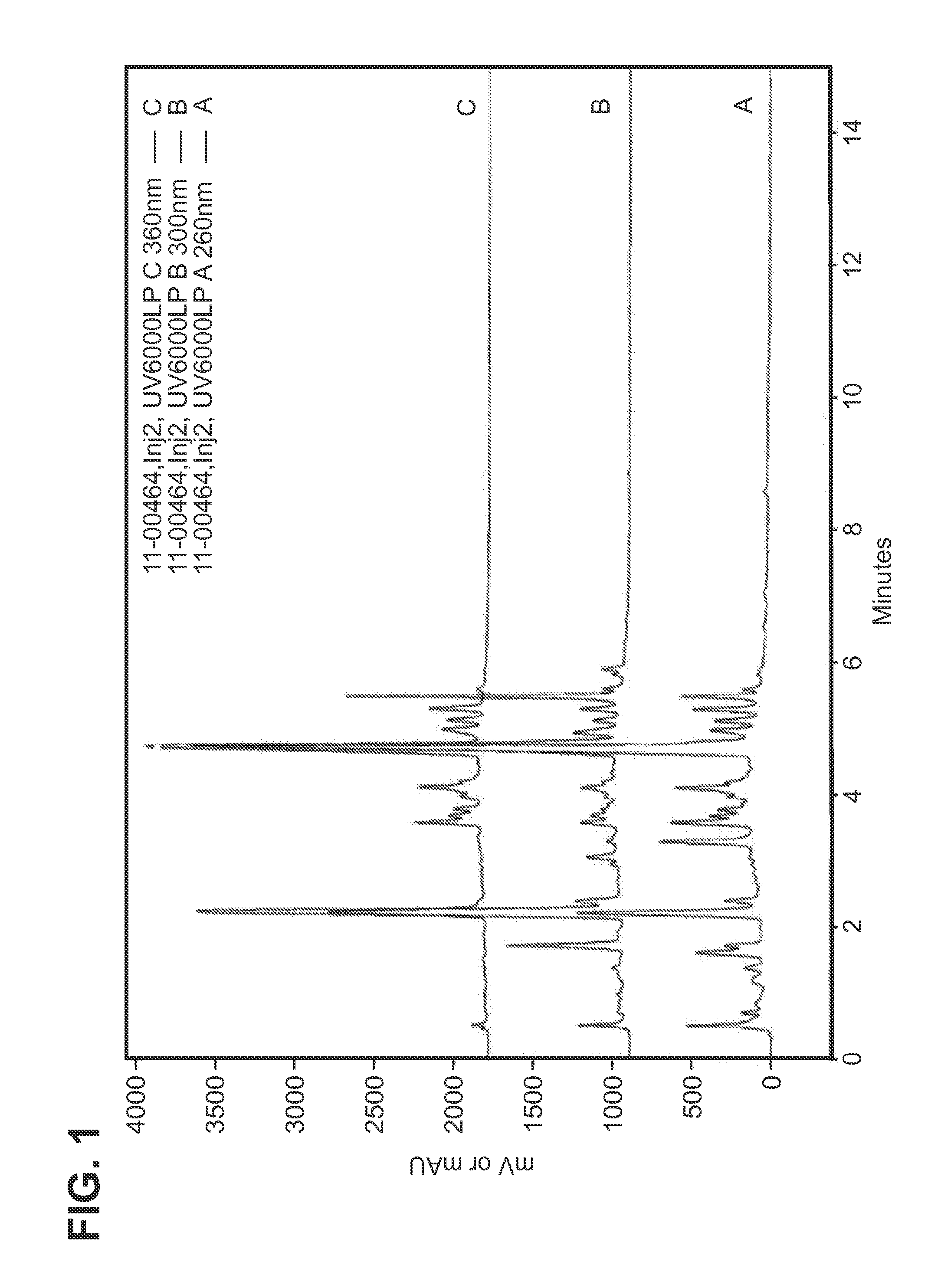
[0124]Extracts were generally characterized by high performance liquid chromatography. A sample size of approximately 5 mg / mL was dispersed in 25 / 75 MeOH / H2O and sonicated. The characterization was performed on a Zorbax SBC-18 column (7.5 cm×4.6 mm, 3.5 um particle size) and detection was achieved using diode array UV absorbance, 260 nm 300 nm and 360 nm, with lines on FIG. 1 depicted in ascending order and 260 nm on bottom. Operating conditions were flow rate 1.5 ml / min; temperature, 40° C.; sample injection volume, 20 μL, and time of run, 19 minutes. The mobile phase gradient used was as follows. In one embodiment, the extracted composition of a compound, in substantial isolation, exhibits an HPLC profile substantially similar to that depicted herein in FIG. 1.
example 3
MTT Growth Assay
[0125]Fibroblast (2.0×10³ cells / well), A2058 human melanoma cells and B16F10 mouse melanoma cells (1.0×10³ cells / well) were plated in 96 well plates in 100 μL medium, and incubated before sample treatment at 37° C. for 24 hours. After 24 hours, various concentrations of candidate substances were added in medium (100 μL) and incubated for another 48 hours. The metabolic activity of each well was determined by the MTT assay and related to untreated cells. After removal of 100 μL medium, MTT stock dye solution was added (15 μL / 100 μL medium) to each well, the plate was incubated at 37° C. in 5% CO2 atmosphere. After 4 hours, the solubilization / stop solution 100 μL was added to each well mixed thoroughly to dissolve the dye crystals. The absorbance was measured by using ELISA plate reader at 540 nm with a reference wavelength of 630 nm, and the extract of the present invention had no adverse effect on cell growth as assessed using this protocol.
PUM
 Abstract
Description
Claims
Application Information
 - R&D
- Intellectual Property
- Life Sciences
- Materials
- Tech Scout
- Unparalleled Data Quality
- Higher Quality Content
- 60% Fewer Hallucinations
Browse by: Latest US Patents, China's latest patents, Technical Efficacy Thesaurus, Application Domain, Technology Topic, Popular Technical Reports.
© 2025 PatSnap. All rights reserved.Legal|Privacy policy|Modern Slavery Act Transparency Statement|Sitemap|About US| Contact US: help@patsnap.com

