Nanopipette device and method for subcellular analysis

Active Publication Date: 2016-02-04
RGT UNIV OF CALIFORNIA
View PDF2 Cites 11 Cited by
  • Summary
  • Abstract
  • Description
  • Claims
  • Application Information

AI Technical Summary

Benefits of technology

[0038]In certain aspects of the present invention, the intracellular content from a cell, extracted in native form by the present method, comprises either organelles or nucleic acids. In some aspects, the extracted nucleic acids include genomic DNA, RNA (including mRNA and rRNA from a ribosome), or mitochondrial DNA. In other aspects, the organelle is a mitochondrion. In addition, one may extract portions of a cellular cytoplasm or vesicle containing proteins, small molecules, metabolites of cellular processes, etc.
[0039]In certain aspects, the present invention also pertains to a method for mechanically extracting intracellular content from a cell, comprising (a) prepar

Problems solved by technology

This assumption arose not from experimental evidence but from the lack of tools capable of analyzing individual cells.
The initial capture of the single cell, however, remains a major technical issue (1).
However, these methods either cannot examine cells in their native environment, or as with the original micropipette, they are limited to probing isolated cells (8).
The cylindrical shape of these nanoendoscopes allows the probing of organelles deep inside the cell but these technologies lack any automati

Method used


Image

  • Nanopipette device and method for subcellular analysis
  • Nanopipette device and method for subcellular analysis
  • Nanopipette device and method for subcellular analysis

Examples


example 1

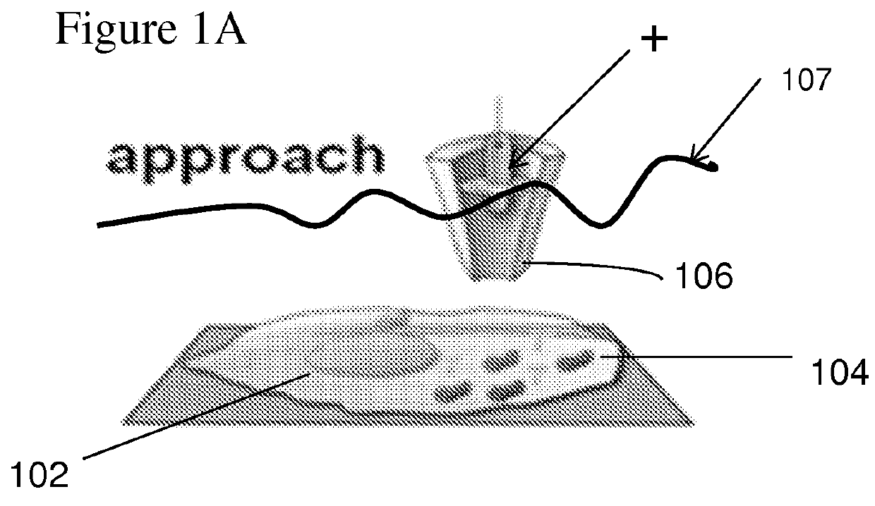
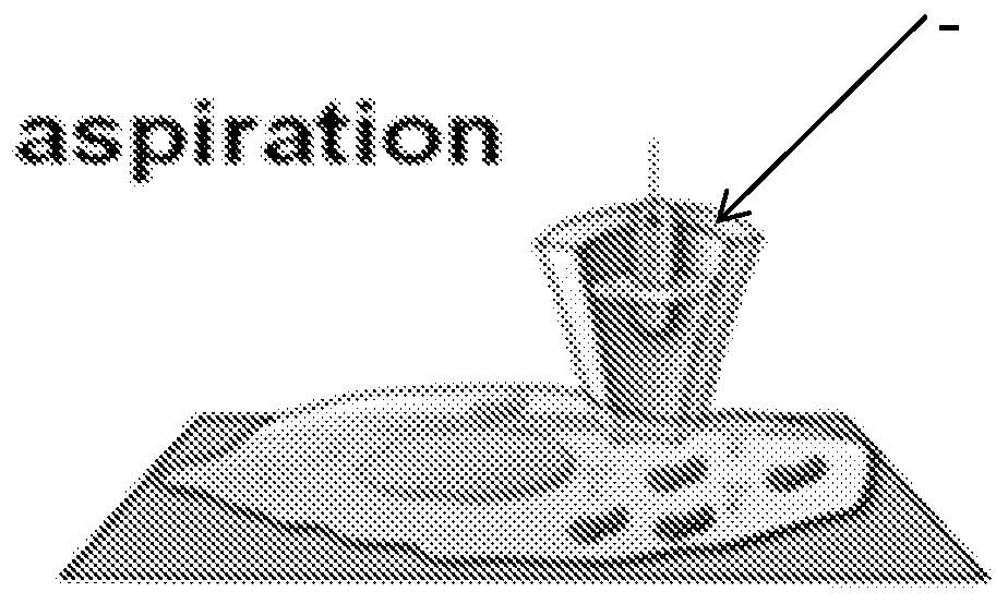
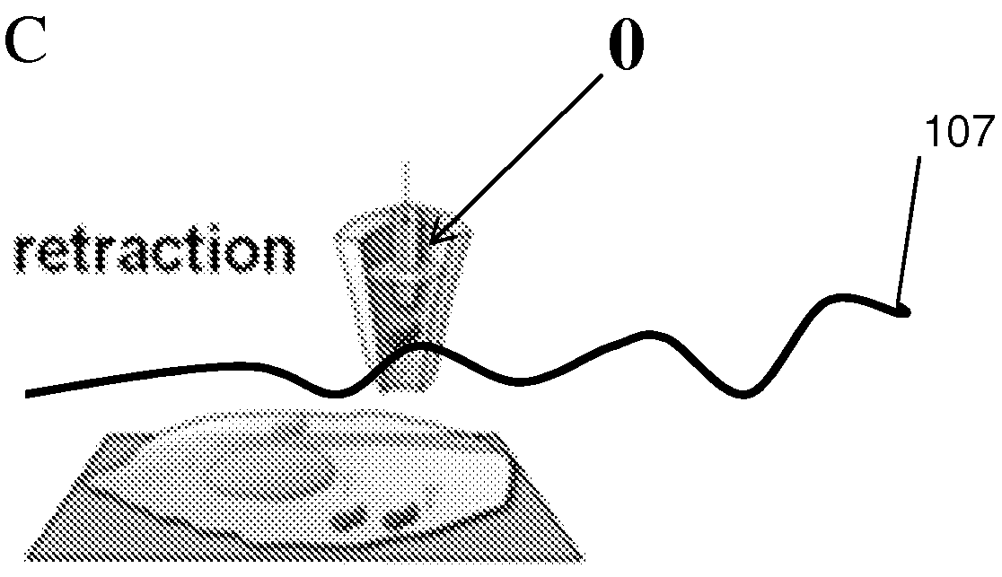
Nanobiopsy Platform Setup

[0120]Quartz nanopipettes with pore diameters of 115±15 nm (images not shown) were fabricated using a conventional laser-puller (P2000, Sutter Instruments) and filled with a solution of 1-2 dichlorethane (DCE) containing 10 mM tetrahexylammonium tetrakis(4-chlorophenyl)borate (THATPBCl).

[0121]To adapt the SICM as a single-cell biopsy platform, the nanopipette was filled with a 10 mM THATPBCl solution in DCE and fitted with a silver wire coated with silver tetrakis(4-chlorophenyl)borate (AgTBACl) in the barrel of the nanopipette. When a DCE-filled nanopipette is immersed into an aqueous solution a liquid-liquid interface is formed at the nanopore lumen due to the hydrophobic nature of DCE. The application of a voltage across this interface induces a change in the DCE surface tension. The application of a voltage across the interface between the hydrophobic liquid in the nanopipette and the electrolyte solution external to the nanopipette induces a change in t...

example 2

Biopsy of Green Fluorescent Protein Transcripts

[0125]We performed biopsies on HeLa cells expressing Green Fluorescent Protein (GFP) and used PCR methods to validate the success of the protocol by targeting selective amplification of GFP transcripts (FIG. 4). We performed cDNA synthesis on the content aspirated from within a single cell using iScript™ cDNA Synthesis Kit (BioRad). This process reverse-transcribed all the RNA present in the biopsy into cDNA. Real time-PCR is then performed on the cDNA to confirm the presence of GFP transcripts. In a real time-PCR experiment the fluorescence remains at background levels (cycles 1-18, FIG. 5) until enough amplified product accumulates to yield a detectable fluorescence signal. The cycle number at which this occurs is called the quantification cycle, or Cq. A difference of X in Cq values of two reactions reflects a 2X difference in amount of starting material.

[0126]Real-time PCR amplification plots show a Cq value of 20 for the positive c...

example 3

Biopsy of Human BJ Fibroblast Mitochondria

[0128]The nanobiopsy platform is by no means limited to RNA extraction from single cells but it can be employed to sampling of cellular organelles as well. Human BJ fibroblasts (skin fibroblast cells, ATCC CRL 2522) were stained using mitotracker (Invitrogen) (data not shown), a fluorescent label for mitochondria. The nanopipette was placed above a region with an abundance of mitochondria, and a biopsy was performed with the same protocol described before (data not shown). Fluorescence within the tip of the nanopipette indicates the successful aspiration of mitochondria (data not shown). PCR analysis of the biopsied mitochondria was used to confirm the presence of mitochondrial DNA. The mitochondrial DNA was amplified using primers specific to the mitochondrial genome, and gel electrophoresis of the amplified mitochondrial DNA shows that all 5 samples taken show bands at the correct length for human mitochondrial DNA (FIG. 8).


PUM

PropertyMeasurementUnit
Fractionaaaaaaaaaa
Timeaaaaaaaaaa
Timeaaaaaaaaaa

Abstract

Described herein are devices and methods for extracting cellular material from living cells and then depositing them into to a receptacle in a nanoliter scale. Using a nanopipette integrated into a scanning ion conductance microscope (SICM), extraction of mitochondrial DNA from human BJ fibroblasts and Green Fluorescent Protein (GFP) transcripts from HeLa/GFP cells was achieved with minimal disruption to the cellular milieu and without chemical treatment prior to obtaining the isolated sample. Success of the extraction was confirmed by fluorescence microscopy and PCR analysis of the extracted material. The method and apparatus may be applied to many different cell types and intracellular targets, allowing not only single cell analysis, but single subcellular compartment analysis of materials extracted in their native state.

Description

CROSS-REFERENCE TO RELATED APPLICATIONS[0001]This application claims priority from U.S. Provisional Patent Application No. 61 / 781,841 filed on Mar. 14, 2013, which is hereby incorporated by reference in its entirety.STATEMENT OF GOVERNMENTAL SUPPORT[0002]This invention was made with Government support under contract HG000205 awarded by the National Institutes of Health, and contract U54CA143803 awarded by the National Cancer Institute. The Government has certain rights in this invention.REFERENCE TO SEQUENCE LISTING, COMPUTER PROGRAM, OR COMPACT DISK[0003]The instant application contains a Sequence Listing which has been submitted as an ASCII text file and is hereby incorporated by reference in its entirety. This text file was created on Feb. 18, 2014, is named “482—35—1_PCT_Seq_List.txt” and is 717 bytes in size.BACKGROUND OF THE INVENTION[0004]1. Field of the Invention[0005]The present invention relates to the field of nanodevices for single cell extraction of intracellular conten...

Claims


Application Information

Patent Timeline
IPC IPC(8): C12N15/10C12Q1/68G01N33/573
CPCC12N15/1003C12Q1/6806C12Q1/686C12Q1/6874C12Q2600/158C12Q1/6876G01N2333/91G01N2440/14G01N33/573C12M47/06G01N2035/1034G01Q60/44
Inventor ACTIS, PAOLOMAALOUF, MICHELLE M.POURMAND, NADER
Owner RGT UNIV OF CALIFORNIA
Why Eureka
Try Eureka