Optical scanning systems for in situ genetic analysis

a technology of optical scanning and applied in the field of optical scanning systems for in situ genetic analysis, can solve the problems of increased resolution at the cost of decreased signal intensity, poor confocality, and often required long exposures, and achieve high confocality, high resolution images of samples, and high s/n ratio

Active Publication Date: 2016-04-21
ILLUMINA INC
View PDF5 Cites 5 Cited by
  • Summary
  • Abstract
  • Description
  • Claims
  • Application Information

AI Technical Summary

Benefits of technology

[0005]Systems and methods for performing fluorescence in situ sequencing are disclosed. Namely, one embodiment provided herein is a confocal time delay and integration (TDI) line scan imaging system that has high S / N ratio and high confocality for producing high resolution images of a sample. In one example, the confocal TDI line scan imaging system comprises various pinhole and / or slit aperture mechanisms in front of the image sensor, wherein the various pinhole and / or slit aperture mechanisms are used to reject out-of-focus light. In another example, the confocal TDI line scan imaging system comprises the various pinhole and / or slit aperture mechanisms in an intermediate image plane conjugating to the image sensor.
[0006]Also provided herein are structures that comprise focus tracking features that can be used to maintain focus during imaging. In one example, provided are various configurations of focusing strips on a substrate in contact with the tissue sample to be imaged. In another example, strips are cut into the tissue sample, thereby providing exposed strips of substrate that can be used as focus tracking features.

Problems solved by technology

However, as much of the light from sample fluorescence is blocked at the pinhole, this increased resolution is at the cost of decreased signal intensity—so long exposures are often required.
A drawback of some photoluminescence-based scanning instruments (or imaging systems) that are used in current fluorescence-based sequencing-by-synthesis (SBS) systems is that they have poor confocality (i.e., are semi-confocal at best).
These semi-confocal imaging systems have a low signal-to-noise (S / N ratio) and therefore are not adequate to eliminate out-of-focus features in specimens.
Further, current dithering focus tracking methods are unable to maintain focus during imaging.

Method used


Image

  • Optical scanning systems for in situ genetic analysis
  • Optical scanning systems for in situ genetic analysis
  • Optical scanning systems for in situ genetic analysis

Examples


Embodiment Construction

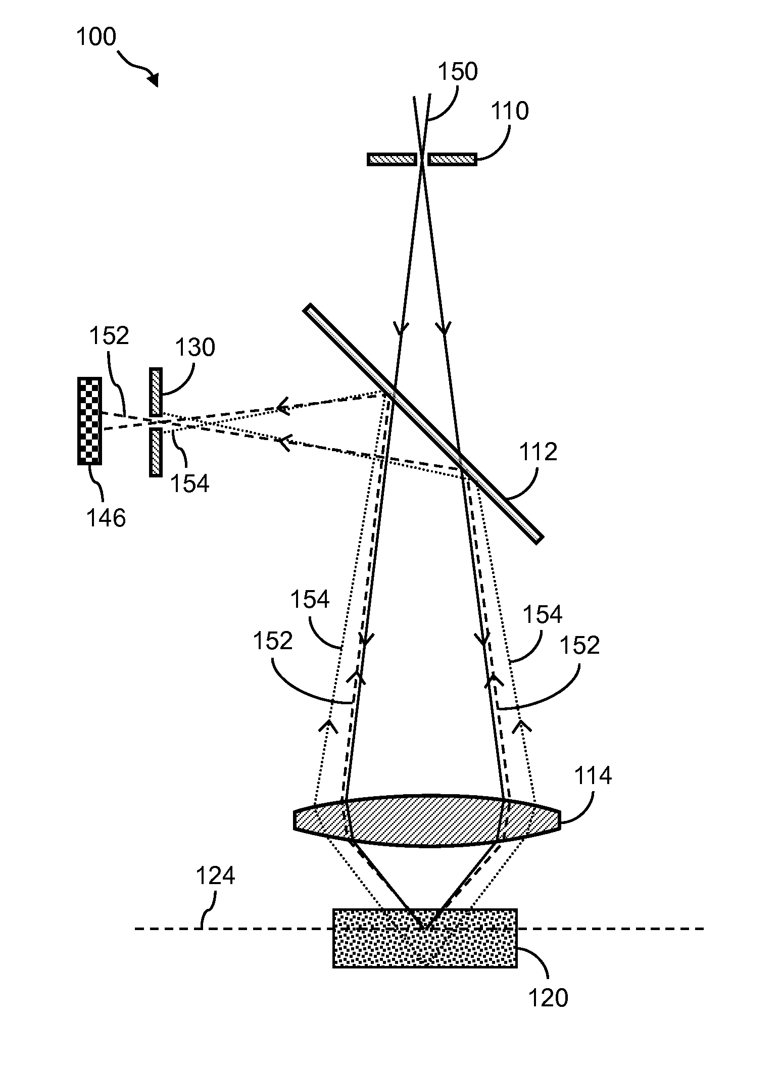
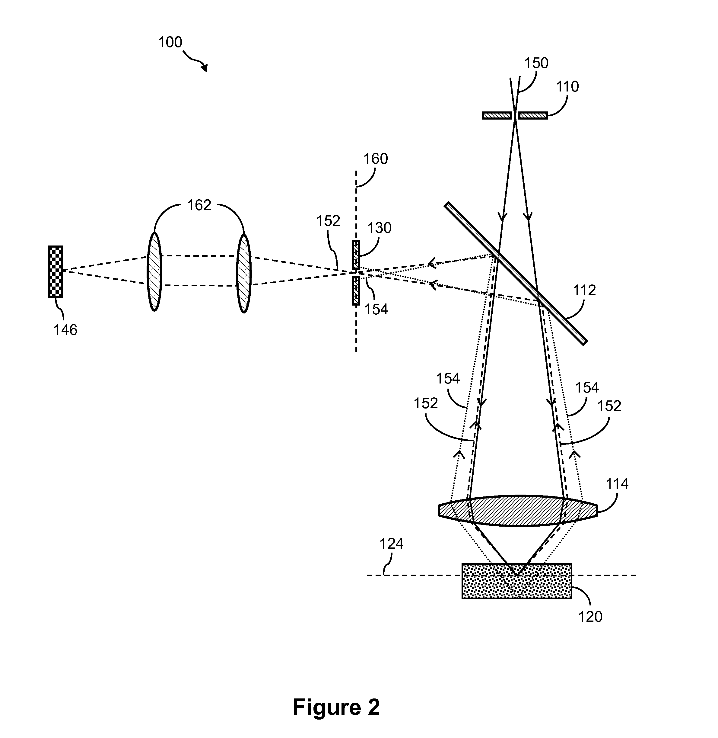
[0028]The detailed description set forth below in connection with the appended drawings is intended as a description of exemplary embodiments of the invention and is not intended to represent the only embodiments in which the invention may be practiced. The term “exemplary” used throughout this description means “serving as an example, instance, or illustration,” and should not necessarily be construed as preferred or advantageous over other exemplary embodiments. The detailed description includes specific details for the purpose of providing a thorough understanding of the exemplary embodiments of the invention. In some instances, some devices are shown in block diagram form.

Sequencing

[0029]Systems and methods described herein can be used in conjunction with a variety of nucleic acid sequencing techniques. These sequencing techniques include, but are not limited to, in situ sequencing techniques for reading sequence information from nucleic acids directly from cells or tissue (Lee,...


PUM

PropertyMeasurementUnit
widthaaaaaaaaaa
widthaaaaaaaaaa
switching frequencyaaaaaaaaaa

Abstract

Presented herein are systems and methods for performing sequencing, including fluorescence in situ sequencing. In one embodiment, a confocal time delay and integration (TDI) line scan imaging system may include various pinhole and/or slit aperture mechanisms in front of the image sensor. The system may also include structures with focusing strips on a substrate in contact with the tissue sample to be imaged. Alternatively, these strips may be cut into the tissue sample. The system may also include configurations and methods of placing a tissue sample inside a reaction chamber of a flow cell during the assembly of the flow cell and then performing chemistry operations on the tissue sample. The flow cells may use an open container for performing chemistry operations on the tissue sample.

Description

CROSS REFERENCE TO RELATED APPLICATIONS[0001]Any and all applications for which a foreign or domestic priority claim is identified in the Application Data Sheet as filed with the present application are hereby incorporated by reference under 37 CFR 1.57.BACKGROUND[0002]In a conventional (i.e., wide-field) fluorescence microscope, the entire specimen is flooded evenly in light from a light source. All parts of the specimen in the optical path are excited at the same time and the resulting fluorescence is detected by the microscope's photodetector or camera including a large unfocused background part. In contrast, a confocal microscope uses point illumination and a pinhole in an optically conjugate plane in front of the detector to eliminate out-of-focus signal. As only light produced by fluorescence very close to the focal plane can be detected, the image's optical resolution, particularly in the sample depth direction, is much better than that of wide-field microscopes. However, as ...

Claims


Application Information

Patent Timeline
Patent Type & Authority Applications(United States)
IPC IPC(8): G02B21/36H04N5/225H04N5/232
CPCG02B21/361G02B21/362H04N5/2254H04N5/2253H04N5/232G02B21/006G02B21/0076G02B21/008H04N23/54H04N23/55C12Q1/6841C12Q2543/101C12Q2563/179C12Q2565/514C12Q2565/629
Inventor FENG, WENYI
Owner ILLUMINA INC
Why Eureka
Try Eureka