Method for separation of sporadic cells from body fluids, and apparatus for carrying out said method

a technology for sporadic cells and body fluids, which is applied in the field of methods for separating sporadic cells from body fluids, and the apparatus for carrying out said methods, can solve the problems of poor prognosis, difficult selection of authentic and viable ctcs/dtcs, and failure to perform molecular characterization of ctcs, etc., and achieves the effect of increasing the success rate of filtration

Inactive Publication Date: 2016-05-05
METACELL
View PDF4 Cites 6 Cited by
  • Summary
  • Abstract
  • Description
  • Claims
  • Application Information

AI Technical Summary

Benefits of technology

[0035]The filters with sporadic cells may be stored for longer period frozen, dried or fixed by conventional methods in use to be analyzed later.
[0036]As already stated, especially in the case where the subsequent culturing of sporadic cells is planned

Problems solved by technology

Despite their great clinical significance, a molecular characterization of CTCs is still not performed.
The population of cancer stem cells has the ability to self-regeneration and is resistant to treatment, meaning a worse prognosis.
These methods are specific to cells and more gentle than pressure filtration methods, but upon binding the antibody interacts with receptors and antigens on the cell membrane, and thus authentic and viable CTCs/DTCs are difficult to select.
The presence of sporadic cells may also be detected indirectly by RT-PCR, but this approach is often limited by a low expression of tumor-specific markers and the non-specificity of the antigens typical for CTCs and DTCs (Andreopoulou 2012).
Additionally, a high degree of discrepancy in expression of surface antigens between primary tumors and CTCs/DTCs has been proved, suggesting great unreliability in the use of these markers for the CTCs/DTCs separation.
The common disadvantage

Method used


Image

  • Method for separation of sporadic cells from body fluids, and apparatus for carrying out said method
  • Method for separation of sporadic cells from body fluids, and apparatus for carrying out said method
  • Method for separation of sporadic cells from body fluids, and apparatus for carrying out said method

Examples


example 1

Determination of Circulating Tumor Cells in Prostate Cancer from the Peripheral Blood of Patients Undergoing Surgical Removal of the Primary Tumor

[0063]5-10 ml of peripheral blood is collected during surgery into a collecting tube with EDTA (e.g. Vacuette K3E (REF 456 036), S-monovette K3E (REF 02,1066,001). Blood is processed immediately or stored at 4° C. for a maximum of 24 hours.

[0064]Peripheral blood is gradually applied by the pipette to the container 3 of the apparatus for performing the filtration. The blood present on the first side of the membrane 1 is filtered through the filter membrane 1 of the diameter of 25 mm consisting of a polycarbonate membrane (PCTE) having a pore size of 8 microns (GE, Polycarbonate, 8.0 Micron, 25 mm), wherein the filter membrane 1 is in an intimate contact with the absorbent material 2, the pulp (Pur-Zellin Hartmann). The filtration process takes ca 5 min, depending on the volume of the filtered blood and the blood viscosity.

[0065]Further, it ...

example 2

Determination of Disseminated Tumor Cells in the Metastatic Ovarian Cancer of the Pleural Effusion

[0073]Disseminated tumor cells from the pleural effusion removed from a patient with metastatic ovarian cancer were separated as follows: the effusion was applied by a pipette into the container 3 of the filtration apparatus. The effusion on the first side of the membrane 1 is filtered through the filter membrane 1 of 25 mm diameter made of polycarbonate membrane (PCTE) having the pore size of 8 μm as in Example 1, wherein the absorbent material 2, pulp (Pur-Zellin, Hartmann) is immediately adjacent to the filter membrane 1. The cells were fixed to the filter and analysed as hematological blood stains, stained by standard May-Grünwald staining protocol and evaluated under the microscope.

example 3

Apparatus for Performing Filtration

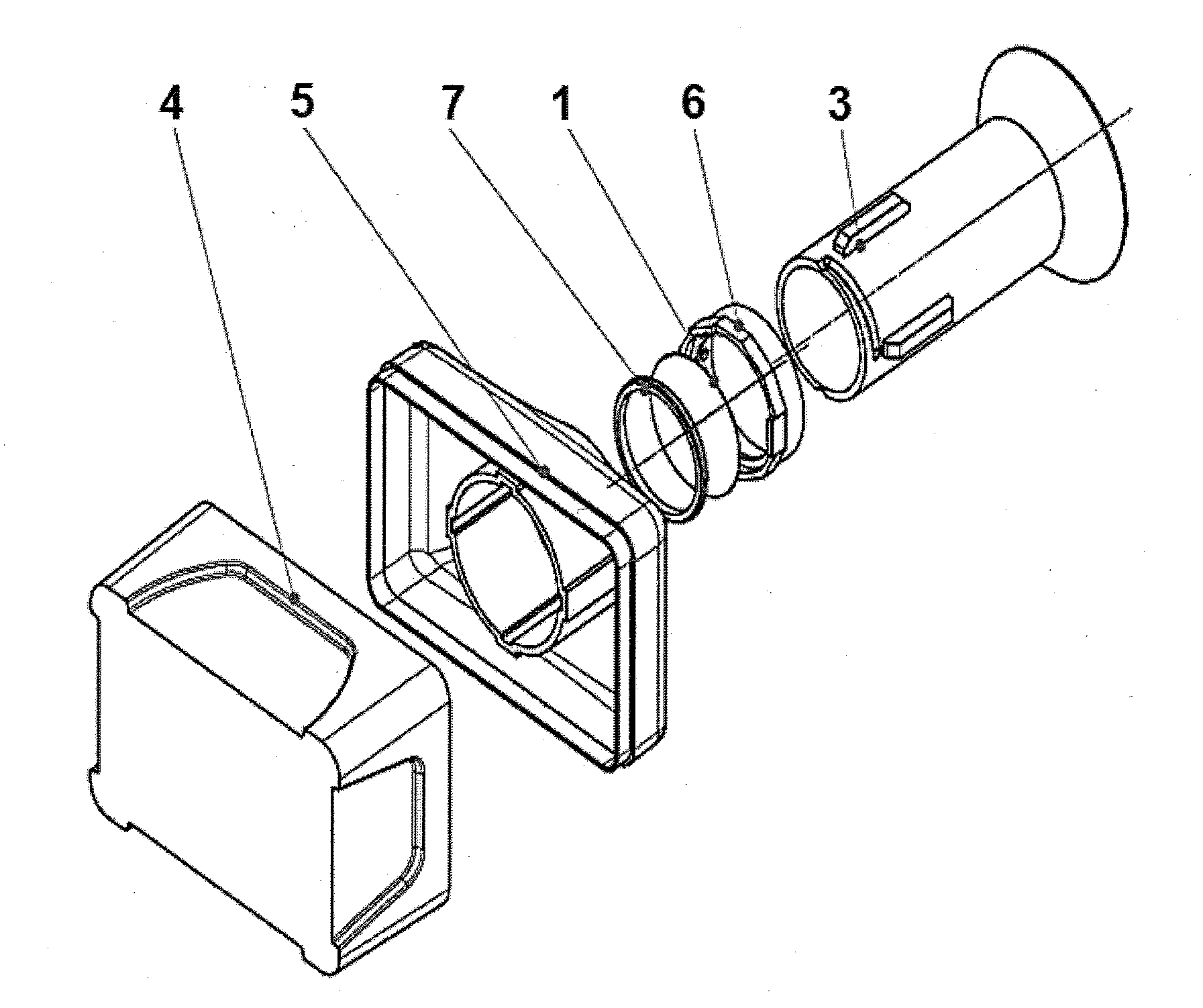
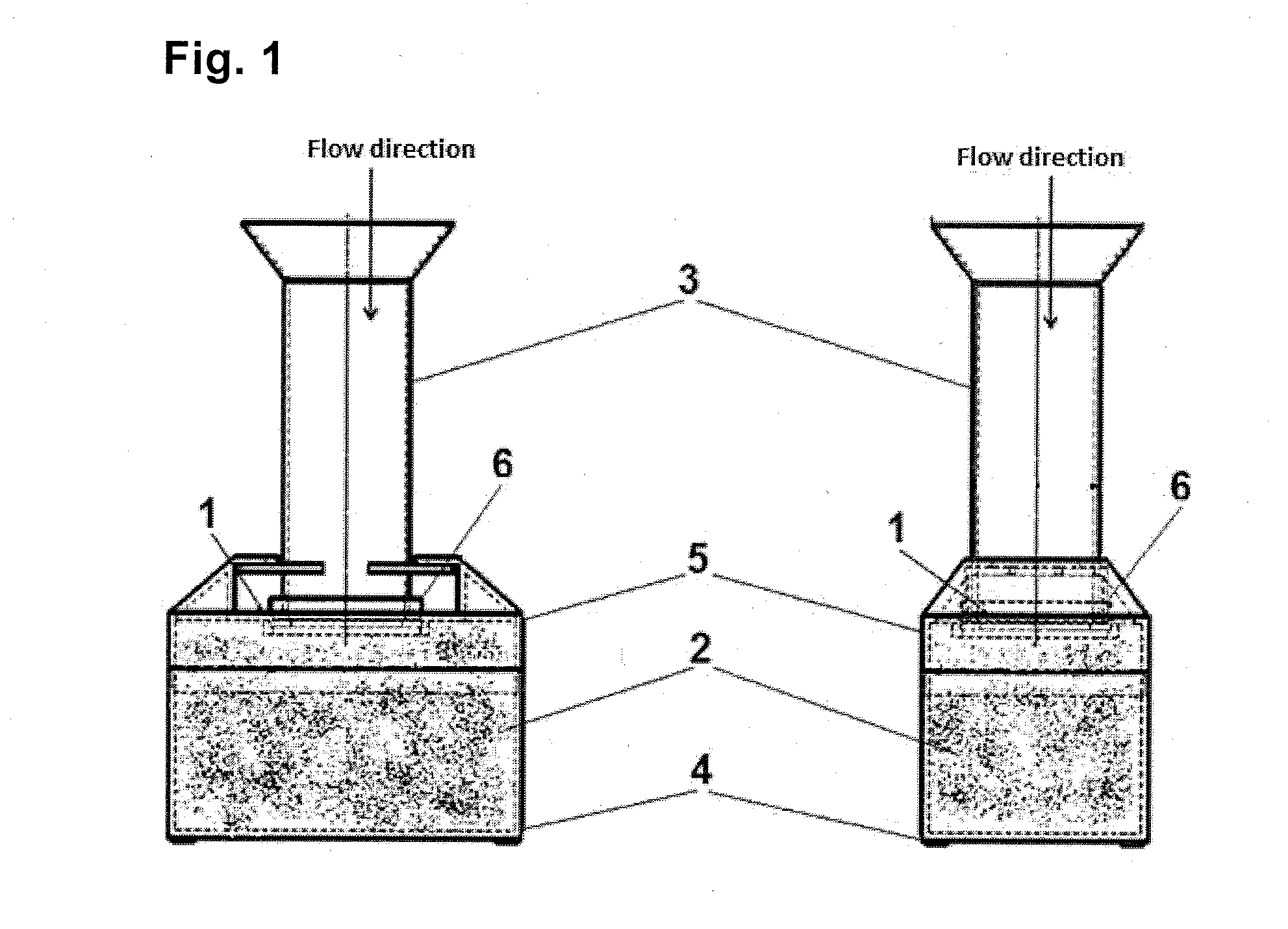
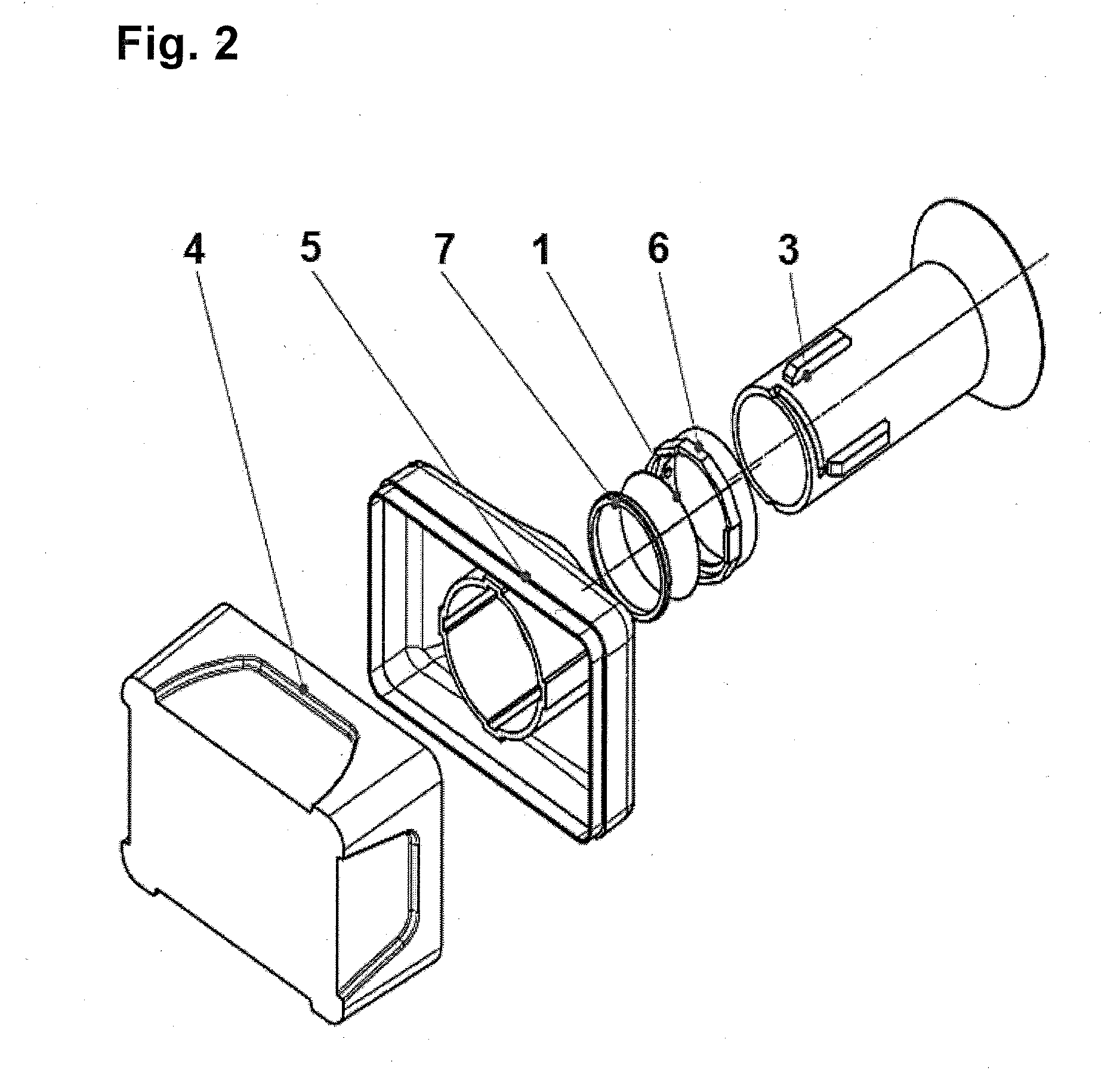
[0074]One embodiment of the apparatus for performing the filtration shown in FIGS. 1-3 comprises a polycarbonate filter membrane 1, with a pore size of 8 μm, which the absorbent material 2 consisting of pulp is in an intimate contact with, from the bottom side of said membrane. The apparatus further includes a top- and bottom-open hollow container 3 for receiving a mixture of sporadic cells present in the body fluid. For easier pouring of liquids the upper edge of the container 3 is broadened so as to form a funnel. The lower periphery of the container 3 is in tight contact with the upper first side of the membrane 1, and the lower second side of the membrane 1 is in an intimate contact with the absorbent material 2, so that the membrane 1 separates the inner space of the container 3 from said absorbent material 2.

[0075]The absorbent material 2 is arranged in a collecting base container 4 provided with an upper lid 5, into which lid a circumferenti...


PUM


Abstract

Method for gentle separation of viable sporadic cells from body fluids such as blood, from malignant effusions, bronchoalveolar lavage fluid, peritoneal lavage fluid and amniotic fluid, based on a filter membrane which is in an intimate contact with an absorbent material. Using the present method it is possible to isolate for example circulating and disseminated tumor cells, endometrial cells and circulating trophoblast cells, allowing subsequent detection, quantification, characterization and especially culturing of said cells. An apparatus for carrying out the method is further disclosed.

Description

FIELD OF THE INVENTION[0001]The present invention provides a method of gentle separation of viable sporadic cells (rare cells) from body fluids such as blood, from malignant effusions (ascites, pleural effusion), bronchoalveolar lavage fluid, peritoneal lavage fluid and amniotic fluid. Using the present method it is possible to isolate for example circulating (CTCs, circulating tumor cells) and disseminated tumor cells (DTCs, disseminated tumor cells), endometrial cells and circulating trophoblast cells (CFTC, circulating fetal trophoblast cells), allowing subsequent detection, quantification, characterization and especially culturing.BACKGROUND OF THE INVENTION[0002]Metastatic lesions are the most common cause of death in patients with carcinomas (Birchmeier, 1996). During metastasizing tumor cells detach from the primary tumor and enter the blood stream directly or through the lymphatic system, migrating into secondary organs and developing a metastatic deposit. Detection of CTCs ...

Claims


Application Information

Patent Timeline
IPC IPC(8): C12M1/00
CPCC12M47/04C12M47/02
Inventor BOBEK, VLADIMIRKOLOSTOVA, KATARINA
Owner METACELL
Why Eureka
Try Eureka