Method for detecting precancerous lesions

a technology for cancer and lesions, applied in combinational chemistry, biochemistry apparatus and processes, library screening, etc., can solve the problems of poor results in cancer therapy, limited early diagnosis, and cancer cells significantly different from normal cells, and are highly complex and variabl

Active Publication Date: 2016-06-02
GENOMICTREE
View PDF3 Cites 5 Cited by
  • Summary
  • Abstract
  • Description
  • Claims
  • Application Information

AI Technical Summary

Benefits of technology

[0016]It is a main object of the present invention to provide a methylation biomarker which can be effectively used for detection of precancerous lesions corresponding to a cell proliferative disorder of colon tissue, and to provide information for early detecting precancerous lesions using the methylation biomarker.

Problems solved by technology

Such poor results in cancer therapy are due not only to the problem of therapeutic methods, but also to the fact that it not easy to diagnose cancer at an early stage and to accurately diagnose progressed cancer and to carry out the follow-up of cancer patients after cancer therapy.
Meanwhile, tumor markers for monitoring substances that are directly or indirectly produced from cancers are used in cancer monitoring after the treatment of the cancer, but they have limitations in early diagnosis, since up to about half thereof appear normal even in the presence of cancer, and they often appear positive even in the absence of cancer.
The reason why the early diagnosis and treatment of cancer are difficult is that cancer cells significantly differ from normal cells and are highly complex and variable.
Cancer cells grow excessively and continuously, invading surrounding tissue and metastasize to distal organs leading to death.
However, this method has the deficiency that it can be applied only to some blood cancers.
However, this method has shortcomings in that it can be applied only to some cancers, including prostate cancer and melanoma, has a high false positive rate.
In addition, it is difficult to standardize detection and reading in this method, and its utility is also limited (Kopreski, M. S. et al., Clin.
However, since these gene mutations occur in some cancer patients, there is a limitation in accurately performing the early diagnosis of cancers.
However, KR 10-1142131 B does not disclose the use of DNA methylation to detect hyperplastic cells and polyps in the clone in a pre-cancerous stage by use of a cell-free DNA sample.

Method used


Image

  • Method for detecting precancerous lesions
  • Method for detecting precancerous lesions
  • Method for detecting precancerous lesions

Examples


example 1

Selection of Optimum Methylation Region of SDC2 Gene for Detection of Colorectal Precancerous Lesion

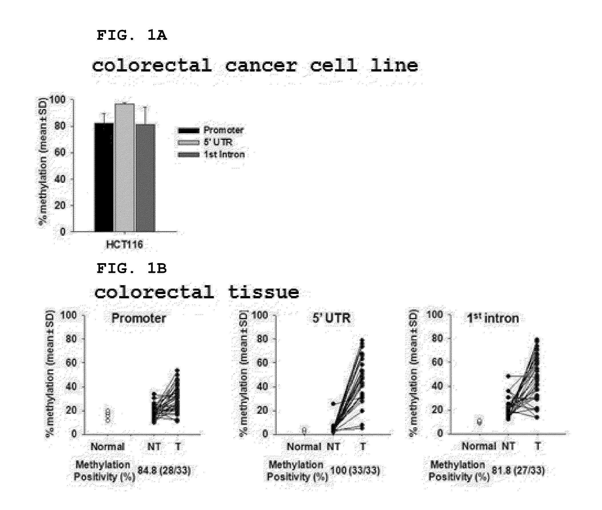
[0190]In order to select the optimum methylation region for detection of a colorectal precancerous lesion and to confirm the methylation state of the SDC2 methylation biomarker gene, pyrosequencing of the CpG islands of the SDC2 genes was performed.

[0191]In order to modify non-methylated cytosine to uracil using bisulfite, total genomic DNA was isolated from each of the colorectal cancer cell line HCT116 (Korea Cell Line Bank (KCLB) No. 10247), normal colorectal tissue (Biochain), and colorectal cancer tissue and a normal tissue adjacent thereto (the Biobank, the Yonsei University Medical Center), and 200 ng of the genomic DNA was treated with bisulfite using the EZ DNA methylation-gold kit (Zymo Research, USA). When the DNA was treated with bisulfite, non-methylated cytosine was modified to uracil, and the methylated cytosine remained without changes. The DNA treated with bisulfite w...

example 2

Verification of Methylation of SDC2 Methylation Marker Gene in Colorectal Precancerous Lesions

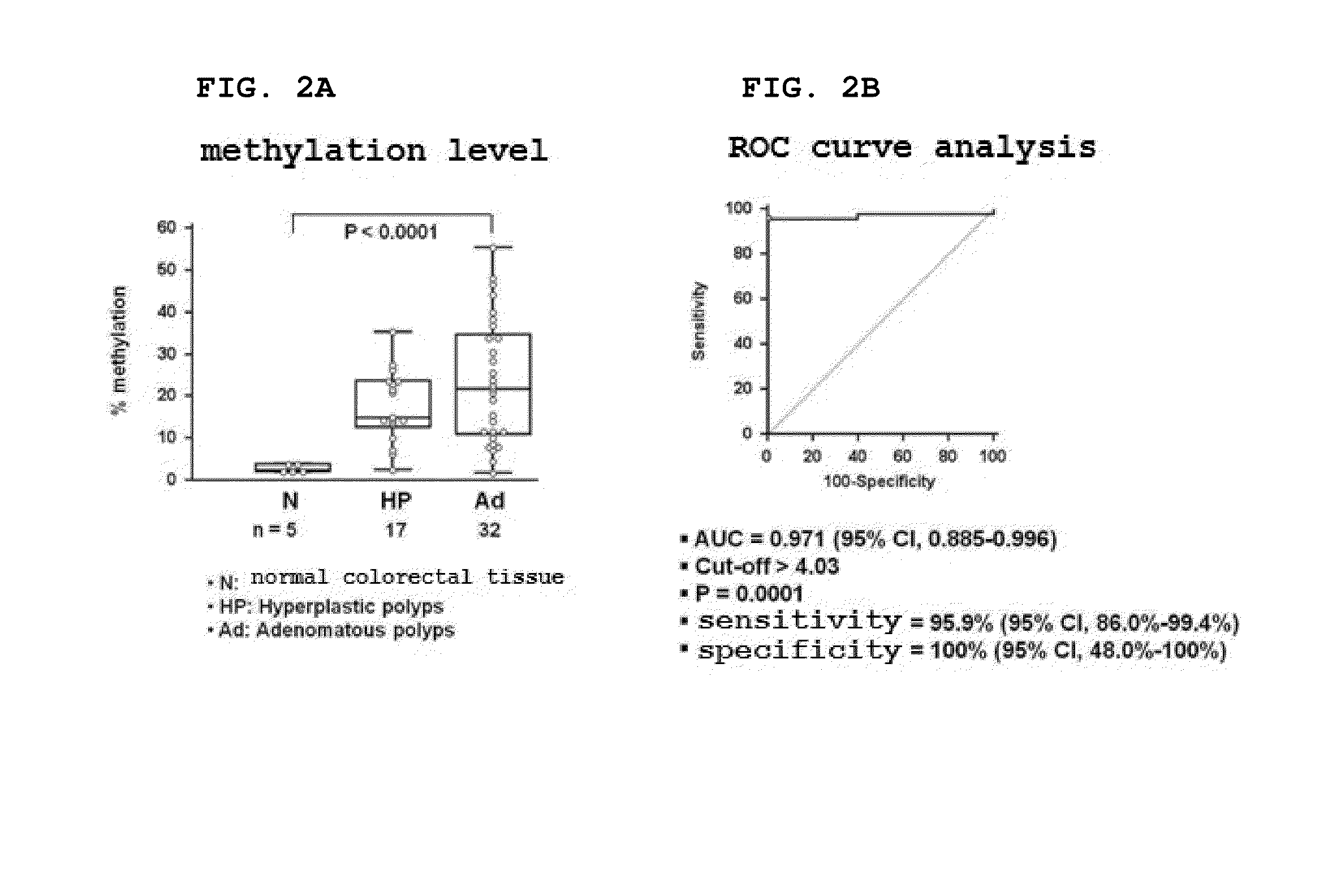
[0196]In order to evaluate the ability of the SDC2 methylation marker gene to detect a colorectal polyp that is a precancerous lesion, pyrosequencing of the 5′ regulatory region confirmed to have the best diagnostic performance in Example 1 was performed. As a result, it was shown that the 5′ regulatory region was not substantially methylated (less than 5%) in normal colorectal tissue (N), but was methylated at very high levels in a hyperplastic polyp (HP) and an adenomatous polyp (Ad) (P program, Belgium) (FIG. 2A)

[0197]In order to evaluate the ability of the SDC2 methylation marker gene to detect colorectal polyps, the sensitivity and specificity of the SDC2 gene for colorectal polyp diagnosis were measured by ROC curve analysis (MedCalc Program, Belgium) (FIG. 2B). As shown in FIG. 2B, it was shown that the sensitivity of the SDC2 methylation marker gene for polyp diagnosis was 95.9% (47...

example 3

Verification of SDC2 Methylation Gene in Sera of Colorectal Precancerous Lesion Patients by Use of Set 1 Primer Pair and Probe

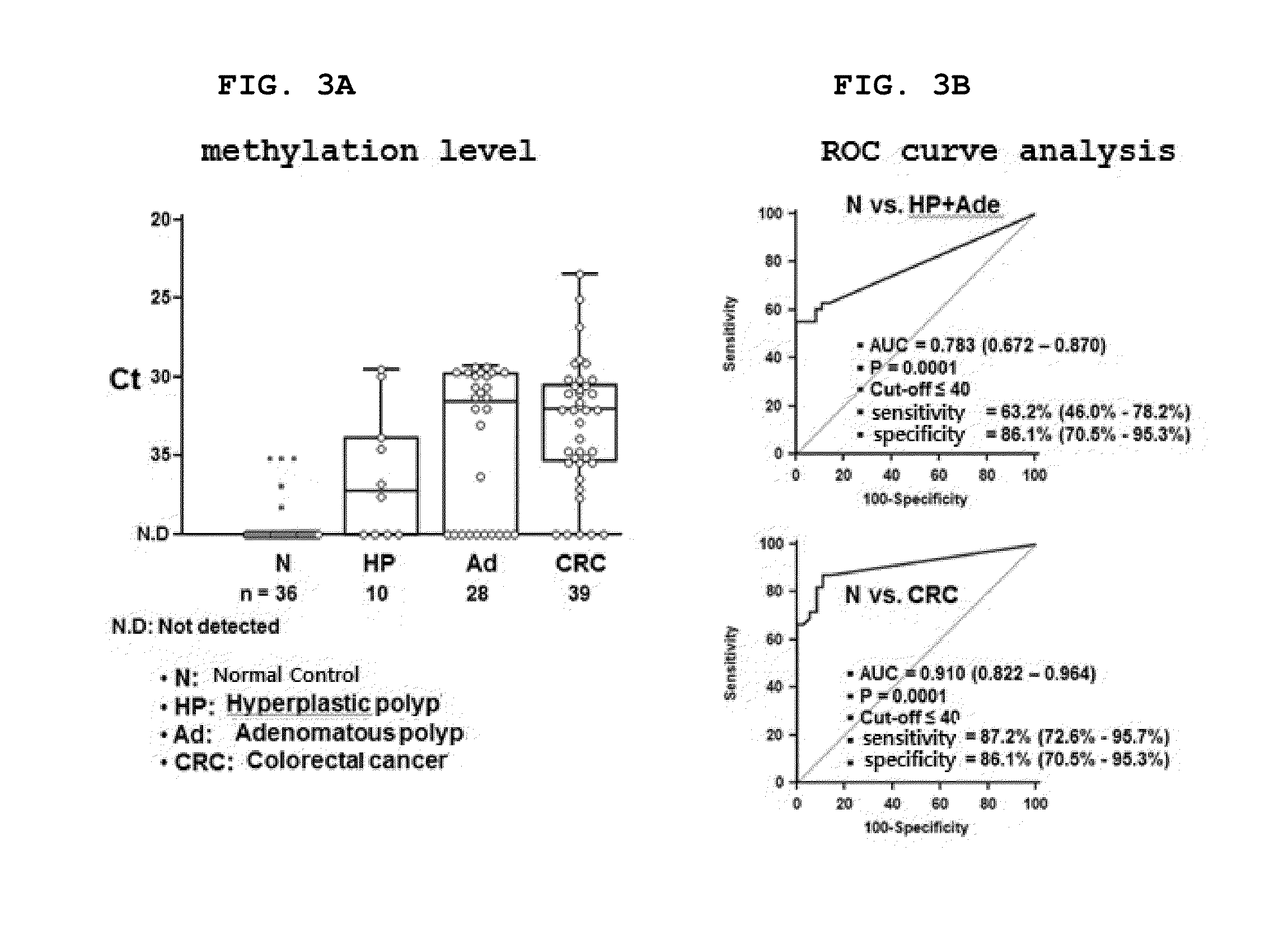
[0198]In order to verify whether the SDC2 methylation biomarker gene is useful for detection of the precancerous lesion colorectal polyp in serum DNA sample, genomic DNA was isolated from the each of the sera of 36 colonoscopy-confirmed normal control sera (BioServe, USA), 10 hyperplastic polyp patient sera (BioServe, USA), 28 colorectal adenomatous polyp patient sera (the Biobank, the Yonsei University Medical Center) and 39 colorectal cancer patient sera (the Biobank, the Yonsei University Medical Center). Genomic DNA was isolated from 1.0 mL of each of the sera using the ChargeSwitch gDNA 1 mL Serum Kit (Invitrogen, USA).

[0199]The isolated genomic DNA was treated with bisulfite using the EZ DNA methylation-Gold kit (Zymo Research, USA), and then eluted with 10 μl of sterile distilled water and subjected to methylation-specific real-time PCR (qMSP; Methylat...


PUM

PropertyMeasurementUnit
diameteraaaaaaaaaa
volumeaaaaaaaaaa
volumeaaaaaaaaaa

Abstract

The present invention relates to the novel use of syndecan-2 (SDC2) gene as a CpG methylation biomarker for detecting precancerous lesions, and more particularly, to the use of the SDC2 gene as a biomarker to early diagnose colorectal precancerous lesions by measuring the degree of methylation of the SDC2 gene. The present invention provides a method of providing information for diagnosis of precancerous lesions by detecting methylation of the CpG islands of the SDC2 gene. In addition, the use of the methylation detection and kit according to the present invention makes it possible to diagnose colorectal precancerous lesions at an early transformation stage, thus enabling the early diagnosis of colorectal precancerous lesions. Furthermore, the methylation detection method and kit of the present invention enables colorectal precancerous lesions to be effectively diagnosed in an accurate and rapid manner compared to conventional methods.

Description

TECHNICAL FIELD[0001]The present invention relates to the use of syndecan-2 (SDC2) gene as a CpG methylation biomarker which is used to detect precancerous lesions, which correspond to a cell proliferative disorder of colorectal tissue, based on an aberrant methylation event that occurs in a specific gene region when normal cells transform into hyperplastic cells. More particularly, the present invention relates to a method of early detecting precancerous lesions by measuring the degree of methylation of a specific region of the SDC2 gene.BACKGROUND ART[0002]Even at the present time when medical science has advanced, the 5-year survival rate of cancer patients, particularly solid tumor patients (other than blood cancer patients) who account for the majority of cancer patients is less than 50%. About two-thirds of all cancer patients are diagnosed at a progressed stage, and most of them die within 2 years after cancer diagnosis. Such poor results in cancer therapy are due not only to...

Claims


Application Information

Patent Timeline
Patent Type & Authority Applications(United States)
IPC IPC(8): C12Q1/68
CPCC12Q1/6886C12Q2600/112C12Q2600/154C12Q2600/118
Inventor AN, SUNGWHANOH, TAE JEONG
Owner GENOMICTREE
Why Eureka
Try Eureka