Biomimetic Cell Culture Substrates

Active Publication Date: 2016-08-04
ULTRA SMALL FIBERS
View PDF3 Cites 13 Cited by
  • Summary
  • Abstract
  • Description
  • Claims
  • Application Information

AI Technical Summary

Benefits of technology

[0012]Disclosed herein are biomimetic substrates with hierarchical architecture created by hot-pressing PCL into patterned laser-ablated nanopore molds. When extracted, these PCL substrates include polymer nanofibers patterned on the micron scale over square centimeters of culture substrate surface. The structures are similar in size and morphology to collagen fibrils universally found within mammalian cells [20, 24, 27]. The attachment of fibers at the substrate base mimics the basement membrane where collagen fibrils are in contact with a highly cross-lin

Problems solved by technology

However, many synthetic culture substrates resulting from these research efforts embody properties that are not completely biomimetic.
Additionally, the minimal flexibility of modifying cell-adhesive surfaces of existing mass-produced culture platforms complicates the ability to probe and understand stem cell behavior as it relates to developmental and regenerative processes [13-14].
Despite the contributions made by the research community, these materials do have limitations.
First, it is difficult to uncouple gel properties, which limits

Method used


Image

  • Biomimetic Cell Culture Substrates
  • Biomimetic Cell Culture Substrates
  • Biomimetic Cell Culture Substrates

Examples


Example

Example 1

[0065]This example employed novel cell culture substrates created by hot-pressing poly(ε-caprolactone) films in femtosecond laser-ablated nanopore molds to form patterned polymer nanofiber matrices on flat PCL substrates. Quantitative real-time polymerase chain reaction and immunocytochemistry were used to show that these polymer nanofiber matrices increased expression of several critical self-renewal factors and markers of cell-cell interaction, thereby maintaining stemness of hMSCs cultured thereon.

Materials and Methods

[0066]All reagents were purchased from Sigma-Aldrich (St. Louis, Mo.) at the highest available quality unless otherwise noted.

[0067]Culture Substrate Fabrication

[0068]For this work, a one millimeter thick, double side polished fused silica wafer (Mark Optics, Santa Ana, Calif.) was diced into 22 mm by 22 mm square chips. Each chip was patterned with a single 10 mm by 10 mm array of nanopores using the femtosecond laser machining system [29]. Each array was ...

Example

Example 2

[0091]This example employed the novel hot-pressing method to identify additional thermoplastic polymers suitable for use in the fabrication of patterned polymer nanofiber matrices from laser ablated nanopore molds.

[0092]Fabrication of Polymer Fiber Substrates from Additional Polymers

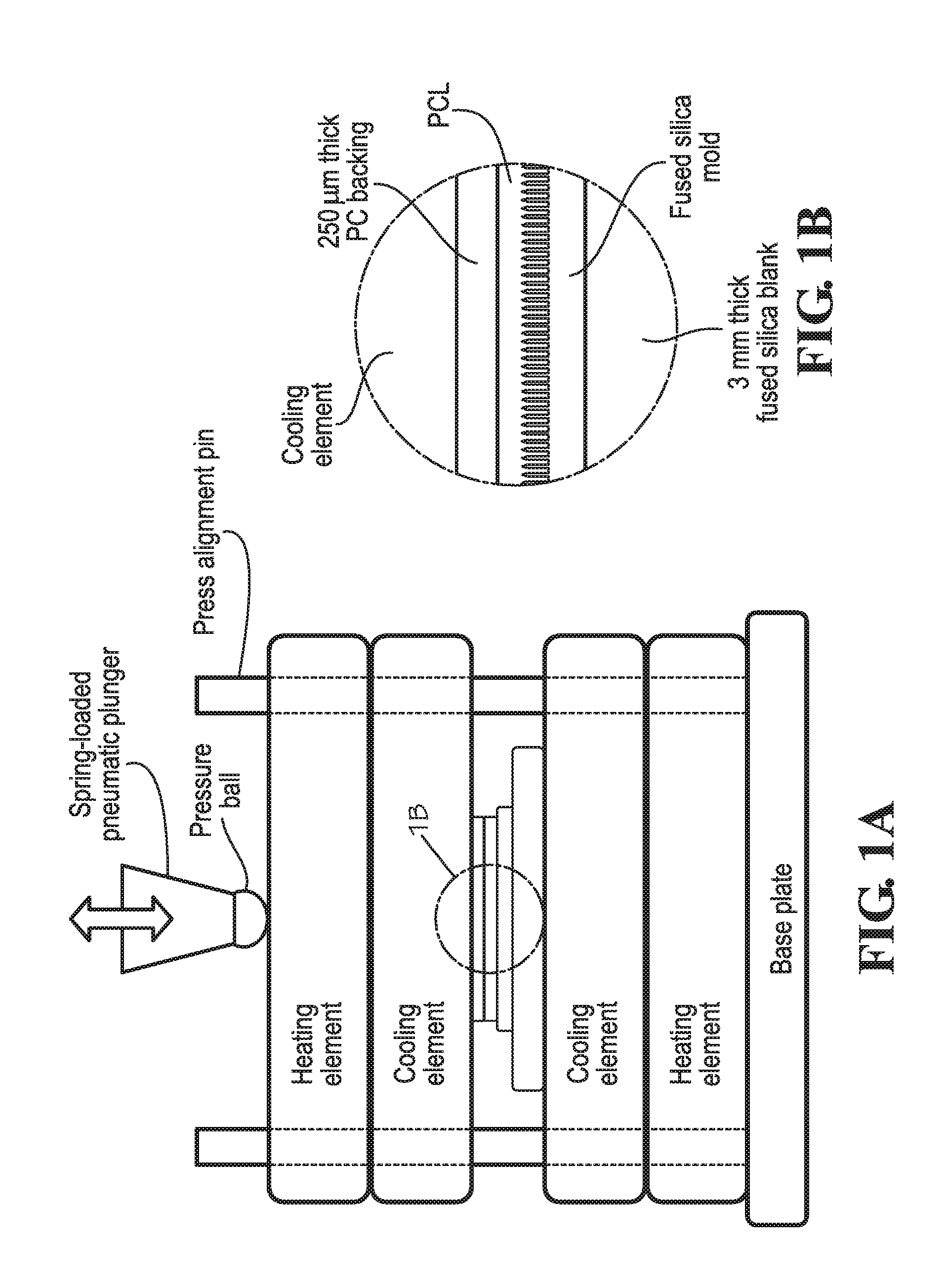
[0093]The technique of forming arrays of polymer nanofibers by hot-pressing thermoplastic polymers into patterned femtosecond laser-ablated nanopore molds using the hot-pressing system depicted in FIGS. 1A-B was successfully demonstrated for each of the polymers listed in Table 2, which provides the glass transition, TG, and melting, TM, temperatures of each polymer. The specific hot-pressing processing conditions used for each polymer and the average recorded pile height and length dimensions for nanofibers formed from these polymers are given in Table 3. All nanofibers were hot-pressed using the applicable temperature and pressure parameters listed in Table 3, and the temporal hold conditions ...


PUM

PropertyMeasurementUnit
Lengthaaaaaaaaaa
Lengthaaaaaaaaaa
Diameteraaaaaaaaaa

Abstract

Embodiments of the presently disclosed subject matter provide biomimetic cell culture substrates comprising highly tunable patterned polymer nanofiber matrices capable of modulating expression of critical self-renewal factors and markers of cell-cell interaction to maintain stemness of human mesenchymal stem cells in vitro. Embodiments of the presently-disclosed subject matter also provide scalable, highly repeatable methods of making biomimetic cell culture substrates by hot pressing thermoplastic polymer films into femtosecond laser-ablated nanopore molds to form patterned polymer nanofiber matrices on flat thermoplastic substrates.

Description

CROSS-REFERENCES TO RELATED APPLICATIONS[0001]This application claims priority to U.S. Provisional Patent Application Ser. No. 62 / 111,551 entitled PATTERNED NANOFIBRE MATRIX PROMOTES STEMNESS AND CELL-CELL INTERACTION OF ADULT STEM CELLS, filed Feb. 3, 2015, the entire disclosure of which is hereby incorporated herein by this reference.STATEMENT REGARDING FEDERALLY SPONSORED RESEARCH OR DEVELOPMENT[0002]Subject matter described herein was made with government support under Grant Numbers HL091465 and 1UH2 TR000491 awarded by the National Institutes of Health and Grant Number CAREER CBET 1056046 awarded by the National Science Foundation. The government has certain rights in the subject matter disclosed herein.REFERENCE TO SEQUENCE LISTING OR COMPUTER PROGRAM LISTING APPENDIX[0003]Not Applicable.TECHNICAL FIELD[0004]The present disclosure relates generally to cell culture substrates and more specifically to tunable arrays of patterned polymer nanofiber matrices that promote stemness a...

Claims


Application Information

Patent Timeline
IPC IPC(8): C12N5/00B29C67/20
CPCC12N5/0068B29C67/20B29K2105/256B29K2067/04C12N2533/30B29K2101/12C12N2535/10
Inventor HOFMEISTER, WILLIAM HUDSONHOFMEISTER, LUCASTEREKHOV, ALEXANDER YURYEVICHDA COSTA, JOSE LINO VASCONCELOS
Owner ULTRA SMALL FIBERS
Why Eureka
Try Eureka