Corneal endothelial cell marker

a corneal endothelial cell and marker technology, applied in the cell field, can solve the problems of corneal endothelial cell number decline, corneal endothelial cell ability to regenerate, severe visual impairment, etc., and achieve the effect of treating or preventing corneal endothelial diseas

Active Publication Date: 2016-09-15
ACTUALEYES INC +2
View PDF2 Cites 8 Cited by
  • Summary
  • Abstract
  • Description
  • Claims
  • Application Information

AI Technical Summary

Benefits of technology

[0095]The present invention can identify the cell differentiation capability of corneal endothelial cells to identify cells that are highly functional. As a result, it is possible to treat or prevent corneal endothelial disease or disorder that was previously impossible or difficult. Further, a method of determining purity when making a sample comprising a cell, a method of purifying a corneal endothelial cell, and a method of preparing a purified corneal endothelial cell are also provided.

Problems solved by technology

In addition, it is known that a decrease in the number of corneal endothelial cells from damage due to trauma, disease or the like to the cells leads to severe visual impairment.
However, once damaged, corneal endothelial cells do not have the ability to regenerate.
Corneal endothelial dystrophy or bullous keratopathy induced by dysfunction of the corneal endothelium due to various causes results in edema or turbidity of the cornea, leading to significant deterioration in vision.
However, cornea donation is insufficient in Japan.

Method used


Image

  • Corneal endothelial cell marker
  • Corneal endothelial cell marker
  • Corneal endothelial cell marker

Examples


reference example 1

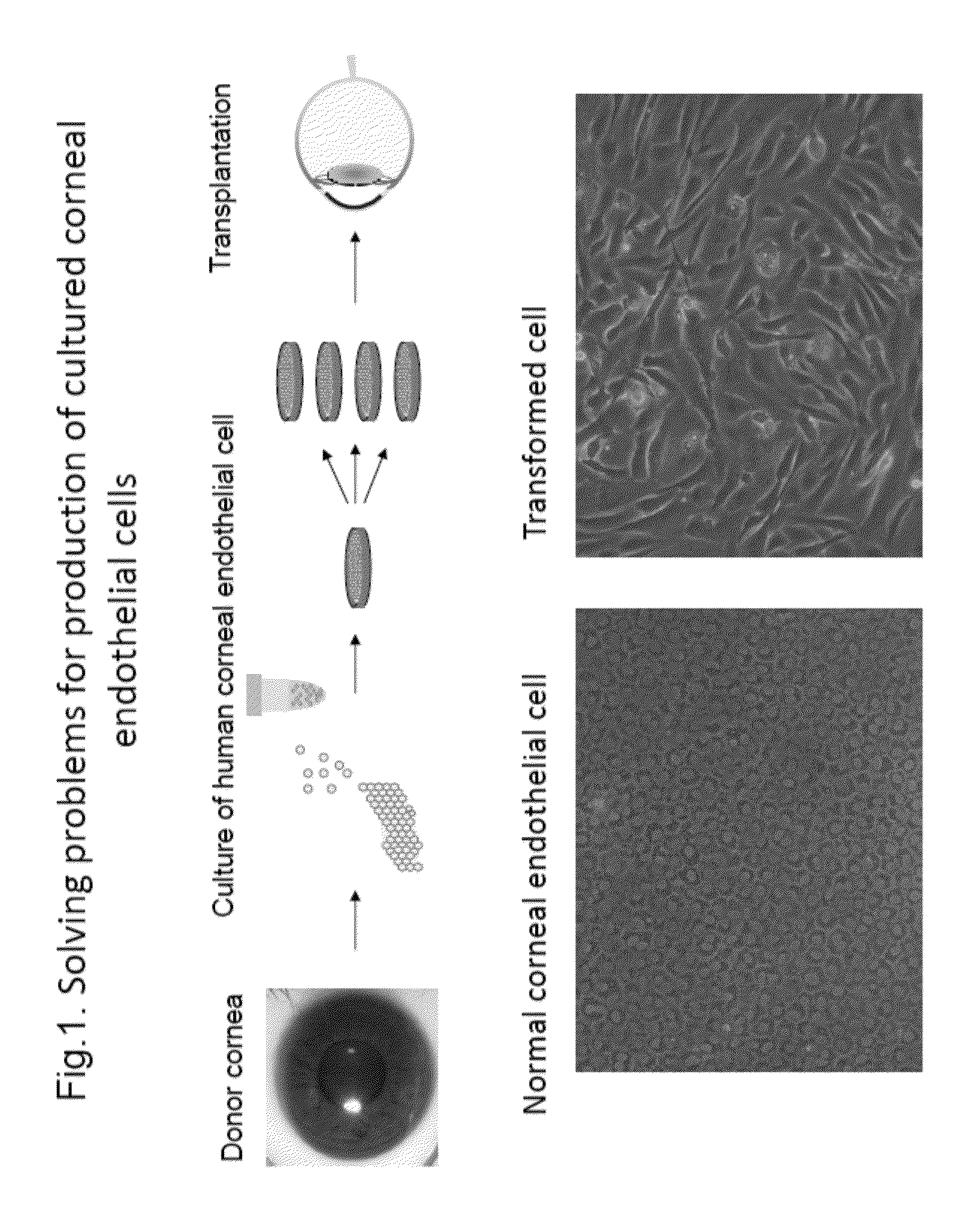
[0301]As shown in FIG. 1, the state of the cells when human corneal endothelial cells are separated from a donor cornea and cultured was observed in order to study the issues in conventional cultured corneal endothelial cell production. The experimental observation conditions are as described in the above-described section of (Experimental materials and methods).

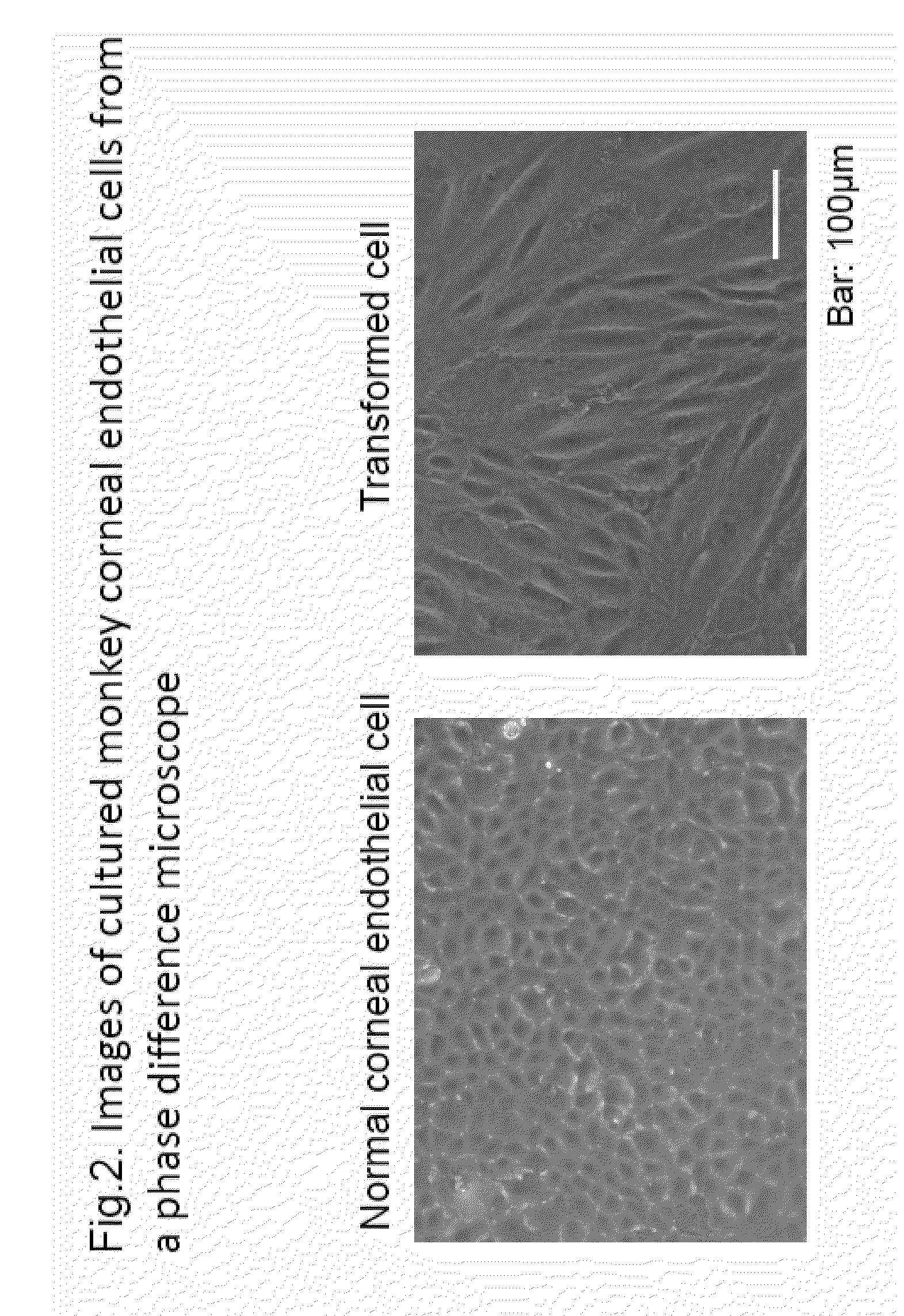
[0302]As shown in FIG. 1, the left side shows corneal endothelium that could be cultured while maintaining a normal polygonal cellular form. However, when separated and cultured by a common method, the cells readily transform and change into a fibroblast-like form to lose cellular function as shown in the bottom right.

[0303]Production of corneal endothelial cells free of transformed cells is essential for clinical applications of cultured corneal endothelial cell transplantation. The current state of ophthalmic science is such that production thereof cannot be achieved with conventional techniques. In this regard, a normal o...

preparation example

Production of Immortalized Strain of Normal Corneal Endothelial Cells and Transformed Corneal Endothelial Cells

[0304]In the present example, immortalized strains of normal corneal endothelial cells and transformed corneal endothelial cells were produced.

[0305](Culture Method)

[0306]Corneal endothelial cells were mechanically peeled off with a basal membrane from a corneal for research purchased from the Seattle Eye Bank. After collagenase was used to detach and collect the corneal endothelial cell from the basal membrane, the cells were subjected to primary culture. As a medium for human cells, Opti-MEM I Reduced-Serum Medium, Liquid (INVITROGEN catalog No.: 31985-070) to which 8% fetal bovine serum (FBS) (BIOWEST, catalog No.: S1820-500), 200 mg / ml CaCl2.2H2O (SIGMA catalog No.: C7902-500G), 0.08% chondroitin sulfate (SIGMA catalog No.: C9819-5G), 20 μg / ml ascorbic acid (SIGMA catalog No.: A4544-25G), 50 μg / ml gentamicin (INVITROGEN catalog No.: 15710-064) and 5 ng / ml EGF (INVITROGE...

example 1

Flow Cytometry in Transformed Corneal Endothelial Cell and Normal Corneal Endothelial Cell

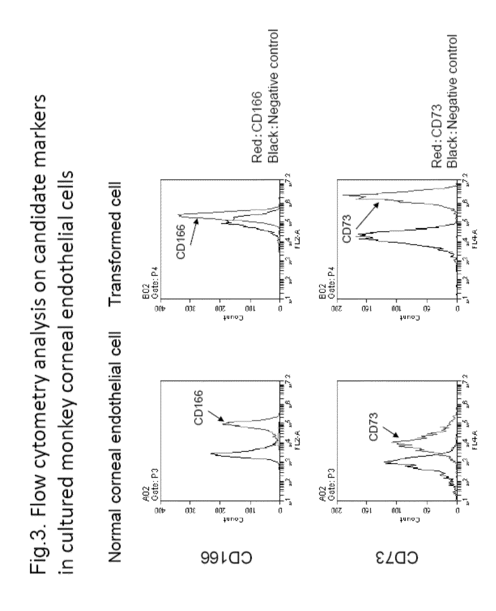
[0309]The inventors screened surface markers of normal human corneal endothelial cells and transformed cells by high-throughput analysis using flow cytometry to proceed with selection of candidate markers.

[0310]Human corneal endothelial cells were cultured by the method described above in (Experimental materials and methods). Cells with normal cellular form and transformed cells were subjected to high-throughput analysis by flow cytometry using Human Cell Surface Marker Screening Panel (BD Lyoplate™, BD Bioscience). Expression of surface antigens in each of normal cells and transformed cells was compared, and markers with enhanced expression in normal cells were used as marker candidates for normal cells (Table 2). Meanwhile, markers with enhanced expression in transformed cells were similarly used as markers of transformed cells (Table 3).

(Table 2 Surface Antigens with Enhanced Expression in N...


PUM

PropertyMeasurementUnit
molecular weightaaaaaaaaaa
pHaaaaaaaaaa
fluorescence maximum wavelengthaaaaaaaaaa

Abstract

The purpose of the present invention is to provide a method of purification and preparation of cultured corneal endothelial cells, and in particular, to provide cell surface markers for use in corneal endothelial cells not including transformed cells. Provided are cell markers for distinguishing normal cells and transformed cells, in particular normal and transformed corneal endothelium cells. These cell markers relate to specific cell surface markers, for example, to a normal corneal endothelial surface marker such as CD166, and a transformed cell surface marker such as CD73. By using the transformed cell surface marker such as CD73 to remove transformed cells by sorting, it becomes possible to improve purity of a normal cultured corneal endothelium. By using normal corneal endothelial surface marker such as CD166, or by combined use with the transformed cell surface marker, it becomes possible to provide a means for verifying the purity of a prepared corneal endothelium.

Description

TECHNICAL FIELD[0001]The present invention relates to a cell, particularly a differentiated marker in the ophthalmological region, and particularly a corneal endothelial cell marker.BACKGROUND ART[0002]Visual information is recognized when light transmitted into the cornea, which is a transparent tissue at the front-most part of an eye ball, reaches the retina and excites nerve cells of the retina and a generated electric signal is transmitted through the optic nerve to the visual cortex of the cerebrum. To attain good vision, it is necessary that the cornea is transparent. The transparency of the cornea is maintained by maintaining constant water content with pumping and barrier functions of corneal endothelial cells.[0003]The cornea is a transparent tissue positioned in the front side of an eyeball and having a three layer structure of mainly a corneal epithelial cell layer, a corneal stroma layer, and a corneal endothelial layer. The corneal endothelial layer is a single-layer ce...

Claims


Application Information

Patent Timeline
Patent Type & Authority Applications(United States)
IPC IPC(8): G01N33/569A61K35/30
CPCG01N33/56966G01N2333/70596A61K35/30A61P27/02C12N5/0621G01N2333/705
Inventor KOIZUMI, NORIKOOKUMURA, NAOKIHIRANO, HIROATSUKINOSHITA, SHIGERUUENO, MORIO
Owner ACTUALEYES INC
Why Eureka
Try Eureka