
Drug for treating corneal endothelium by promoting cell proliferation or inhibiting cell damage

Active Publication Date: 2016-11-17
DOSHISHA CO LTD
View PDF7 Cites 5 Cited by
  • Summary
  • Abstract
  • Description
  • Claims
  • Application Information

AI Technical Summary

Benefits of technology

The present invention is about a technique to prevent or treat a corneal endothelial disorder, which was difficult to achieve before. By inhibiting p38 MAP kinase, the invention suppresses cellular disorder and promotes recovery from wounds and other forms of corneal damage. It is particularly useful as eye drops and can also stop the progression of a disease affecting the corneal endothelium.

Problems solved by technology

However, once damaged, corneal endothelial cells do not have the ability to regenerate.
Corneal endothelial dystrophy or bullous keratopathy induced by corneal endothelial dysfunction due to various causes results in edema or turbidity of the cornea, leading to significant deterioration in vision.
However, there is a shortage of cornea donation in Japan.
In addition, it is known that a decrease in the number of corneal endothelial cells from damage due to trauma, disease or the like to the cells leads to severe visual impairment.

Method used


Image

  • Drug for treating corneal endothelium by promoting cell proliferation or inhibiting cell damage
  • Drug for treating corneal endothelium by promoting cell proliferation or inhibiting cell damage
  • Drug for treating corneal endothelium by promoting cell proliferation or inhibiting cell damage

Examples


example 1

Suppression of Cyclin Dependent Kinase Inhibiting Factors and Transition in Cell Cycle of Corneal Endothelial Cells by p38 MAP Kinase Signal Inhibition

[0099]The present Example shows that p38 MAP kinase signal inhibition suppresses cyclin dependent kinase inhibiting factors and transitions cell cycle of corneal endothelial cells.

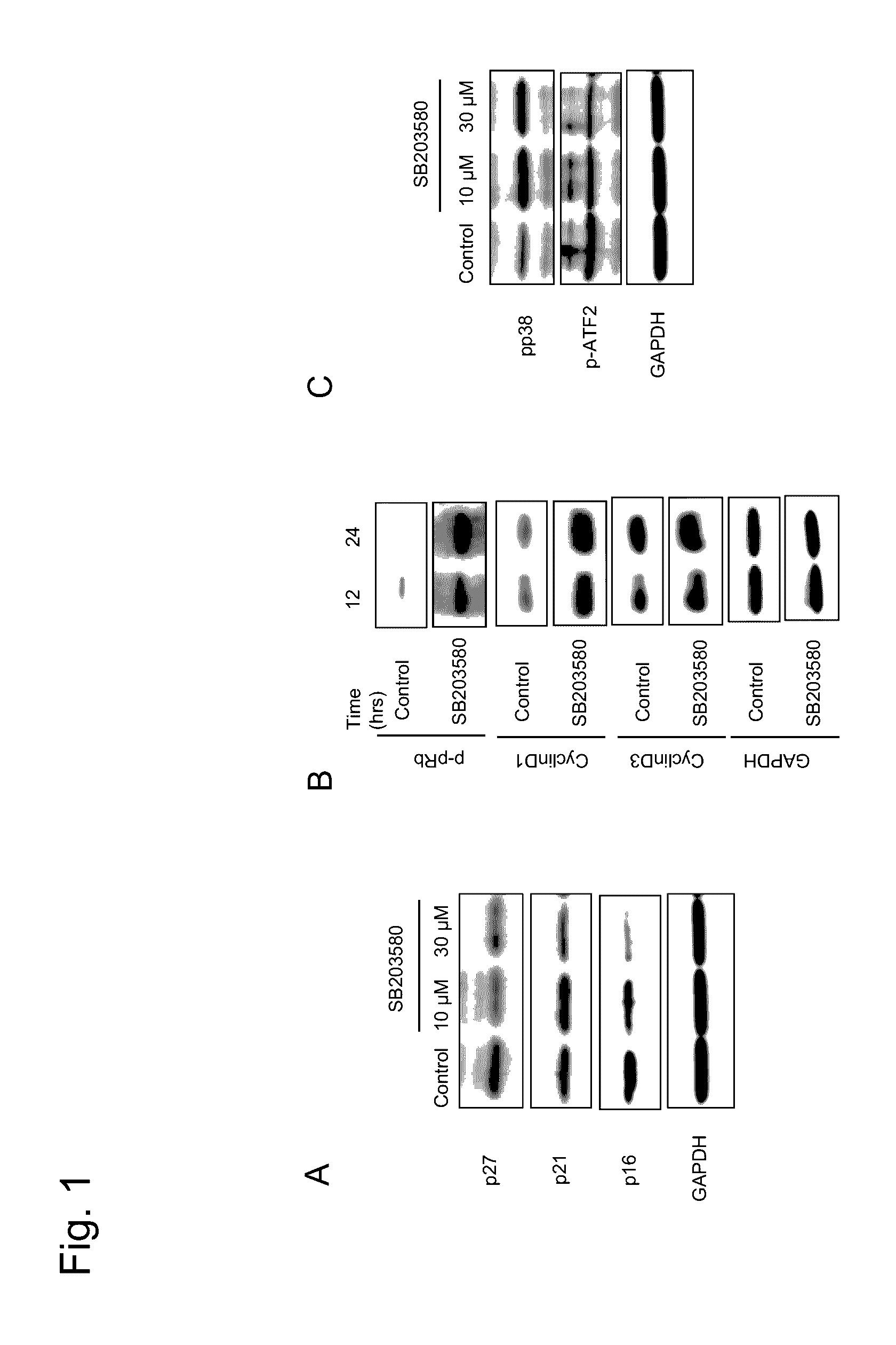
[0100]In accordance with the aforementioned preparation examples, corneal endothelial cells cultured from a cornea for research imported from the Seattle Eye Bank were cultured for use in the following study. A p38 MAP kinase inhibitor SB203580 (13067, Cayman) was added to a cell culture medium. After 20 days, the expression of cyclin dependent kinase inhibiting factors p27, p21, and p16 was studied by Wester blotting. Each of p27, p21, and p16 was suppressed by the addition of SB203580 (FIG. 1 A shows, from the top, p27, p21, p16 and GAPDH. The left lane shows the control, the middle shows 10 μM of SB203580, and the right lane shows 30 μM of SB203580). It i...

example 2

Promotion of Cell Proliferation of Corneal Endothelial Cells by p38 MAP Kinase Inhibitor

[0102]The present Example demonstrate that a p38 MAP kinase inhibitor promotes cell proliferation of corneal endothelial cells.

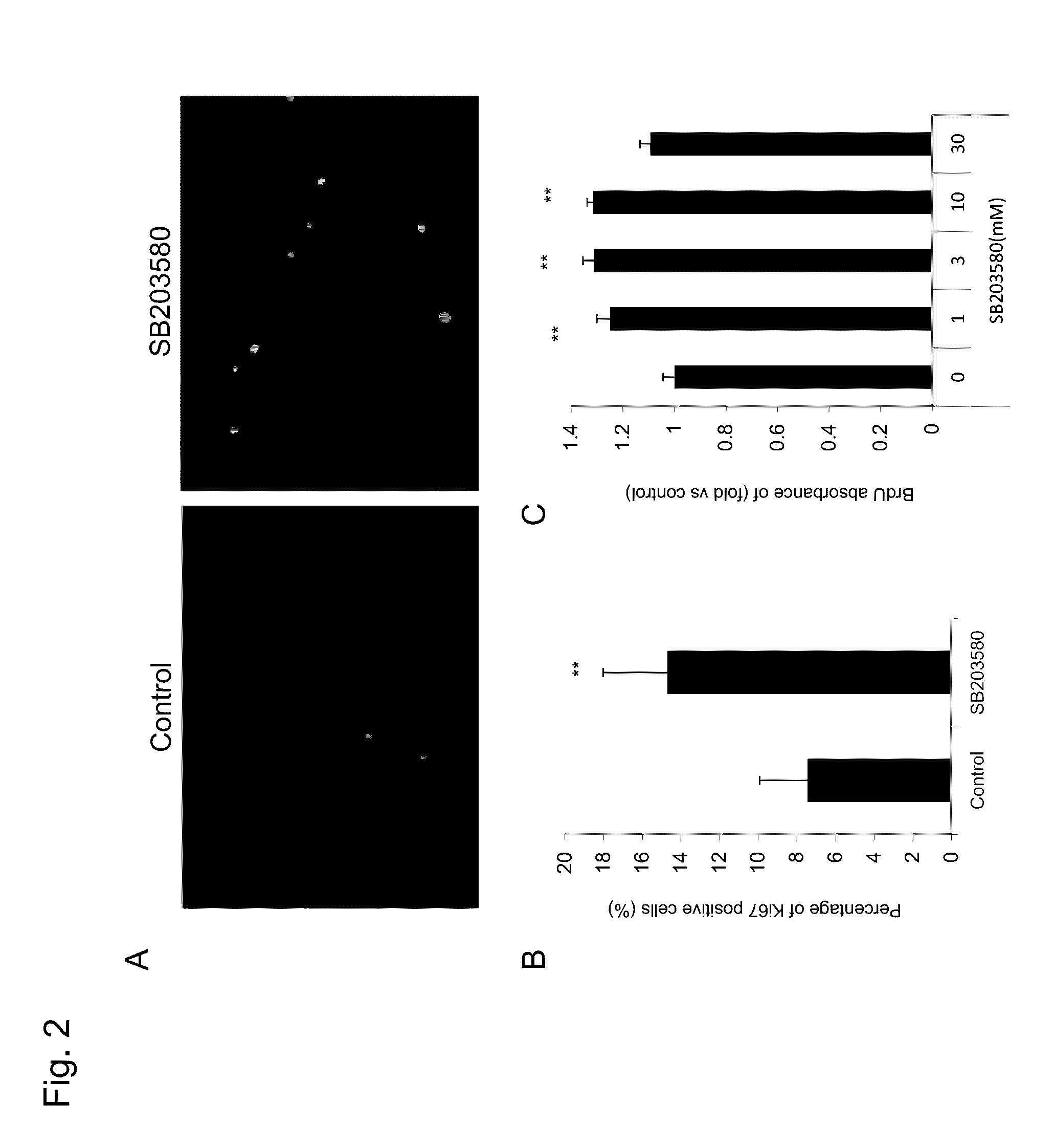
[0103]Cultured human corneal endothelial cells were stimulated with a p38 MAP kinase inhibitor SB203580. After three days, immunostaining was applied with a cell proliferation marker Ki67 (Dako, M7240) based on the experimental methodology discussed herein. The methodology was as discussed above.

[0104]The results are shown in FIG. 2. Expression of Ki67 was observed in significantly more cells by a stimulation with SB203580 (A, B). Similarly, as a cell proliferation marker, uptake of BrdU was studied after 3 days by ELISA as an indicator. BrdU uptake was significantly promoted by a stimulation with SB203580 (C). The above results show that p38 MAP kinase signal inhibition promotes cell proliferation of corneal endothelial cells.

example 3

Promotion of Corneal Endothelial Cell Proliferation with p38 MAP Kinase Inhibitor in a Partial Rabbit Corneal Endothelial Disorder Model

[0105]The present Example studied whether p38 MAP kinase signal inhibition promotes corneal endothelial cell proliferation in a living organism by using a partial rabbit corneal endothelial disorder model.

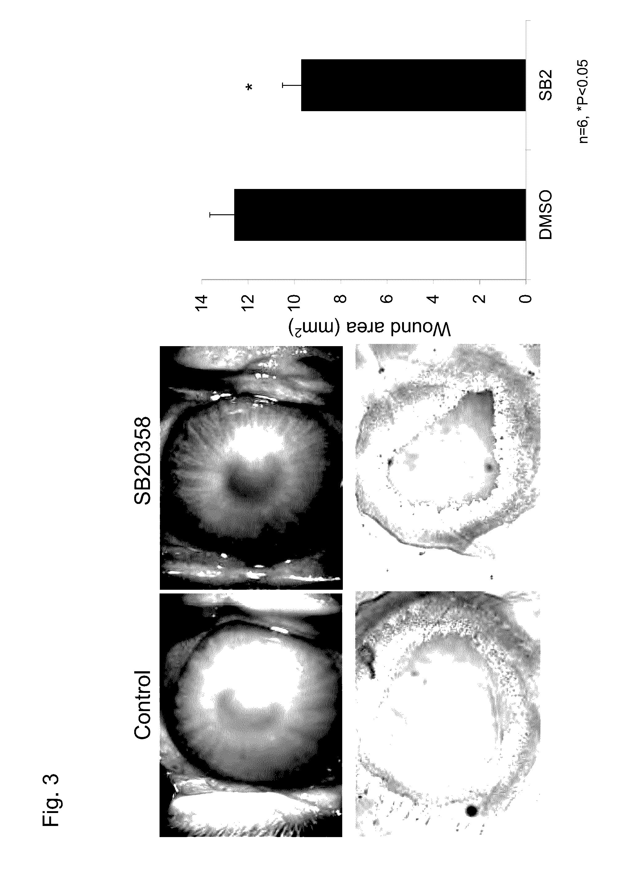
[0106]A stainless steel chip with a diameter of 7 mm was immersed in liquid nitrogen and cooled. The chip was contacted with the center portion of a corneal of a Japanese white rabbit (obtained from Oriental Bioservice) for 15 seconds under general anesthesia, such that corneal endothelial cells at the center portion partially fell off. Eye drops of SB203580 adjusted to 10 mM was then administered 4 times a day for 2 days at 50 μl per dose. As a control, eye drops of a base agent were administered in an eye in which a partial corneal endothelial disorder was similarly created, and a picture was taken. Further, the rabbit was euthanized and the corn...


PUM

PropertyMeasurementUnit
Densityaaaaaaaaaa
Solubility (mass)aaaaaaaaaa
Cell proliferation rateaaaaaaaaaa

Abstract

Provided is an agent for treating or preventing a corneal endothelial disorder wherein cell proliferation is required. More specifically, provided is an agent for treating or preventing a corneal endothelial disorder, wherein cell proliferation is required, said agent comprising a p38MAP kinase inhibitor. In a preferred embodiment, the corneal endothelial disorder is a wound. In a preferred embodiment, the p38MAP kinase inhibitor is soluble in water. The p38MAP kinase inhibitor may comprise 4-[4-(4-fluorophenyl)-2-(4-methylsulfinylphenyl)-1H-imidazole-5-yl]pyridine (SB203580) or a salt thereof.

Description

TECHNICAL FIELD[0001]The present invention relates to a technique and a method for culturing corneal endothelial cells in a normal state and an agent and a medium therefor.BACKGROUND ART[0002]Visual information is recognized when light transmitted into the cornea, which is a transparent tissue at the front-most part of an eye ball, reaches the retina and excites nerve cells of the retina, and a generated electric signal is transmitted through the optic nerve to the visual cortex of the cerebrum. To attain good vision, it is necessary that the cornea is transparent. The transparency of the cornea is maintained by maintaining constant water content with pumping and barrier functions of corneal endothelial cells.[0003]Human corneal endothelial cells are present at a density of about 3000 cell per 1 mm2 at birth. However, once damaged, corneal endothelial cells do not have the ability to regenerate. Corneal endothelial dystrophy or bullous keratopathy induced by corneal endothelial dysf...

Claims


Application Information

Patent Timeline
IPC IPC(8): A61K31/4439A61K31/437A61K31/4418A61K31/44A61K31/167A61K31/5377
CPCA61K31/4439A61K31/167A61K31/437A61K31/4418A61K31/44A61K31/5377A61K31/4155A61P27/02A61P43/00
Inventor KOIZUMI, NORIKOOKUMURA, NAOKIKINOSHITA
Owner DOSHISHA CO LTD
Learn More