Self-assembling two-dimensional protein arrays

Inactive Publication Date: 2016-12-22
HOWARD HUGHES MEDICAL INST +1
View PDF0 Cites 4 Cited by
  • Summary
  • Abstract
  • Description
  • Claims
  • Application Information

AI Technical Summary

Benefits of technology

[0009]As described herein, protein homo-oligomers can be placed into a 2D layer group and used to form 2D protein arrays mediated by noncovalent protein-protein interfaces. The 2D protein array described herein provides new avenues for processes requiring a 2D array of proteins never before afforded by traditional methods of crystallography, design or fusions. The ease of use afforded by these methods and materials allows for the crystal structure of any small monomeric protein to be obtained in a matter of

Problems solved by technology

DNA origami methods (Seeman, Annual review of biochemistry 79, 65-87 (2010); Rothemund, Nature 440, 297-302 (2006)) have been used to generate a wide var

Method used


Image

  • Self-assembling two-dimensional protein arrays
  • Self-assembling two-dimensional protein arrays
  • Self-assembling two-dimensional protein arrays

Examples


Example

Example 1

Design of Ordered Two-Dimensional Arrays Mediated by Noncovalent Protein-Protein Interfaces

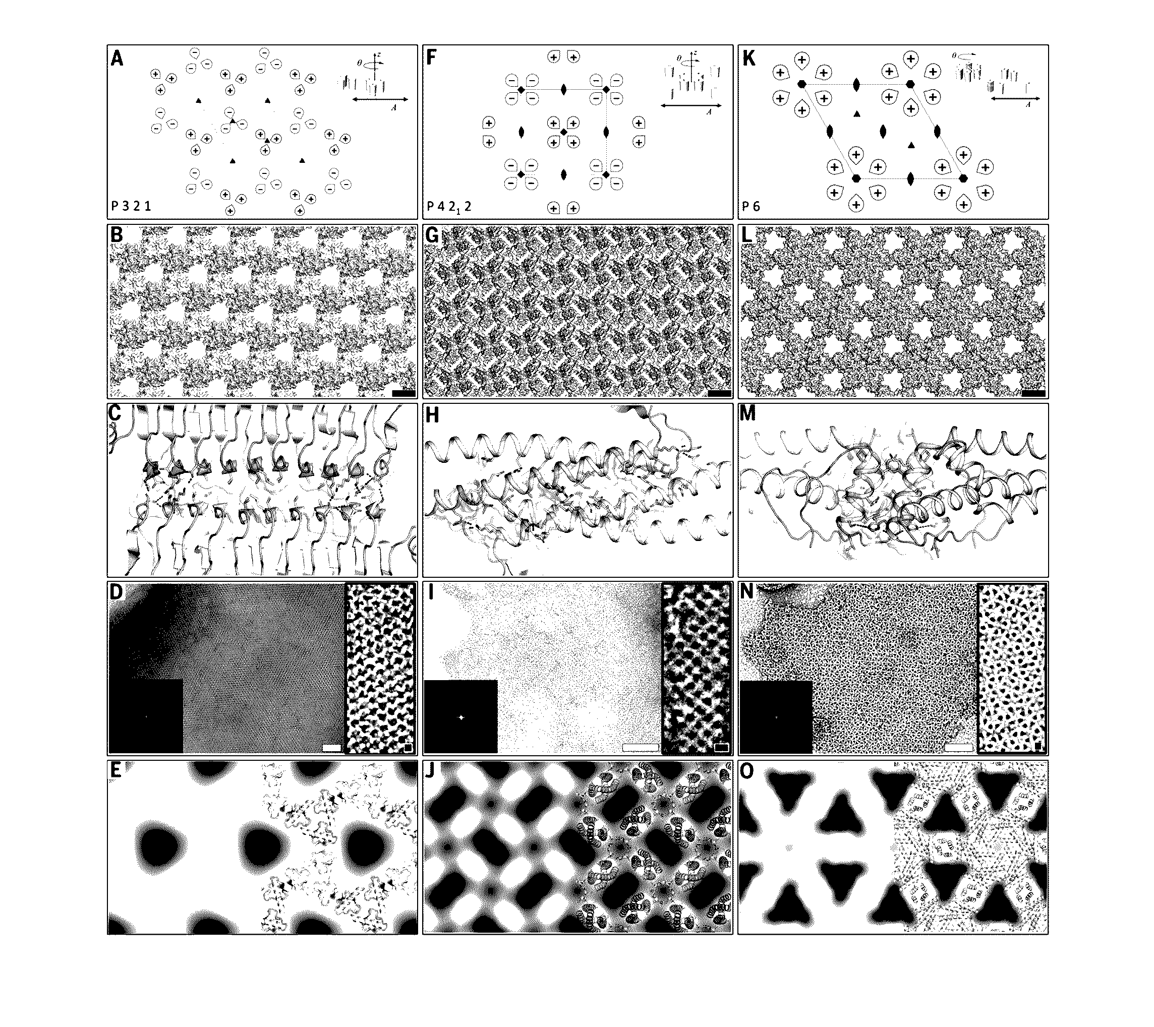
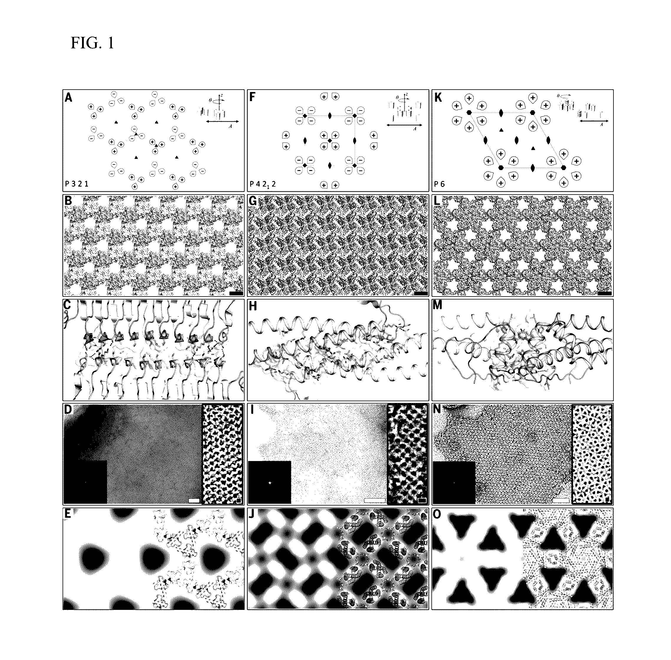
[0061]Ordered two-dimensional arrays mediated by designed protein-protein interfaces stabilized by extensive non-covalent interactions were designed. Symmetric arrays were focused on as symmetry reduces the number of distinct protein interfaces required to stabilize the lattice. There are seventeen distinct ways (layer groups) in which three-dimensional objects can come together to form periodic two-dimensional layers (Nannenga et al., “Overview of electron crystallography of membrane proteins: crystallization and screening strategies using negative stain electron microscopy.” Coligan et al. (Eds.) Current Protocols in Protein Science Chapter 17, Unit 17 15 (2013)). In some layer groups there are only two unique interfaces between identical subunits, in others, three or four. Layer groups involving only two unique interfaces, and building blocks with internal point symmetry (which alr...

Example

Example 2

Atomic Patterning of Proteins and Fluorescent Dyes Using Designed Two-Dimensional Protein Arrays

[0095]Proteins of interest were genetically fused to the N- or C-terminus of each of the array monomers using small linkers made of Glycine-Serine and Glycine-Glycine repeats (6-8 amino acid residues total), whereby the designed residues will drive self-assembly of both proteins (FIG. 8B). Based on the results obtained in the original study, design p4Z-9 had the smallest unit cell size (˜5 nm repeats) and was made up of very small proteins (˜12 kDa) and design p6-9H was shown to be both slow to form an array in vivo and highly soluble in vitro unless concentrated to a very high concentration. p3Z-42 is made up of large building blocks (˜25 kDa) and was shown to assemble into arrays at a very fast rate, both by in vivo and in vitro expression and would be well suited for fusion arrays.

[0096]Synthetic genes of each fusion were obtained and protein was expressed in Escherichia coli ...


PUM

PropertyMeasurementUnit
Lengthaaaaaaaaaa
Lengthaaaaaaaaaa
Lengthaaaaaaaaaa

Abstract

This document relates to two dimensional (2D) protein arrays can be used in biotechnology applications, as well as methods of making and using 2D protein arrays. In some cases, a 2D protein array can be used to evaluate (e.g., image) a structure (e.g., a three dimensional (3D) structure) of a protein of interest. In some cases, a 2D protein array can be used to evaluate (e.g., characterize) protein-protein interactions (e.g., stable interactions vs. transient interactions). In some cases, a 2D protein array can be used to evaluate a binding domain in a protein of interest. In some cases, a 2D protein array can be used to evaluate (e.g., identify) binding targets and/or partners of a protein of interest.

Description

CROSS-REFERENCE TO RELATED APPLICATIONS[0001]This application claims the benefit of U.S. Provisional Application No. 62 / 182,368, filed Jun. 19, 2015. The disclosure of the prior application is incorporated by reference in its entirety.STATEMENT REGARDING FEDERAL FUNDING[0002]This invention was made with government support under grant no. FA9550-12-1-0112, awarded by the Air Force Office of Scientific Research, and under grant no. N00024-10-D-6318 / 002, awarded by the Defense Threat Reduction Agency. The government has certain rights in the invention.SEQUENCE LISTING[0003]The instant application includes a sequence listing in electronic format submitted to the United States Patent and Trademark Office via the electronic filing system. The ASCII text file, which is incorporated-by-reference herein, is titled “30872-0012001_ST25.txt,” was created on Jun. 20, 2016, has a size of 48 kilobytes.BACKGROUND[0004]1. Technical Field[0005]This document relates to methods and materials for making...

Claims


Application Information

Patent Timeline
IPC IPC(8): C12N15/10G06F19/16G01N33/68G16B15/30
CPCC12N15/1037G01N33/6803G01N2610/00G01N33/6845G06F19/16G01N33/542G16B15/00G16B15/30
Inventor GONEN, TAMIRGONEN, SHANELIONNET, TIMOTHEEBAKER, DAVIDDIMAIO, FRANKENGLISH, BRIANROUAULT, HARVE
Owner HOWARD HUGHES MEDICAL INST
Why Eureka
Try Eureka