Novel Method

Inactive Publication Date: 2017-05-18
CAMBRIDGE ENTERPRISE LTD
0 Cites 20 Cited by

AI-Extracted Technical Summary

Problems solved by technology

These approaches are limited, however, by requirements for additional individualized reagents and/or leave cells coated with residual antibody-antigen complexes.
All of these techniques rely on the use of ant...

Abstract

The invention relates to a method of cell selection by using a nucleic acid molecule comprising a first nucleic acid sequence encoding a streptavidin binding peptide and a second nucleic acid sequence encoding a cell surface protein. The invention also relates to nucleic acid molecules, vectors, cells and kits for use with said method.

Application Domain

Polypeptide with localisation/targeting motifVectors +5

Technology Topic

Molecular biologyNucleic acid molecule +6

Image

  • Novel Method
  • Novel Method
  • Novel Method

Examples

  • Experimental program(2)

Example

Example 1: Development of Antibody-Free Magnetic Cell Sorting
Materials and Methods
Antibodies and Reagents
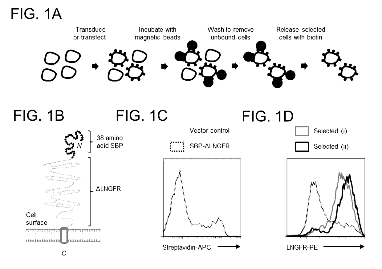
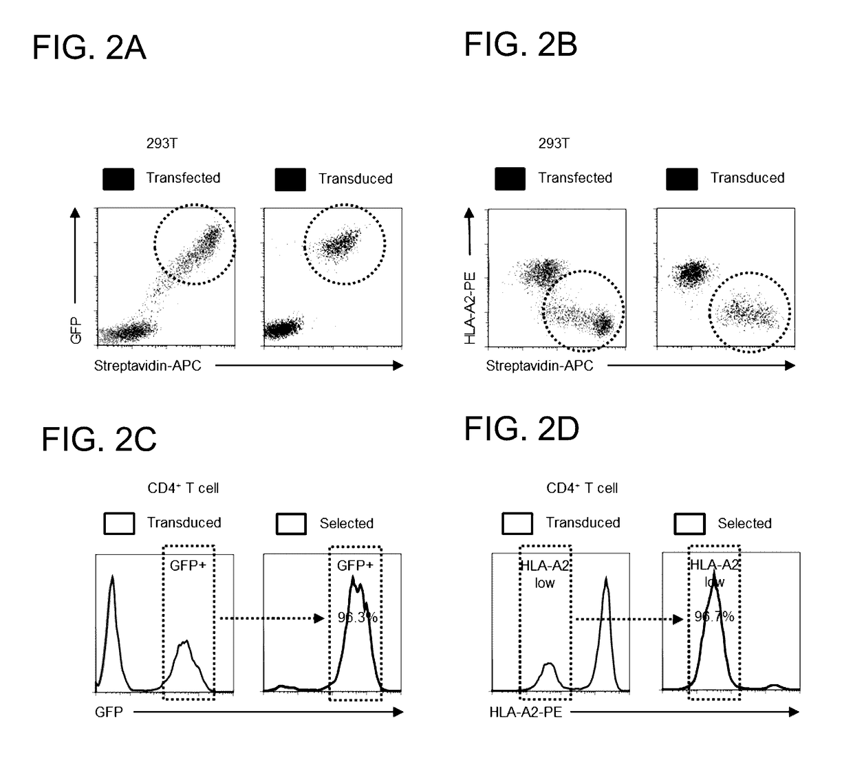
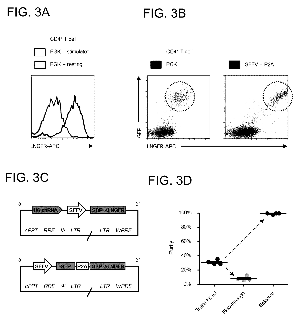
[0096]The following fluorescent conjugates were used for flow cytometry: ME20.4 anti-LNGFR-PE/APC (BioLegend); BB7.2 anti-HLA-A2-PE (BioLegend); W6/32 anti-MHC-I-AF647 (BioLegend); and streptavidin-APC (eBioscience). Bovine Serum Albumin (BSA) Cohn fraction V (A4503; Sigma) which does not contain free biotin was used for Antibody-Free Magnetic Cell Sorting.
Cell Culture
[0097]HEK 293T cells (293 Ts) were cultured in Dulbecco's Modified Eagle Medium (DMEM) supplemented with 10% Fetal Calf Serum (FCS) and 1% penicillin/streptomycin. Primary human CD4+T cells were isolated from peripheral blood by density gradient centrifugation using Lympholyte-H (Cedarlane Laboratories) followed by negative selection using the Dynabeads Untouched Human CD4 T Cells Kit (Invitrogen) according to the manufacturer's instructions. Cells were cultured in RPMI-1640 supplemented with 10% FCS and 1% penicillin/streptomycin and activated within 48 hours using Dynabeads Human T-Activator CD3/CD28 beads (Invitrogen) according to the manufacturer's instructions. Purity was assessed by flow cytometry for CD3 and CD4 and typically found to be ≧95%.
Plasmids
[0098]The lentiviral expression construct pHRSIN-HA-HLA-A2 (encoding HLA-A2 with an N-terminal hemagglutinin (HA) tag and a murine immunoglobulin signal peptide) has been previously described (Burr, van den Boomen et al. (2013) PNAS 108(5): 2034-2039). Overlapping DNA oligomers encoding the 38 amino acid Streptavidin Binding Peptide (SBP) (Keefe, Wilson et al. (2001) Protein Expr. Purif. 23(3): 440-446; Wilson, Keefe et al. (2001) PNAS 98(7): 3750-3755) were synthesised (Sigma), ligated and inserted using EcoRI/XhoI sites to generate pHRSIN-HA-SBP-HLA-A2. The truncated Low-affinity nerve growth factor receptor (LNGFR) was then amplified by PCR from the retroviral vector pZLRS-IRES-ΔLNGFR (Hassink, Barel et al. (2006) J. Biol. Chem. 281(4): 30063-30071) and inserted using XhoI/NotI sites in place of HLA-A2 to generate the pHRSIN-HA-SBP-ΔLNGFR construct utilised for pilot experiments in 293 Ts (FIG. 1).
[0099]To generate bicistronic lentiviral vectors (FIGS. 2 and 3), a codon-optimised SBP-ΔLNGFR fusion protein construct was synthesised in pUC57 (Genscript). For co-expression with an exogenous gene of interest, this construct was subcloned into a self-inactivating lentiviral vector derived from pHRSIN-cPPT-SEW kindly provided by Yasuhiro Ikeda (Demaison, Parsley et al. (2002) Hum. Gene Ther. 13(7): 803-813) to generate pHRSIN-SE-PGK-SBP-ΔLNGFR-W (encoding SFFV-EGFP and PGK-SBP-ΔLNGFR with a distal Woodchuck Hepatitis Virus post-transcriptional regulatory element [WPRE]). The phosphoglycerate kinase (PGK) promoter was replaced with a Porcine teschovirus-1 2A (P2A) sequence (Kim, Lee et al. (2011) PLoS One 6(4): e18556) synthesised in pUC57 (Gencsript) to generate pHRSIN-SE-P2A-SBP-ΔLNGFR-W. BamHI and NotI sites flanking EGFP allow substitution of alternative Genes Of Interest (GOI) for co-translation as GOI-P2A-SBP-ΔLNGFR. For co-expression with an shRNA of interest, the SBP-ΔLNGFR construct was subcloned into a self-inactivating lentiviral vector derived from pCSRQ kindly provided by Greg Towers (Schaller, Ocwieja et al. (2011) PLoS Pathol. 7(12): e1002439) to generate pHRSIREN-PGK-SBP-ΔLNGFR-W (encoding a U6-shRNA cassette and PGK-SBP-ΔLNGFR with a distal WPRE). The PGK promoter was replaced with a spleen focus-forming virus (SFFV) promoter PCR-amplified from pHRSIN-cPPT-SEW to generate pHRSIREN-S-SBP-ΔLNGFR-W. BamHI and EcoRI sites allow insertion of alternative shRNAs of interest into the U6-shRNA cassette as described for the pSIREN-RetroQ vector (Clontech). For Clustered Regularly Interspaced Short Palindromic Repeats (CRISPR)/Cas9 genome editing, P2A-SBP-ΔLNGFR was subcloned from pHRSIN-SE-P2A-SBP-ΔLNGFR-W into pSpCas9(BB)-2A-Puro (PX459; Addgene) to generate pSpCas9(BB)-P2A-SBP-ΔLNGFR (encoding a U6-guide RNA (gRNA) cassette and human codon-optimized S. pyogenes Cas9 (SpCas9) co-translated with SBP-ΔLNGFR via a P2A peptide linker). BbsI sites allow insertion of site-specific gRNAs identified using the CRISPR Design Tool (http://crispr.mit.edu) (Hsu et al. (2013) Nat. Biotechnol. 31(9): 827-832) according to protocols kindly supplied by Feng Zhang (http://www.genome-engineering.org) (Cong et al. (2013) Science 339(6121): 819-823).
[0100]The final nucleotide and amino acid sequences of the codon-optimised SBP-ΔLNGFR construct are shown (FIG. 5). For knockdown of β2-microglobulin (β2m), the following shRNA target sequence was used: 5′-GAATGGAGAGAGAATTGAA-3′ (SEQ ID NO: 19) (Burr, van den Boomen et al. (2013) PNAS 108(5): 2034-2039). For knockout of β2m, the following gRNA target sequence was kindly selected and subcloned by Dick van den Boomen: 5′-GGCCGAGATGTCTCGCTCCG-3′ (SEQ ID NO: 20).
Transfection and Lentiviral Transduction
[0101]FuGENE 6 (Promega; lentiviral production) or TransIT-293 (Mirus; general transfections) were used for plasmid DNA transfections in 293 Ts. To generate pseudotyped lentiviral stocks, 293 Ts were co-transfected with pHRSIN-/pHRSIREN-based lentivector, pCMVR8.91 and pMD.G, media changed at 24 hours and viral supernatant harvested and filtered (0.45 μm) at 48 hours prior to concentration using Lenti-X Concentrator (Clontech) or storage at −80° C. Transduction of primary human CD4+T cells 6-24 hours after activation was performed by spinoculation at 800 g for 1-2 hours in a benchtop centrifuge.
Flow Cytometry
[0102]293 Ts were harvested with enzyme-free cell dissociation buffer and Dynabeads Human T-Activator CD3/CD28 beads were removed from primary human CD4+T cells using a DynaMag-2 magnet (Invitrogen). Typically 2×10⁵ washed cells were incubated for 30 minutes in 100 μL PBS with the indicated fluorochrome-conjugated antibody or streptavidin-APC. All steps were performed on ice or at 4° C. and stained cells were analysed immediately or fixed in PBS/1% paraformaldehyde.
Antibody-Free Magnetic Cell Sorting
[0103]For pilot experiments in transfected 293 Ts (FIG. 1), washed cells were harvested with enzyme-free dissociation buffer and filtered (50 μm) to remove clumps. For selection using Dynabeads Biotin Binder (Invitrogen) cells were resuspended in Incubation Buffer (IB; PBS without calcium/magnesium, 2 mM EDTA, 0.1% BSA) at 107 cells/ml and incubated with Dynabeads at a bead-to-total cell ratio of 4:1 for 30 minutes at 4° C. Bead-bound cells were selected using a DynaMag-2 (Invitrogen) then released from the beads by incubation in IB supplemented with 2 mM biotin for 15 minutes at room temperature (RT) and analysed by flow cytometry. For selection using Streptavidin MicroBeads (Miltenyi) cells were resuspended in IB at 2.5×10⁷ cells/ml and incubated with MicroBeads at a bead-to-total cell ratio of 10 μl:107 cells for 30 minutes at 4° C. Bead-bound cells were selected using an MS Column and MACS Separator (Miltenyi) and analysed by flow cytometry without MicroBead removal. For selection of transduced primary human CD4+T cells, Dynabeads Human T-Activator CD3/CD28 beads were first removed according to the manufacturer's instructions. An optimised protocol for Antibody-Free Magnetic Cell Sorting using Dynabeads Biotin Binder is shown in Example 2.
Results and Discussion
[0104]The 38 Amino Acid SBP May be Displayed at the Cell Surface by Fusion with the Truncated LNGFR.
[0105]The 38 amino acid SBP is a high-affinity streptavidin-binding peptide tag previously used for purification of recombinant proteins and, more recently, as an affinity tag in live cells for the synchronisation of secretory traffic (Keefe, Wilson et al. (2001) Protein Expr. Purif. 23(3): 440-446; Wilson, Keefe et al. (2001) PNAS 98(7): 3750-3755; Boncompain, Divoux et al. (2012) Nat. Methods 9(5): 493-498). To express the 38 amino acid SBP at the cell surface, it was fused it to the N-terminus of the truncated LNGFR (SBP-ΔLNGFR; FIG. 1b). 293 Ts transfected with this construct were readily stained with streptavidin-APC in the absence of permeabilisation, indicating expression of SBP-ΔLNGFR at the plasma membrane and accessibility for streptavidin binding (FIG. 1c). SBP-ΔLNGFR was also readily detected using an LNGFR-specific antibody (FIG. 1d). LNGFR is a 399 amino acid Type I transmembrane cell surface glycoprotein member of the Tumour Necrosis Factor Receptor superfamily (Rogers, Beare et al. (2008) J. Biol. Regul. Homeost. Agents 22(1): 1-6). The truncated LNGFR, which lacks a cytoplasmic domain, has been previously used as a non-functional cell surface marker for antibody-based cell selection, including in vitro and in vivo for purification of transduced human lymphocytes in the setting of allogenic bone marrow transplantation (Bonini, Ferrari et al. (1997) Science 276(5319): 1719-1724; Ruggieri, Aiuti et al. (1997) Hum Gene Ther. 8(13): 1611-1623). The level of cell surface streptavidin-binding peptide expression achieved was critically dependent on the fusion protein chosen, since preliminary experiments using the 38 amino acid SBP fused to the HLA-A2 heavy chain, or the streptavidin-binding Nano-tag peptide fused to a membrane-targeted red fluorescent protein construct (Lamla and Erdmann (2004) Protein Expr. Purif. 33(1): 39-47; Winnard, Kluth et al. (2007) Cancer Biol. Ther. 6(12): 1889-1899), showed poor staining at the surface of transfected cells.
Cells Expressing SBP-ΔLNGFR May be Selected Using Streptavidin-Conjugated Magnetic Beads.
[0106]To test whether SBP-ΔLNGFR could be used for cell selection, transfected 293 Ts were incubated with streptavidin-conjugated magnetic beads. Bead-bound cells were washed, and then either analysed directly by flow cytometry, or released from the beads by incubation with excess biotin. Selected cells were markedly enriched for SBP-ΔLNGFR expression, and comparable results were achieved using streptavidin-conjugated beads from 2 different manufacturers (FIG. 1d). Dynabeads Biotin Binder were used for subsequent experiments at an optimised bead-to-target cell ratio of 10:1. Although the 38 amino acid SBP interacts strongly with streptavidin (nanomolar Kd, comparable to a strong antibody-antigen interaction), it is readily out-competed by biotin (femtomolar Kd, one of the strongest non-covalent interactions known) (Green (1990) Methods Enzymol. 184: 51-67; Brent (2001) Curr. Protoc. Protein Sci. Chapter 19: Unit 19 11; Keefe, Wilson et al. (2001) Protein Expr. Purif. 23(3): 440-446; Boncompain, Divoux et al. (2012) Nat. Methods 9(5): 493-498). In practice, bound cells could be completely released from streptavidin-conjugated beads by incubation with 2 mM biotin for as little as 15 minutes. Magnetic selection of cells expressing cell surface streptavidin (using bead-bound anti-streptavidin antibody) or co-expressing a cell surface biotin-acceptor peptide with the E. coli biotin ligase BirA (using streptavidin-conjugated beads) has been previously described (Gotoh and Matsumoto (2007) Gene 389(2): 146-153; Han, Liu et al. (2011) PLoS One 6(11): e26380; Lee and Lufkin (2012) J. Biomol. Tech. 23(2): 69-77), as has FACS of cells expressing a cell surface biotin-mimetic peptide (using fluorochrome-conjugated streptavidin) (Helman, Toister-Achituv et al. (2014) Cytometry A 85(2): 162-168). Conversely, this is the first report of the use of a cell surface streptavidin binding peptide for magnetic cell sorting, combining the advantages of bead-based cell isolation with the ability to release beads from selected cells by competition with biotin.
SBP-ΔLNGFR Affinity Purification May be Used to Isolate Cells Expressing an shRNA or Exogenous Gene of Interest.
[0107]To select genetically modified mammalian cells using SBP-ΔLNGFR affinity purification, the fusion protein was co-expressed with an exogenous gene or shRNA on the same lentiviral construct. As proof of principle, SBP-ΔLNGFR was subcloned into lentiviral vectors encoding either GFP or an shRNA to β2-microbglobulin (β2m). (β2m is an essential subunit of MHC class I molecules and its depletion may therefore be detected by reduction of cell surface MHC class I alleles such as HLA-A2 (Burr, Cano et al. (2011) PNAS 108(5): 2034-2039). Co-expression of SBP-ΔLNGFR with GFP (FIG. 2a) or shRNA to (β2m (FIG. 2b) was confirmed by transient transfection of 293 Ts, and similar results were obtained using VSVg-pseudotyped lentiviral particles (FIGS. 2a and 2b). The selection of cells genetically modified ex vivo remains a significant methodological challenge for human gene therapy. As well as the treatment of monogenic disorders such as ADA-SCID (adenosine deaminase deficiency resulting in severe combined immunodeficiency) major research efforts have focussed on cancer immunotherapy using engineered T cells expressing tumour-specific T cell receptor α and β chains (αβTCRs) or chimeric antigen receptors (CARs), and the production of HIV-resistant CD4+T cells through, for example, disruption or downregulation of the CCR5 HIV co-receptor (Kalos and June (2013) Immunity 39(1): 49-60; Kaufmann, Buning et al. (2013) EMBO Mol. Med. 5(11): 1642-1661; Peterson, Younan et al. (2013) Gene Ther. 20(7): 695-702). It was therefore tested whether magnetic selection for SBP-ΔLNGFR could be used to purify genetically modified primary human CD4+T lymphocytes expressing an exogenous gene or shRNA of interest. Indeed, following lentiviral transduction and SBP-ΔLNGFR affinity purification, pure populations of cells either high in GFP or low in HLA-A2 were successfully isolated (FIGS. 2c and 2d).
Antibody-Free Magnetic Cell Sorting Yields Greater than 99% Pure Populations of Primary Human CD4+T Cells in Less than 1 Hour.
[0108]Expression of SBP-ΔLNGFR from the PGK promoter was noted to vary markedly according to the activation state of transduced T cells (FIG. 3a). PGK encodes the glycolytic enzyme phoshpoglycerokinase, and glycolysis is known to be highly regulated in T cells (MacIver, Michalek et al. (2013) Annu. Rev. Immunol. 31: 259-283). To optimise the system for selecting primary human lymphocytes, the SFFV promoter was introduced to drive expression of SBP-ΔLNGFR either as a single cistron (pHRSIREN-S-SBP-ΔLNGFR-W) or co-translated with an exogenous gene of interest via a P2A “self-cleaving” peptide linker for bicistronic expression (pHRSIN-SE-P2A-SBP-ΔLNGFR-W). These modifications increased SBP-ΔLNGFR expression without compromising levels of the co-expressed gene or shRNA of interest (FIG. 3b). Expression levels could be increased depending on both the WPRE and the promoter strategy used, with inferior results obtained using the EF1a promoter, ECMV IRES or dual SFFV promoter systems, or when the WPRE was absent or alternatively located. The SFFV promoter is known to provide high-level transgene expression in primary human haematopoietic cells (Demaison, Parsley et al. (2002) Hum. Gene Ther. 13(7): 803-813) and 2A peptides have been shown to enable stoichiometric co-expression of multiple cistrons across different organisms and cell types (Szymczak, Workman et al. (2004) Nat. Biotechnol. 22(5): 589-594; Kim, Lee et al. (2011) PLoS One 6(4): e18556). These small viral peptide sequences are co-translationally “cleaved” in a process known as “ribosomal skipping” in which formation of a glycyl-prolyl peptide bond at the C-terminus of the 2A peptide is “skipped” without interrupting translation of the downstream polypeptide (Donnelly, Hughes et al. (2001) J. Gen. Virol. 82(Pt.5): 1027-1041). To test the optimised vectors (FIG. 3c), primary human CD4+T cells were transduced using 4 different constructs (expressing 2 different shRNAs and 2 different exogenous genes). From a starting purity of 31.0%, the average purity of selected cells was 99.2% (FIG. 3d). No deficit in cell viability or function in a wide range of downstream applications has been observed, and the Antibody-Free Magnetic Cell Sorting procedure (from incubation with magnetic beads through release with biotin) may be readily completed (including multiple samples) in less than 1 hour.
Antibody-Free Magnetic Cell Sorting Allows Isolation of Cells Following CRISPR/Cas9 Genome Editing.
[0109]The type II bacterial CRISPR “immune system” has recently been re-purposed to allow facile site-specific genome engineering in mammalian cells by co-expression of the Cas9 nuclease with a short gRNA (Cho et al. (2013) Nat. Biotechnol. 31(3): 230-232; Cong et al. (2013) Science 339(6121): 819-823; Mali et al. (2013) Science 339(6121): 823-826). Complementary base-pairing through the gRNA recruits the gRNA/Cas9 complex to target sequences in the genomic DNA, where it introduces double-strand DNA breaks. Repair of these breaks by non-homologous end joining frequently introduces short insertions and deletions (InDels), leading to frameshifts and/or premature stop codons in the open reading frame (ORF) of the targeted gene (knock-out). Alternatively, where an exogenous DNA repair template is also supplied, homology-directed repair copies the sequence of this template to the cut target sequence, allowing the introduction of specific nucleotide changes (knock-in). To test whether Antibody-Free Magnetic Cell Sorting could be used to select cells following CRISPR gene editing, 293 Ts were transfected with a vector encoding a gRNA targeting the 5′ end of the β2m gene and SBP-ΔLNGFR co-translated with Cas9 via a P2A peptide linker (FIG. 4). As with shRNA knockdown, disruption of the β2m gene may be detected by reduction in cell surface MHC class I (MHC-I). Following Antibody-Free Magnetic Selection, MHC-I low cells were markedly enriched.
Conclusions
[0110]Antibody-Free Magnetic Cell Sorting is a novel, efficient way to select transfected or transduced mammalian cells. Selection is readily scalable to almost any cell number and may be completed in less than 1 hour (plus cell washes). No antibody is required, allowing rapid one-step affinity purification and making the process extremely cost-effective. Enrichment to greater than 99% purity is routinely achieved and, following release with biotin, cells are left “untouched” by residual beads or antibody-antigen complexes. As well as providing a useful tool for life sciences research, the system may be used to select genetically modified cells for human gene therapy applications. Genetic modifications need not be limited to expression of shRNAs, exogenous genes of interest or CRISPR/Cas9 genome editing. For example, vectors may be developed for one-step magnetic selection of cells infected with an HIV reporter virus (Zhang, Zhou et al. (2004) J. Virol. 78(4): 1718-1729), or expression of SBP-ΔLNGFR may be used as a reporter gene for selection of cells in which a promoter of interest is active in vitro or in vivo.

Example

Example 2: Protocol for Antibody-Free Magnetic Cell Sorting
[0111]The following protocol has been optimised for Antibody-Free Magnetic Cell Sorting of transduced primary human CD4+ T cells to maximum purity using Dynabeads Biotin Binder. It may be readily scaled for almost any cell number and adapted for other transfected or transduced cell types. It is important to note that: [0112] Adherent cells must be harvested with enzyme-free dissociation buffer [0113] All cells must be washed thoroughly to avoid carry-over of biotin from culture media [0114] Where indicated by the manufacturer, streptavidin-conjugated beads must be washed before use to remove preservative and/or free (unconjugated) streptavidin
Materials
[0115]
Incubation Buffer (IB) PBS without calcium/magnesium, pH 7.4 Pre-cool on ice 2 mM EDTA 0.1% BSA (A4503; Sigma) Release Buffer (RB) Complete media e.g. RPMI-1640 Pre-warm to 37° C. with 10% FCS and 1% pencillin/streptomycin 10 mM HEPES buffer, pH 7.4 2 mM biotin
Protocol
[0116]1. If required, remove Dynabeads Human T-Activator CD3/CD28 beads (Invitrogen) according to the manufacturer's instructions.
2. Wash cells 3 times with cold IB then resuspend in same at 107 cells/ml.
3. Add Dynabeads Biotin Binder at a bead:transduced cell ratio of 10:1 and incubate at 4° C. for 30 minutes with gentle agitation.
4. Place tube on appropriate magnet for 2-3 minutes and remove supernatant containing unbound cells.
5. Gently wash bead-bound cells once with cold IB then return to magnet for 2-3 minutes and remove supernatant containing unbound cells.
6. Resuspend bead-bound cells in pre-warmed RB at no more than 107 cells/ml and incubate at room temperature for 15 minutes with gentle agitation.
7. Place tube on appropriate magnet for 2-3 minutes then transfer supernatant containing released cells to new tube.
8. If desired, to maximise yield, wash beads once with RB then return to magnet for 2-3 minutes and pool supernatants containing released cells.
9. Wash released cells twice with complete media and use as required for downstream applications.
<160> NUMBER OF SEQ ID NOS: 20
<210> SEQ ID NO: 1
<211> LENGTH: 38
<212> TYPE: PRT
<213> ORGANISM: Artificial
<223> OTHER INFORMATION: Synthetic Peptide
<400> SEQENCE: 1
Met Asp Glu Lys Thr Thr Gly Trp Arg Gly Gly His Val Val Glu Gly
1 5 10 15
Leu Ala Gly Glu Leu Glu Gln Leu Arg Ala Arg Leu Glu His His Pro
20 25 30
Gln Gly Gln Arg Glu Pro
35
<210> SEQ ID NO: 2
<211> LENGTH: 114
<212> TYPE: DNA
<213> ORGANISM: Artificial
<223> OTHER INFORMATION: Synthetic Nucleotide
<400> SEQENCE: 2
atggacgaaa agaccacagg atggcgagga ggacacgtgg tcgagggact ggcaggagag 60
ctggaacagc tgcgggctag actggaacac catcctcagg gacagcgaga gcca 114
<210> SEQ ID NO: 3
<211> LENGTH: 15
<212> TYPE: PRT
<213> ORGANISM: Artificial
<223> OTHER INFORMATION: Synthetic Peptide
<400> SEQENCE: 3
Asp Val Glu Ala Trp Leu Asp Glu Arg Val Pro Leu Val Glu Thr
1 5 10 15
<210> SEQ ID NO: 4
<211> LENGTH: 15
<212> TYPE: PRT
<213> ORGANISM: Artificial
<223> OTHER INFORMATION: Synthetic Peptide
<400> SEQENCE: 4
Asp Val Glu Ala Trp Leu Gly Glu Arg Val Pro Leu Val Glu Thr
1 5 10 15
<210> SEQ ID NO: 5
<211> LENGTH: 15
<212> TYPE: PRT
<213> ORGANISM: Artificial
<223> OTHER INFORMATION: Synthetic Peptide
<400> SEQENCE: 5
Asp Val Glu Ala Trp Leu Gly Ala Arg Val Pro Leu Val Glu Thr
1 5 10 15
<210> SEQ ID NO: 6
<211> LENGTH: 9
<212> TYPE: PRT
<213> ORGANISM: Artificial
<223> OTHER INFORMATION: Synthetic Peptide
<400> SEQENCE: 6
Asp Val Glu Ala Trp Leu Asp Glu Arg
1 5
<210> SEQ ID NO: 7
<211> LENGTH: 9
<212> TYPE: PRT
<213> ORGANISM: Artificial
<223> OTHER INFORMATION: Synthetic Peptide
<400> SEQENCE: 7
Asp Val Glu Ala Trp Leu Gly Glu Arg
1 5
<210> SEQ ID NO: 8
<211> LENGTH: 9
<212> TYPE: PRT
<213> ORGANISM: Artificial
<223> OTHER INFORMATION: Synthetic Peptide
<400> SEQENCE: 8
Asp Val Glu Ala Trp Leu Gly Ala Arg
1 5
<210> SEQ ID NO: 9
<211> LENGTH: 101
<212> TYPE: PRT
<213> ORGANISM: Artificial
<223> OTHER INFORMATION: Synthetic Peptide
<400> SEQENCE: 9
Met Asp Glu Lys Thr His Cys Phe His Pro Gly Asp His Leu Val Arg
1 5 10 15
Leu Val Glu Glu Leu Gln Ala Leu Ala Glu Gly Leu Gln Arg Gln Gly
20 25 30
Gly Arg Gln Pro His Arg Leu Pro Arg Arg Arg Pro His His Leu Gln
35 40 45
Leu Leu Leu Asp Glu Ala His Pro Gln Ala Gly Pro Leu Arg Glu Arg
50 55 60
Ala His Gln Val Asp Gly Arg Leu Leu Leu Gln His His Pro Gln Gly
65 70 75 80
Asp Arg Leu Leu Gln Gln Pro Gln Asp His Pro Leu Glu Leu Val Trp
85 90 95
Arg Leu Pro Pro Ser
100
<210> SEQ ID NO: 10
<211> LENGTH: 101
<212> TYPE: PRT
<213> ORGANISM: Artificial
<223> OTHER INFORMATION: Synthetic Peptide
<400> SEQENCE: 10
Met Asp Glu Lys Thr His Cys Thr Ile Ser Met Asn Gly Ala Val Pro
1 5 10 15
Leu Val Pro His His His Pro Gln Gly Asp Pro Leu Arg Leu Leu His
20 25 30
Arg Pro Gln Pro Ala Leu Leu Val Arg His Pro Gln Gly Asp Leu Val
35 40 45
Ala Leu Val Glu His His Glu Gly Val Asp Arg Gly Leu Val Ala Leu
50 55 60
Pro Glu Leu His Ala Glu Glu Leu Gly Glu Pro Val Gly Asp Leu Val
65 70 75 80
Gln Gly Pro Val Glu Gln Val Gln Gly Val Val Asp Ala Leu Val Trp
85 90 95
Arg Leu Pro Pro Ser
100
<210> SEQ ID NO: 11
<211> LENGTH: 102
<212> TYPE: PRT
<213> ORGANISM: Artificial
<223> OTHER INFORMATION: Synthetic Peptide
<400> SEQENCE: 11
Met Asp Glu Lys Thr His Trp Val Asn Val Tyr His Pro Gln Gly Asp
1 5 10 15
Leu Leu Val Arg Gly His Gly His Asp Val Glu Ala Leu His Asp Gln
20 25 30
Gly Leu His Gln Leu Asp Leu Leu Val Gly Pro Pro Pro Glu Val Val
35 40 45
Arg Ala Leu Arg Gly Glu Val Leu Gly Gly Leu Arg Arg Leu Val Pro
50 55 60
Leu Asp His Pro Gln Gly Glu Ala Leu Asp Gln Ala Arg Gln Arg Pro
65 70 75 80
Gln His Leu Leu Glu Leu His His Arg Ala Leu Pro Pro Ala Leu Val
85 90 95
Trp Arg Leu Pro Pro Ser
100
<210> SEQ ID NO: 12
<211> LENGTH: 101
<212> TYPE: PRT
<213> ORGANISM: Artificial
<223> OTHER INFORMATION: Synthetic Peptide
<400> SEQENCE: 12
Met Asp Glu Lys Thr His Trp Leu Glu Asp Leu Lys Gly Val Leu Lys
1 5 10 15
Asp Cys Leu Lys Asp Leu Met Asp Phe Thr Lys Asp Cys Arg Ser Pro
20 25 30
Arg Val Gln Pro Gln Pro Leu Leu His His Asp Arg Gly Glu Pro Val
35 40 45
Pro Leu Leu Arg Glu Ala Gly Arg Asp Leu Gly Gly Leu Gly Pro Arg
50 55 60
Ala Pro Arg Gln Ala Arg Pro Leu His His Gly Arg His Asp Leu His
65 70 75 80
Glu Pro Leu Val Leu Gln Asp His Pro Gln Gly Gly Pro Leu Val Cys
85 90 95
Gly Cys His His His
100
<210> SEQ ID NO: 13
<211> LENGTH: 246
<212> TYPE: PRT
<213> ORGANISM: Artificial
<223> OTHER INFORMATION: Synthetic Peptide
<400> SEQENCE: 13
Lys Glu Ala Cys Pro Thr Gly Leu Tyr Thr His Ser Gly Glu Cys Cys
1 5 10 15
Lys Ala Cys Asn Leu Gly Glu Gly Val Ala Gln Pro Cys Gly Ala Asn
20 25 30
Gln Thr Val Cys Glu Pro Cys Leu Asp Ser Val Thr Phe Ser Asp Val
35 40 45
Val Ser Ala Thr Glu Pro Cys Lys Pro Cys Thr Glu Cys Val Gly Leu
50 55 60
Gln Ser Met Ser Ala Pro Cys Val Glu Ala Asp Asp Ala Val Cys Arg
65 70 75 80
Cys Ala Tyr Gly Tyr Tyr Gln Asp Glu Thr Thr Gly Arg Cys Glu Ala
85 90 95
Cys Arg Val Cys Glu Ala Gly Ser Gly Leu Val Phe Ser Cys Gln Asp
100 105 110
Lys Gln Asn Thr Val Cys Glu Glu Cys Pro Asp Gly Thr Tyr Ser Asp
115 120 125
Glu Ala Asn His Val Asp Pro Cys Leu Pro Cys Thr Val Cys Glu Asp
130 135 140
Thr Glu Arg Gln Leu Arg Glu Cys Thr Arg Trp Ala Asp Ala Glu Cys
145 150 155 160
Glu Glu Ile Pro Gly Arg Trp Ile Thr Arg Ser Thr Pro Pro Glu Gly
165 170 175
Ser Asp Ser Thr Ala Pro Ser Thr Gln Glu Pro Glu Ala Pro Pro Glu
180 185 190
Gln Asp Leu Ile Ala Ser Thr Val Ala Gly Val Val Thr Thr Val Met
195 200 205
Gly Ser Ser Gln Pro Val Val Thr Arg Gly Thr Thr Asp Asn Leu Ile
210 215 220
Pro Val Tyr Cys Ser Ile Leu Ala Ala Val Val Val Gly Leu Val Ala
225 230 235 240
Tyr Ile Ala Phe Lys Arg
245
<210> SEQ ID NO: 14
<211> LENGTH: 290
<212> TYPE: PRT
<213> ORGANISM: Artificial
<223> OTHER INFORMATION: Synthetic Peptide
<400> SEQENCE: 14
Met Asp Glu Lys Thr Thr Gly Trp Arg Gly Gly His Val Val Glu Gly
1 5 10 15
Leu Ala Gly Glu Leu Glu Gln Leu Arg Ala Arg Leu Glu His His Pro
20 25 30
Gln Gly Gln Arg Glu Pro Gly Ser Gly Ala Ile Ala Lys Glu Ala Cys
35 40 45
Pro Thr Gly Leu Tyr Thr His Ser Gly Glu Cys Cys Lys Ala Cys Asn
50 55 60
Leu Gly Glu Gly Val Ala Gln Pro Cys Gly Ala Asn Gln Thr Val Cys
65 70 75 80
Glu Pro Cys Leu Asp Ser Val Thr Phe Ser Asp Val Val Ser Ala Thr
85 90 95
Glu Pro Cys Lys Pro Cys Thr Glu Cys Val Gly Leu Gln Ser Met Ser
100 105 110
Ala Pro Cys Val Glu Ala Asp Asp Ala Val Cys Arg Cys Ala Tyr Gly
115 120 125
Tyr Tyr Gln Asp Glu Thr Thr Gly Arg Cys Glu Ala Cys Arg Val Cys
130 135 140
Glu Ala Gly Ser Gly Leu Val Phe Ser Cys Gln Asp Lys Gln Asn Thr
145 150 155 160
Val Cys Glu Glu Cys Pro Asp Gly Thr Tyr Ser Asp Glu Ala Asn His
165 170 175
Val Asp Pro Cys Leu Pro Cys Thr Val Cys Glu Asp Thr Glu Arg Gln
180 185 190
Leu Arg Glu Cys Thr Arg Trp Ala Asp Ala Glu Cys Glu Glu Ile Pro
195 200 205
Gly Arg Trp Ile Thr Arg Ser Thr Pro Pro Glu Gly Ser Asp Ser Thr
210 215 220
Ala Pro Ser Thr Gln Glu Pro Glu Ala Pro Pro Glu Gln Asp Leu Ile
225 230 235 240
Ala Ser Thr Val Ala Gly Val Val Thr Thr Val Met Gly Ser Ser Gln
245 250 255
Pro Val Val Thr Arg Gly Thr Thr Asp Asn Leu Ile Pro Val Tyr Cys
260 265 270
Ser Ile Leu Ala Ala Val Val Val Gly Leu Val Ala Tyr Ile Ala Phe
275 280 285
Lys Arg
290
<210> SEQ ID NO: 15
<211> LENGTH: 22
<212> TYPE: PRT
<213> ORGANISM: Artificial
<223> OTHER INFORMATION: Synthetic Peptide
<400> SEQENCE: 15
Met Gly Trp Ser Cys Ile Ile Leu Phe Leu Val Ala Thr Ala Thr Gly
1 5 10 15
Val His Ser Gln Val Gln
20
<210> SEQ ID NO: 16
<211> LENGTH: 317
<212> TYPE: PRT
<213> ORGANISM: Artificial
<223> OTHER INFORMATION: Synthetic Peptide
<400> SEQENCE: 16
Met Gly Trp Ser Cys Ile Ile Leu Phe Leu Val Ala Thr Ala Thr Gly
1 5 10 15
Val His Ser Gln Val Gln Leu Glu Gly Ser Gly Met Asp Glu Lys Thr
20 25 30
Thr Gly Trp Arg Gly Gly His Val Val Glu Gly Leu Ala Gly Glu Leu
35 40 45
Glu Gln Leu Arg Ala Arg Leu Glu His His Pro Gln Gly Gln Arg Glu
50 55 60
Pro Gly Ser Gly Ala Ile Ala Lys Glu Ala Cys Pro Thr Gly Leu Tyr
65 70 75 80
Thr His Ser Gly Glu Cys Cys Lys Ala Cys Asn Leu Gly Glu Gly Val
85 90 95
Ala Gln Pro Cys Gly Ala Asn Gln Thr Val Cys Glu Pro Cys Leu Asp
100 105 110
Ser Val Thr Phe Ser Asp Val Val Ser Ala Thr Glu Pro Cys Lys Pro
115 120 125
Cys Thr Glu Cys Val Gly Leu Gln Ser Met Ser Ala Pro Cys Val Glu
130 135 140
Ala Asp Asp Ala Val Cys Arg Cys Ala Tyr Gly Tyr Tyr Gln Asp Glu
145 150 155 160
Thr Thr Gly Arg Cys Glu Ala Cys Arg Val Cys Glu Ala Gly Ser Gly
165 170 175
Leu Val Phe Ser Cys Gln Asp Lys Gln Asn Thr Val Cys Glu Glu Cys
180 185 190
Pro Asp Gly Thr Tyr Ser Asp Glu Ala Asn His Val Asp Pro Cys Leu
195 200 205
Pro Cys Thr Val Cys Glu Asp Thr Glu Arg Gln Leu Arg Glu Cys Thr
210 215 220
Arg Trp Ala Asp Ala Glu Cys Glu Glu Ile Pro Gly Arg Trp Ile Thr
225 230 235 240
Arg Ser Thr Pro Pro Glu Gly Ser Asp Ser Thr Ala Pro Ser Thr Gln
245 250 255
Glu Pro Glu Ala Pro Pro Glu Gln Asp Leu Ile Ala Ser Thr Val Ala
260 265 270
Gly Val Val Thr Thr Val Met Gly Ser Ser Gln Pro Val Val Thr Arg
275 280 285
Gly Thr Thr Asp Asn Leu Ile Pro Val Tyr Cys Ser Ile Leu Ala Ala
290 295 300
Val Val Val Gly Leu Val Ala Tyr Ile Ala Phe Lys Arg
305 310 315
<210> SEQ ID NO: 17
<211> LENGTH: 342
<212> TYPE: PRT
<213> ORGANISM: Artificial
<223> OTHER INFORMATION: Synthetic Peptide
<400> SEQENCE: 17
Ala Ala Ala Gly Ser Gly Ala Thr Asn Phe Ser Leu Leu Lys Gln Ala
1 5 10 15
Gly Asp Val Glu Glu Asn Pro Gly Pro Met Gly Trp Ser Cys Ile Ile
20 25 30
Leu Phe Leu Val Ala Thr Ala Thr Gly Val His Ser Gln Val Gln Leu
35 40 45
Glu Gly Ser Gly Met Asp Glu Lys Thr Thr Gly Trp Arg Gly Gly His
50 55 60
Val Val Glu Gly Leu Ala Gly Glu Leu Glu Gln Leu Arg Ala Arg Leu
65 70 75 80
Glu His His Pro Gln Gly Gln Arg Glu Pro Gly Ser Gly Ala Ile Ala
85 90 95
Lys Glu Ala Cys Pro Thr Gly Leu Tyr Thr His Ser Gly Glu Cys Cys
100 105 110
Lys Ala Cys Asn Leu Gly Glu Gly Val Ala Gln Pro Cys Gly Ala Asn
115 120 125
Gln Thr Val Cys Glu Pro Cys Leu Asp Ser Val Thr Phe Ser Asp Val
130 135 140
Val Ser Ala Thr Glu Pro Cys Lys Pro Cys Thr Glu Cys Val Gly Leu
145 150 155 160
Gln Ser Met Ser Ala Pro Cys Val Glu Ala Asp Asp Ala Val Cys Arg
165 170 175
Cys Ala Tyr Gly Tyr Tyr Gln Asp Glu Thr Thr Gly Arg Cys Glu Ala
180 185 190
Cys Arg Val Cys Glu Ala Gly Ser Gly Leu Val Phe Ser Cys Gln Asp
195 200 205
Lys Gln Asn Thr Val Cys Glu Glu Cys Pro Asp Gly Thr Tyr Ser Asp
210 215 220
Glu Ala Asn His Val Asp Pro Cys Leu Pro Cys Thr Val Cys Glu Asp
225 230 235 240
Thr Glu Arg Gln Leu Arg Glu Cys Thr Arg Trp Ala Asp Ala Glu Cys
245 250 255
Glu Glu Ile Pro Gly Arg Trp Ile Thr Arg Ser Thr Pro Pro Glu Gly
260 265 270
Ser Asp Ser Thr Ala Pro Ser Thr Gln Glu Pro Glu Ala Pro Pro Glu
275 280 285
Gln Asp Leu Ile Ala Ser Thr Val Ala Gly Val Val Thr Thr Val Met
290 295 300
Gly Ser Ser Gln Pro Val Val Thr Arg Gly Thr Thr Asp Asn Leu Ile
305 310 315 320
Pro Val Tyr Cys Ser Ile Leu Ala Ala Val Val Val Gly Leu Val Ala
325 330 335
Tyr Ile Ala Phe Lys Arg
340
<210> SEQ ID NO: 18
<211> LENGTH: 298
<212> TYPE: PRT
<213> ORGANISM: Artificial
<223> OTHER INFORMATION: Synthetic Peptide
<400> SEQENCE: 18
Gln Val Gln Leu Glu Gly Ser Gly Met Asp Glu Lys Thr Thr Gly Trp
1 5 10 15
Arg Gly Gly His Val Val Glu Gly Leu Ala Gly Glu Leu Glu Gln Leu
20 25 30
Arg Ala Arg Leu Glu His His Pro Gln Gly Gln Arg Glu Pro Gly Ser
35 40 45
Gly Ala Ile Ala Lys Glu Ala Cys Pro Thr Gly Leu Tyr Thr His Ser
50 55 60
Gly Glu Cys Cys Lys Ala Cys Asn Leu Gly Glu Gly Val Ala Gln Pro
65 70 75 80
Cys Gly Ala Asn Gln Thr Val Cys Glu Pro Cys Leu Asp Ser Val Thr
85 90 95
Phe Ser Asp Val Val Ser Ala Thr Glu Pro Cys Lys Pro Cys Thr Glu
100 105 110
Cys Val Gly Leu Gln Ser Met Ser Ala Pro Cys Val Glu Ala Asp Asp
115 120 125
Ala Val Cys Arg Cys Ala Tyr Gly Tyr Tyr Gln Asp Glu Thr Thr Gly
130 135 140
Arg Cys Glu Ala Cys Arg Val Cys Glu Ala Gly Ser Gly Leu Val Phe
145 150 155 160
Ser Cys Gln Asp Lys Gln Asn Thr Val Cys Glu Glu Cys Pro Asp Gly
165 170 175
Thr Tyr Ser Asp Glu Ala Asn His Val Asp Pro Cys Leu Pro Cys Thr
180 185 190
Val Cys Glu Asp Thr Glu Arg Gln Leu Arg Glu Cys Thr Arg Trp Ala
195 200 205
Asp Ala Glu Cys Glu Glu Ile Pro Gly Arg Trp Ile Thr Arg Ser Thr
210 215 220
Pro Pro Glu Gly Ser Asp Ser Thr Ala Pro Ser Thr Gln Glu Pro Glu
225 230 235 240
Ala Pro Pro Glu Gln Asp Leu Ile Ala Ser Thr Val Ala Gly Val Val
245 250 255
Thr Thr Val Met Gly Ser Ser Gln Pro Val Val Thr Arg Gly Thr Thr
260 265 270
Asp Asn Leu Ile Pro Val Tyr Cys Ser Ile Leu Ala Ala Val Val Val
275 280 285
Gly Leu Val Ala Tyr Ile Ala Phe Lys Arg
290 295
<210> SEQ ID NO: 19
<211> LENGTH: 19
<212> TYPE: DNA
<213> ORGANISM: Artificial
<223> OTHER INFORMATION: Synthetic Nucleotide
<400> SEQENCE: 19
gaatggagag agaattgaa 19
<210> SEQ ID NO: 20
<211> LENGTH: 20
<212> TYPE: DNA
<213> ORGANISM: Artificial
<223> OTHER INFORMATION: Synthetic Nucleotide
<400> SEQENCE: 20
ggccgagatg tctcgctccg 20

PUM

PropertyMeasurementUnit
Surface
Affinity

Description & Claims & Application Information

We can also present the details of the Description, Claims and Application information to help users get a comprehensive understanding of the technical details of the patent, such as background art, summary of invention, brief description of drawings, description of embodiments, and other original content. On the other hand, users can also determine the specific scope of protection of the technology through the list of claims; as well as understand the changes in the life cycle of the technology with the presentation of the patent timeline. Login to view more.
Why Eureka
Try Eureka