
Conjugates for delivery to mitochondria

Inactive Publication Date: 2017-08-31
THE FORSYTH INST
View PDF3 Cites 2 Cited by
  • Summary
  • Abstract
  • Description
  • Claims
  • Application Information

AI Technical Summary

Benefits of technology

The patent describes a method for treating mitochondrial disorders or dysfunctions, and the use of a conjugate of a specific peptide called A44 or K39, which can increase or decrease the expression of certain factors that promote cell survival or death. The conjugate can be made more stable and have a longer half-life in the body, which makes it a useful therapeutic tool and helps improve safety and efficacy. The patent also describes methods for preventing or decreasing symptoms of mitochondrial dysfunction and reducing the number of mitochondria undergoing a process called mitochondrial permeability transitioning. Overall, the patent provides a technical solution for treating and preventing mitochondrial disorders and dysfunctions.

Problems solved by technology

However, this number may underestimate the true number of people with mitochondrial disease.
One barrier for developing effective therapeutic, diagnostic and research tools for addressing mitochondrial diseases is the lack of efficient strategies and reagents for targeting mitochondria.
The delivery problem is particularly acute for mitochondria-targeted agents because the mitochondrial membranes are generally highly impermeable.
Accordingly, effective transit across one or both mitochondrial membranes can be difficult to achieve.

Method used


Image

  • Conjugates for delivery to mitochondria
  • Conjugates for delivery to mitochondria
  • Conjugates for delivery to mitochondria

Examples


example 1

ion of A44 / K39 Peptide Expression Plasmids and Expression of Recombinant Peptides

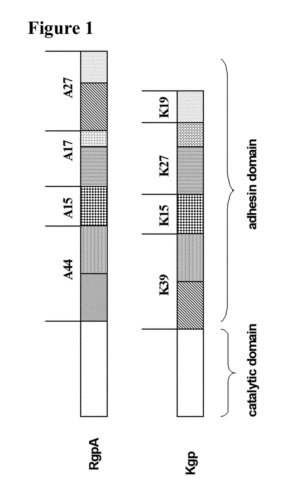
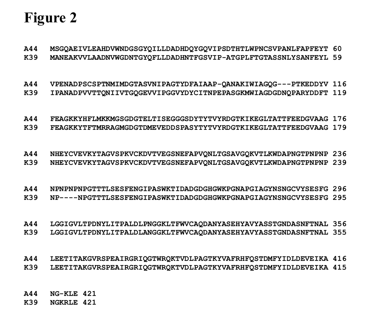
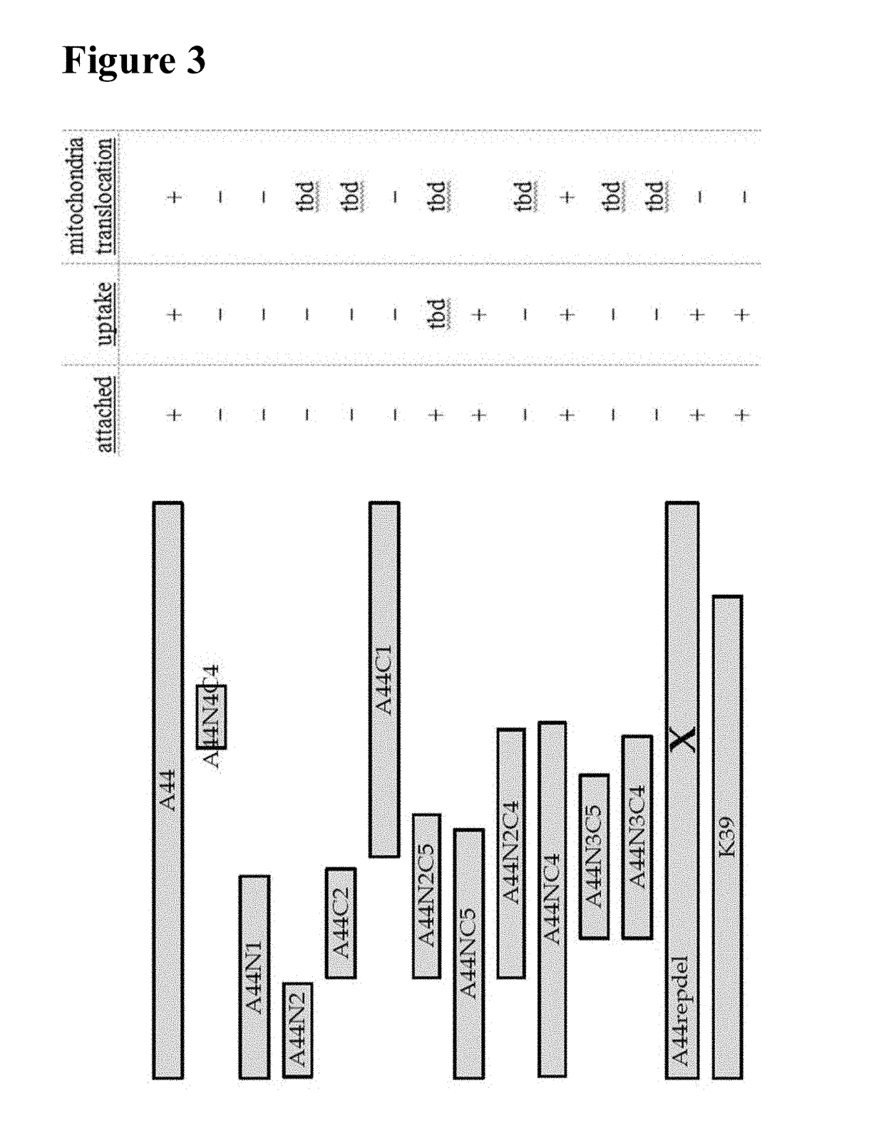
[0212]Construction and purification of the full-length A44 peptide and peptide fragments depicted in FIG. 3 were performed as previously described (Boisvert and Duncan (2008) supra). Briefly, various portions of the A44 peptide-encoding region were amplified from chromosomal DNA of P. gingivalis ATCC 33277 by PCR using various combinations of the forward and reverse primers shown in Table 1. PCR amplicons were ligated directly into pTOPO using the TOPO TA cloning kit (Invitrogen). Resulting pTOPO plasmids were digested with NdeI and XhoI; inserts were gel-purified using the Qiaquick kit (Qiagen) and then ligated with NdeI- and XhoI-digested pET22b vectors. E. coli BL21 (Novagen) were transformed with the ligated vectors and exposed to 1 mM isopropyl-a D-thiogalactopyranoside (IPTG) for 3-4 hours to induce expression of the recombinant peptide. Cells were lysed using 5 ml of lysis buffer per gram wet wei...

example 2

ation of A44 / K39 Peptide Domains Involved in Cellular Untake

[0214]Cellular uptake (i.e., cell penetration) of A44 peptides and fragments thereof was determined by Western blot of acid-washed cell lysates using methods set forth in Boisvert and Duncan (2008) supra. Briefly, 2 mg / ml recombinant A44-based peptide (full length or fragment) was incubated with HEp-2 epithelial cell monolayers for about 60 minutes. Cells were then washed three times with PBS followed by treatment with an acid wash solution (0.2 M acetic acid, 0.5 M NaCl, pH 2.5) to remove any externally attached peptide (5 min, RT). Cells were washed again (3×PBS) and lysed with water. Cellular lysates were harvested and the protein components separated by SDS-PAGE and transferred onto a nitrocellulose membrane. Presence of peptides in lysates was assayed with anti-His-tag antibodies, secondary HRP-antibodies and ECL according to the manufacturers' recommendation (Millipore). Results are summarized in FIG. 3. This is one e...

example 3

ation of A44 / K39 Peptide Domains Involved in Mitochondrial Targeting

[0216]Translocation to mitochondria was determined by fluorescent microscopy and cell fractionation in combination with Western Blots as shown previously (Boisvert and Duncan (2010) supra). Briefly, HEp-2 epithelial cells were incubated with recombinant an A44-derived peptide for about 60 minutes, washed and lysed with saponin buffer (1% (w / v) saponin in 10 mM Tris / HCl (pH 7.5). The supernatant in centrifuged samples contains cytoplasmic proteins. The resulting pellet was resuspended in saponin buffer+0.05% (v / v) Triton-X-100 and centrifuged again (max. speed, 5 min). The supernatant contained mitochondrial proteins as shown by Western Blot with anti-cytochrome C antibodies. Presence of A44-derived peptide in this fraction was visualized as described above. A44-derived peptide identified in this fraction can be further tested by confocal microscopy for co-localization with MitoTracker (Invitrogen) as described previ...


PUM

PropertyMeasurementUnit
Fractionaaaaaaaaaa
Lengthaaaaaaaaaa
Cell deathaaaaaaaaaa

Abstract

The present disclosure relates generally to conjugates and their uses and production. More particularly, it concerns conjugates comprising an A44 or K39 gingipain adhesion peptide, or fragment thereof (e.g., an A44 / K39 peptide portion) covalently attached to a heterologous agent that is capable of entering a cell by endocytosis and translocating to mitochondria.

Description

RELATED APPLICATIONS[0001]This application claims the benefit of priority from U.S. provisional application No. 62 / 063,938, filed Oct. 14, 2014, and U.S. provisional application No. 62 / 063,928, filed Oct. 14, 2014. The disclosures of each of the foregoing applications are hereby incorporated by reference in their entirety.GOVERNMENT SUPPORT[0002]This disclosure was made with government support under Grant Numbers RO1 DE010510 and RO1 DE015931 awarded by the National Institutes of Health. The government has certain rights in the disclosure.FIELD OF THE DISCLOSURE[0003]The present disclosure relates generally to conjugates and their uses and production. More particularly, it concerns conjugates comprising an A44 or K39 gingipain adhesin peptide, or fragment thereof (e.g., an A44 / K39 peptide portion) associated with a heterologous agent that is capable of entering a cell, such as by endocytosis, and translocating to mitochondria. The present disclosure also relates generally to methods...

Claims


Application Information

Patent Timeline
IPC IPC(8): C12N9/52A61K38/48
CPCC12N9/52A61K47/48107A61K38/4873C07K2319/30C07K2319/10C07K2319/31C07K2319/07C07K2319/00A61K38/00A61K47/60A61K47/64A61P3/02A61P39/06
Inventor BOISVERT, HEIKEDUNCAN, MARGARET
Owner THE FORSYTH INST
Learn More