Cancerous lesion identifying method via hyper-spectral imaging technique

a hyper-spectral imaging and lesion technology, applied in the field of cancerous lesion identification methods, can solve the problems of time-consuming data reading, high cost, limited reading speed of spectrum data, etc., and achieve the effect of efficiently increasing the diagnostic rate of clinicians

Active Publication Date: 2017-11-09
NATIONAL CHUNG CHENG UNIV
View PDF3 Cites 13 Cited by
  • Summary
  • Abstract
  • Description
  • Claims
  • Application Information

AI Technical Summary

Benefits of technology

[0010]Accordingly, an objective of the present invention is to provide a cancerous lesion identifying method via hyper-spectral imaging technique to quickly evaluate the probabilities of cancer at various stages.
[0012]By the cancerous lesion identifying method via the hyper-spectral imaging technique in the present invention, the cancerous lesion images are digitized and the diagnosis rate of the clinicians may be efficiently increased to help the patients to start the treatments as soon as possible.

Problems solved by technology

This kind of hyper-spectral imaging system can get best simulating spectrum and spatial resolution, but it is time consuming to read data.
This method can separate materials from the bone marrow cells, but the reading speed of the spectrum data is limited by the effect of the liquid crystal tunable filters.
This hyper-spectral imaging system has a very high image resolution, but it needs to process huge data and the cost is higher.
However, when using the conventional white light endoscopy, the detailed structure of the esophageal endoscopy may not be observed clearly, and biopsy and staining techniques are required for diagnosis.
On the contrary, when the pathological lesion of the mucosa occurs, cells with glycogen will reduce or disappear and it is not easy to stain mucosa brown with iodine solution.
However, according to medical literature, the toluidine blue may cause the damage of the DNA and the toluidine blue staining is time consuming and relied on the operator's experience.
Because of the uneven dye concentration, the improper spraying method or the limitation of the agent, staining difference may occur at the pathological lesions, and the positioning may not be accurate or lesions may be missed.
However, the normal Raman spectra technique has two significant drawbacks: background fluorescence interference is too strong and the efficiency of the Raman scattering is too low.
Therefore, the difficulties in actual clinic practice occur.
However, early stage lesions are not obvious under the white light detection, and the lesions are easy to be ignored and the treatment is delayed.
However, the Lugol's chromoendoscopy will spray dye to have development results, but the dye is not evenly distributed, causing the difficulty of the determination.
In addition, the dye may make the patient's chest feel uncomfortable such as tingling or burning.
For example, when the lesions, such as blending or inflammation, occur, the vision is fuzzy and the resolution of the images is poor.

Method used


Image

  • Cancerous lesion identifying method via hyper-spectral imaging technique
  • Cancerous lesion identifying method via hyper-spectral imaging technique
  • Cancerous lesion identifying method via hyper-spectral imaging technique

Examples


Embodiment Construction

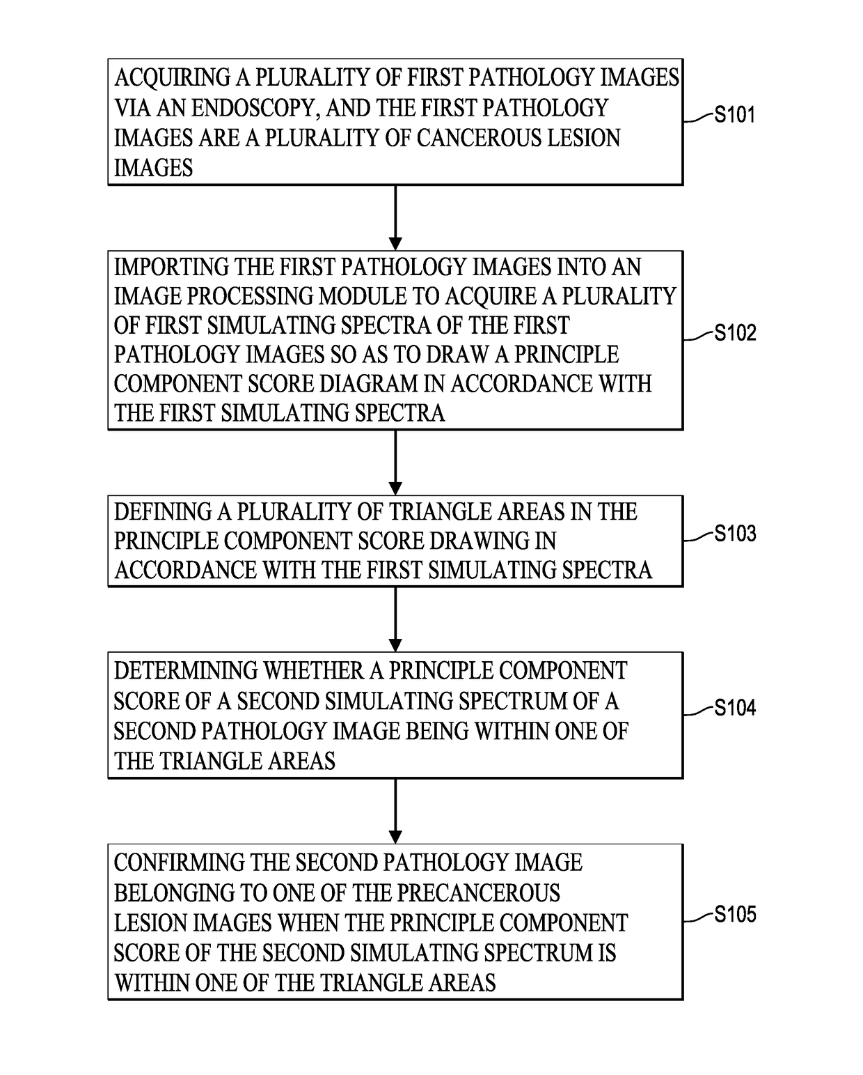
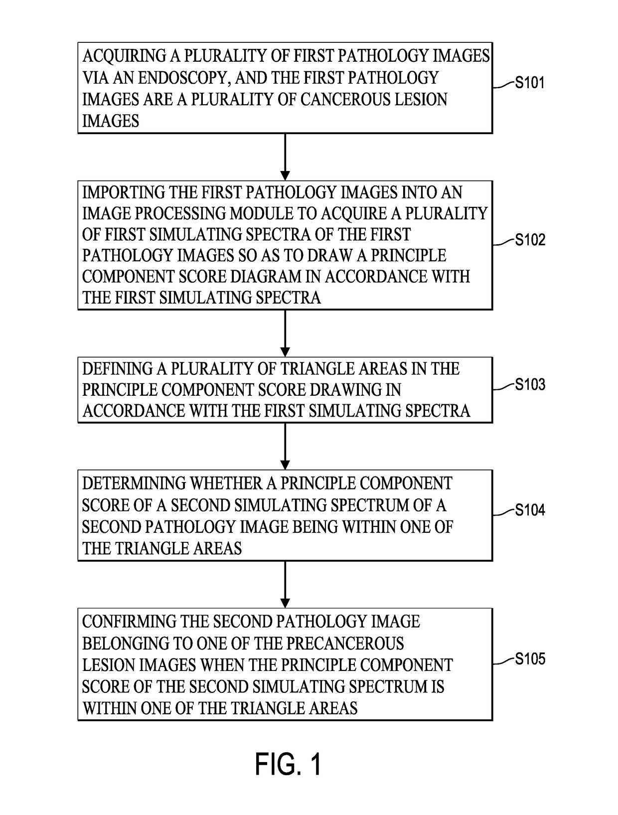
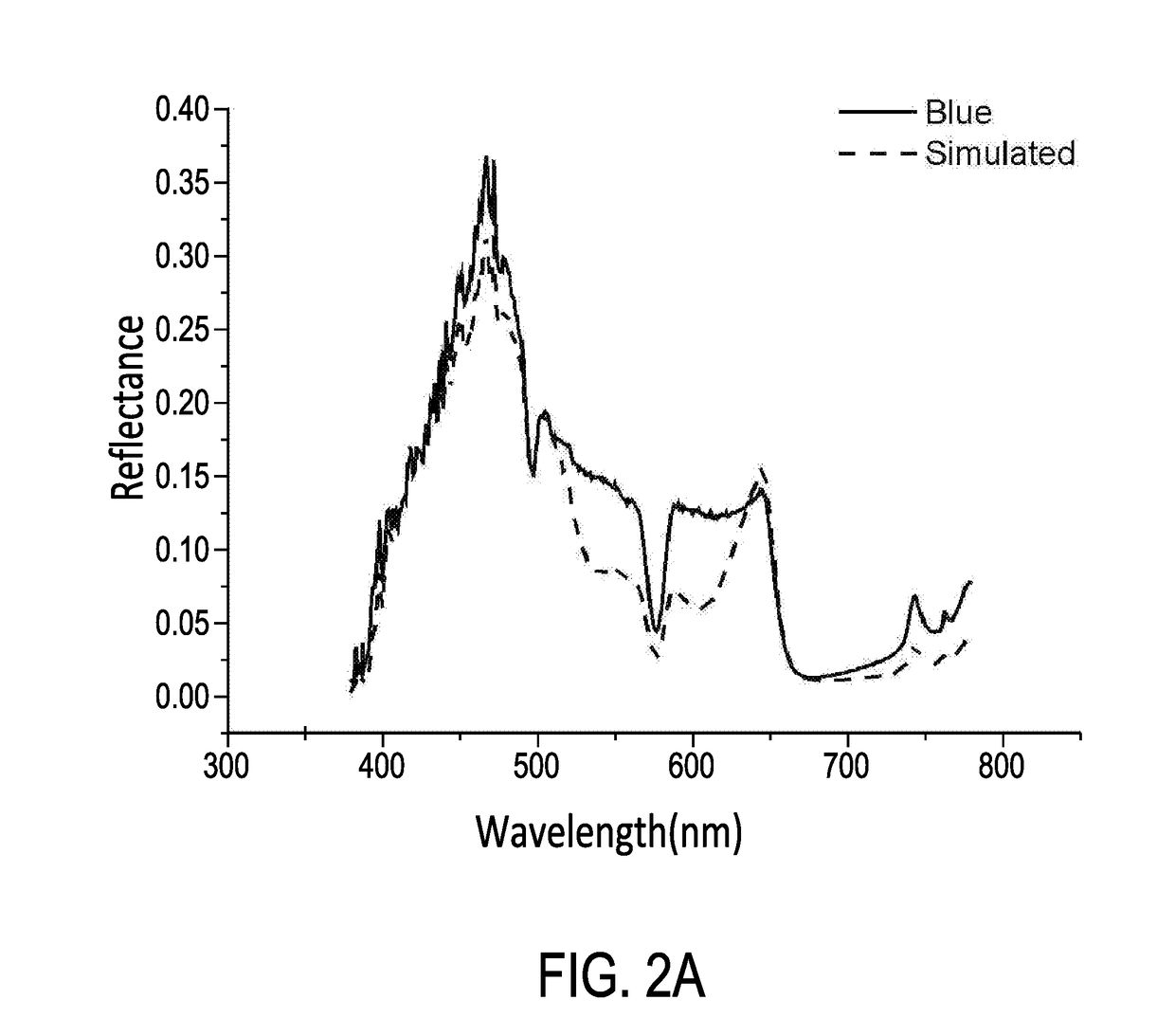
[0025]FIG. 1 is a flow chart of a cancerous lesion identifying method via hyper-spectral imaging technique in an embodiment of the present invention. As shown in FIG. 1, in step S101, a plurality of first pathology images are acquired via an endoscopy and the first pathology images are cancerous lesion images. The hyper-spectral imaging system is built within the endoscopy and a high resolution spectrometer. The hyper-spectral imaging system can acquire image information of 24 mini color checkers. In order to determine cancerous lesions, the spectra at each of the pixels in each of the first pathology images may be acquired and a relationship matrix between the spectrometer and the endoscopy has to be found first. The spectra of the 24 mini color checkers are measured by the spectrometer under the environment of the endoscopy. The range of the spectra is set at the wave band of visible light (380 nm-780 nm). For analytical purposes, each column of the matrix is the corresponding str...


PUM


Abstract

A cancerous lesion identifying method via hyper-spectral imaging technique comprises steps of: acquiring a plurality of first pathology images via an endoscopy, wherein the first pathology images are cancerous lesion images respectively; importing the first pathology images into an image processing module to acquire a plurality of first simulating spectra of the first pathology images so as to generate a principle component score diagram in accordance with the first simulating spectra; defining a plurality of triangle areas in the principle component score diagram in accordance with the first simulating spectra; determining whether a principle component score of a second simulating spectrum of a second pathology image is within any one of the triangle areas; and confirming the second pathology image belongs to one of the cancerous lesion images when the principle component score of the second simulating spectrum is within any one of the triangle areas.

Description

BACKGROUND OF THE INVENTION1. Field of the Invention[0001]The present invention relates to a cancerous lesion identifying method, and more particularly to a cancerous lesion identifying method by implementing hyper-spectral imaging technique and principle component analysis.2. Description of Related Art[0002]With the development of hyper-spectral imaging technique is well developed, the hyper-spectral imaging technique has been implemented in medical examination, such as early stage oral cancer detection, oral lesions detection of enterovirus, rectal mucosa detection, and so on. Different equipments adopt different kinds of hyper-spectral imaging techniques. Accordingly, one of the conventional hyper-spectral imaging systems is to implement a single point spectrum analyzer with a two-dimensional scanning system. This kind of hyper-spectral imaging system can get best simulating spectrum and spatial resolution, but it is time consuming to read data. Another conventional hyper-spectra...

Claims


Application Information

Patent Timeline
Patent Type & Authority Applications(United States)
IPC IPC(8): A61B5/00G06K9/52G06T7/00G06T7/60
CPCA61B5/7282G06T7/0012G06T7/60G06T2207/30096A61B5/0075A61B5/0084G06K9/52A61B5/7264A61B5/1032A61B5/4842A61B1/0005A61B1/043G06V2201/031A61B1/000094
Inventor WANG, HSIANG-CHENCHEN, SHIN-HUAHUANG, SHIH-WEILAI, CHIU-JUNGTING, CHU-CHI
Owner NATIONAL CHUNG CHENG UNIV
Why Eureka
Try Eureka