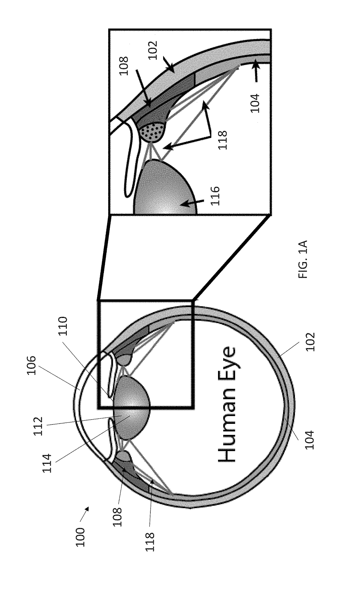
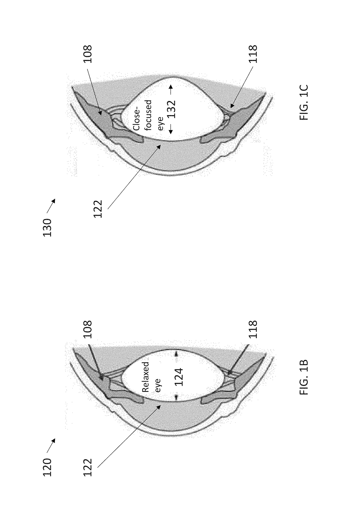
These types of ciliary movements are highly complex and difficult to analyze.
Moreover, it is impossible in experimental
in vivo or
ex vivo to analyze the ciliary muscles individually in order to determine how they
impact the eye functions.
As such, the lack of well-designed intraocular accommodating lenses has prevented these technologies from being successful in living humans and, consequently, the market.
Furthermore, lack of a functional structural whole eye model failed to allow further development of previous technology because this deficiency failed to allow for adequate consideration of the biomechanical impacts, forces, and effects on the
sclera, the
ciliary muscle influences, other extra-lenticular components' influence on the accommodative
system, including the
choroid, zonules, and even the
retina.
In particular, the emphasis on affecting ocular
accommodation to date has typically been focused on identifying and creating changes in ocular lens properties while failing to address underlying
ciliary muscle operations or other extra-lenticular tissues, forces and structures.
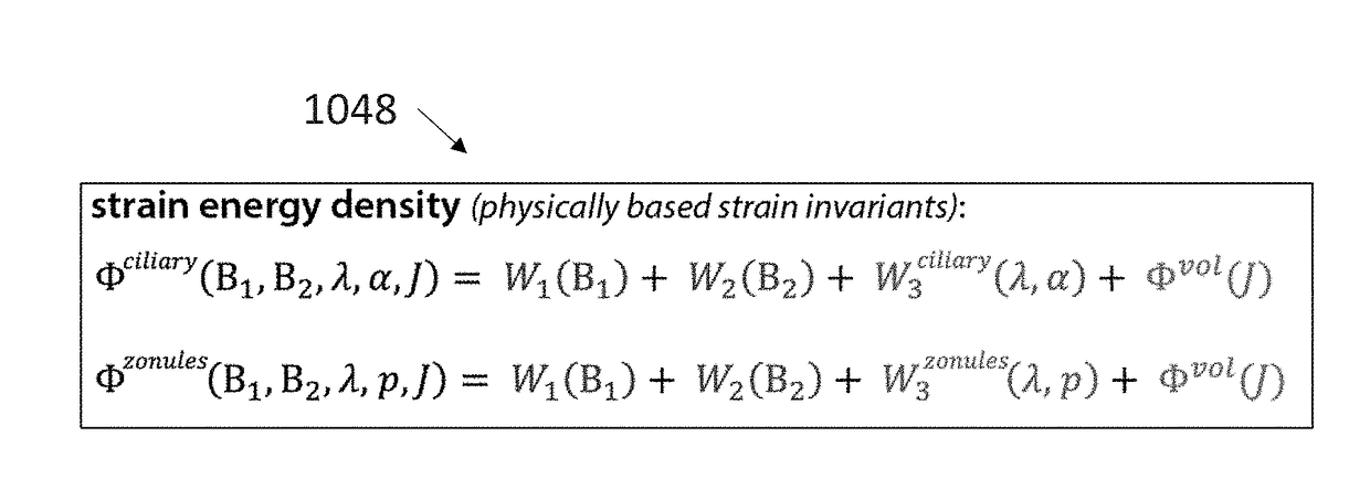
To date, there have been no models established to include a ‘whole eye Finite
Element Model (FEM)’.
Unfortunately, these models depend on a simplified arrangement of zonule attachments and ignore or otherwise
neglect the uniquely complex behaviors of the
ciliary muscle, whose movements are constrained by attachments to the
lens capsule, ocular
sclera and
choroid structures.
Due to the simplification of the ciliary
muscle behaviors as applied in these prior art models, attempts to apply pre-tensioning of zonules prior to ciliary
muscle contraction have not been successful.
This has led not only to a gap in the understanding of the
accommodation mechanism but also to a lack of
effective treatment of in restoring the accommodative functions that the conditions created by
presbyopia and other age-related eye afflictions.
Also contributing to the lack of
effective treatment for deteriorated accommodative function is the fact that there is an overall scarcity of data with respect to the functioning accommodative mechanisms for healthy human eyes, especially
in vivo or
dynamic data.
Since accommodative functioning is difficult to measure because of the delicate nature of the
human eye, most current measurement techniques have relied on data gathered from experimentation on the ocular systems of human cadavers and other primates.
Gathering this data usually requires isolating or disturbing at least a portion of the accommodative ocular
system, making procedures difficult and dangerous for live human test subjects.
As a result of insufficient data regarding the accommodative ocular
system, its underlying mechanisms and the related problem of incomplete modeling, analysis of existing data provides a disjointed and incomplete understanding of ocular accommodation in humans and any implications resulting from age-related changes to ocular structures.
As people age, they develop
presbyopia and lose accommodative ability, leaving people over the age of 50 with an almost complete lack of focusing ability for
near vision.
Although scientists have studied accommodation for centuries the functional mechanism is not well understood.
Without understanding the interactions of the
muscle, lens, and other structures that alter the eye's optic power, treatments for
presbyopia that effectively restore this ability cannot be successfully developed.
This lack of understanding is also in part due to the limited data, especially
in vivo or dynamic, of healthy human eyes; most current measurement techniques require isolating or disturbing some portion of the accommodative system and are limited to cadavers or monkey models.
Despite years of intensive research, there is little understanding of how aqueous drainage resistance is controlled in normal eyes, glaucomatous eyes, and how they compare.
Further, it is generally unknown where
aqueous flow resistance originates.
Accommodation mechanisms are highly complex and difficult to analyze, especially those of the
ciliary body (muscles) which are under emphasized and grossly overlooked and not well characterized to date.
Most prior art accommodation models focus solely on the actions of lenses and zonules in isolation of extralenticular structures and whole eye
biomechanics, and thus, oversimplify ciliary movement as a single muscular displacement.
In particular, the emphasis for ocular accommodation to date has typically been focused on identifying and creating changes in ocular lens properties, while not addressing underlying ciliary muscle operations.
Unfortunately, these models depend on a simplified arrangement of zonule attachments and ignore or otherwise
neglect the uniquely complex behaviors of the ciliary muscle, whose movements are constrained by attachments to the ocular
sclera and
choroid structures.
Due to the simplification of the ciliary muscle behaviors as applied in these prior art models, attempts to apply pre-tensioning of zonules prior to ciliary
muscle contraction have not been successful.
This has led not only to a gap in the understanding of the accommodation mechanism but also to a lack of
effective treatment in restoring the accommodative functions that the conditions created by presbyopia and other age-related eye afflictions, including proper
aqueous flow hydrodynamics and normal
organ function to name a few.
Also contributing to the lack of effective treatment for deteriorated accommodative function is the fact that there is an overall scarcity of data with respect to the functioning accommodative mechanisms for healthy human eyes, especially in vivo or
dynamic data.
Since accommodative functioning is difficult to measure because of the delicate nature of the
human eye, most current measurement techniques have relied on data gathered from experimentation on the ocular systems of human cadavers and other primates.
Gathering this data usually requires isolating or disturbing at least a portion of the accommodative ocular system, making procedures difficult and dangerous for live human test subjects.
As a result of insufficient data regarding the accommodative ocular system, its underlying mechanisms and the related problem of incomplete modeling, analysis of existing data provides a disjointed and incomplete understanding of ocular accommodation in humans and any implications resulting from age-related changes to ocular structures.
Trans-corneal delivery by passive
diffusion is difficult because the
drug needs to have hydrophobic characteristics to pass through the
corneal epithelium and
endothelium and hydrophilic characteristics to pass through the corneal stroma.
However, the scleral stroma is still a significant barrier, and a number of studies have examined the permeability of this tissue.
However, it is known that ciliary
muscle contraction greatly affects the unconventional outflow, and that PGF2a greatly increases unconventional outflow by decreasing the flow resistance of the interstitial spaces in the ciliary muscle.
Although this might be expected if the flow were primarily osmotically driven into the uveal vessels, this would not be the expected characteristic of a trans-scleral flow.


