
Leukocyte and microparticles fractionation using microfluidics

a technology of microfluidics and leukocytes, applied in the field of biochemistry, can solve the problems of inability to efficiently and passively sort the particles, lack of suitable isolation and assay methods, and increasing difficulties

Active Publication Date: 2018-07-05
NANYANG TECH UNIV
View PDF0 Cites 6 Cited by
  • Summary
  • Abstract
  • Description
  • Claims
  • Application Information

AI Technical Summary

Benefits of technology

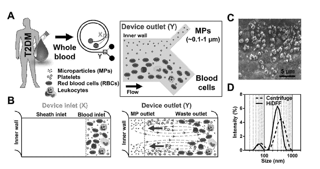
The present invention provides a method for separating blood cells using a technique called Dean Flow Fractionation (DFF). It allows for the purification of blood components, including neutrophils, without the need for centrifugation or labeling with a marker molecule. The method is gentle and fast compared to other methods, resulting in a purification of native neutrophils.

Problems solved by technology

Conventional separation strategies include centrifugation, sedimentation or membrane filtration which are limited by batch-mode processing and increasing difficulties when applied to separation of small micro / nanoparticles (<5 μm).
However, there is a lack of efficient and passive particle sorting methods which advocates the need to develop new separation technologies that can achieve small particle separation of closely spaced sizes.
A main reason behind is the lack of suitable isolation and assay methods to study cellular functions in a simple and reproducible manner.
This is further aggravated by blood sample preparation difficulties using conventional leukocyte isolation methods (density gradient centrifugation and RBCs lysis) which require large blood volume (˜10 mL) and may cause undesirable cell activation [Yang, J., et al., 2002.
In laboratory settings, immunophenotyping by flow cytometry is possible but is limited by cost, equipment access and laborious leukocyte purification procedures.
In type 2 diabetes mellitus (T2DM), patients suffer from impaired glucose metabolism which results in low-grade inflammation and activation of the innate immune system, with an increased risk of cardiovascular complications [Stehouwer, C. D. A. et al.
However, it is non-trivial to isolate neutrophils in their native state from peripheral blood as conventional neutrophil isolation methods are laborious and prone to cell activation [Fukuda, S.
Critically, there are currently no point-of-care (POC) technologies enabling the quantification of neutrophil function related to a low grade inflammatory state.
This inherently limits downstream applications including in vitro cell culture assays and flow cytometry analysis since it is challenging to recover the sorted neutrophils from the device.

Method used


Image

  • Leukocyte and microparticles fractionation using microfluidics
  • Leukocyte and microparticles fractionation using microfluidics
  • Leukocyte and microparticles fractionation using microfluidics

Examples


example 1

dic Neutrophil Purification and Phenotyping in Diabetes Mellitus

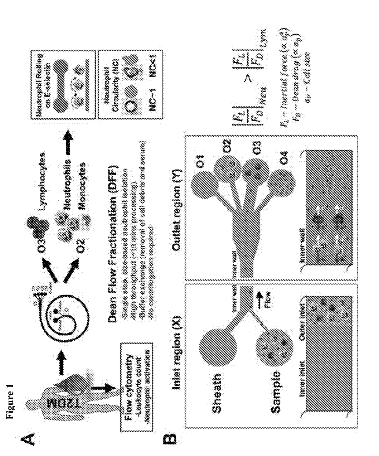
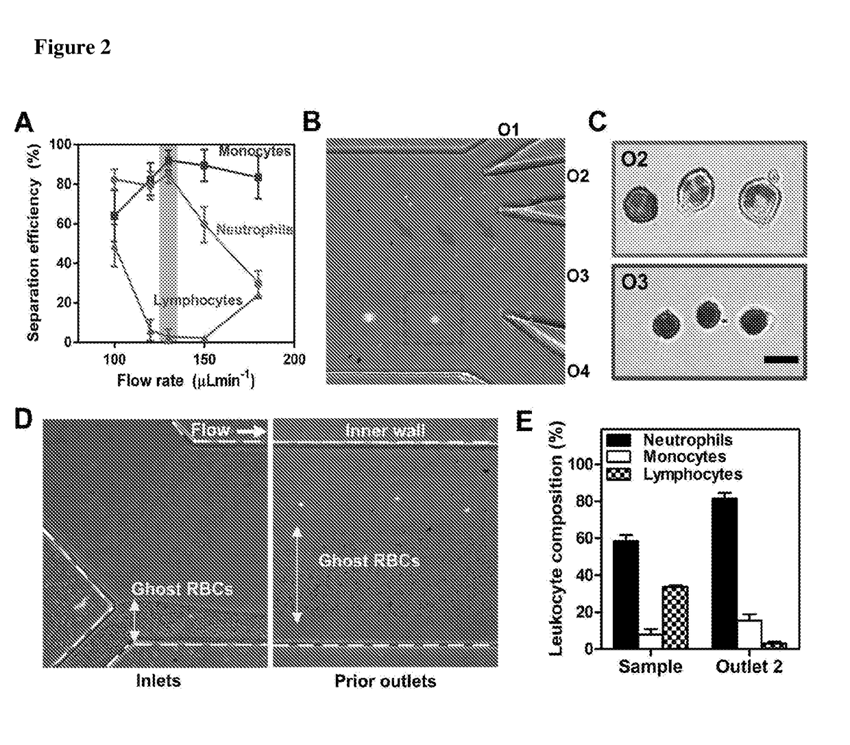
[0124]A microfluidic cell sorting technology termed as Dean Flow Fractionation (DFF) has been developed for size-based separation of diseased cells including circulating tumor cells (CTCs) [Hou, H. W. et al. Sci. Rep. 3 (2013)] and microorganisms [Hou, H. W. et al. Lab Chip 15, 2297-2307 (2015)] from whole blood. Here, it was tested if the subtle cell size difference between leukocyte subtypes may be sufficient for separation by a 4-outlet DFF spiral device to purify neutrophils from lysed whole blood in a single-step manner (FIG. 1).

[0125]The DFF spiral device was fabricated in polydimethylsiloxane (PDMS) and consists of a two-inlet, four-outlet spiral microchannel (500 μm (w)×115 μm (h)) with a total length of ˜10 cm. The channel height was fixed at 115 μm so that only the larger leukocytes (˜8 to 12 μm, ap / h >0.07, where ap is particle size) can experience inertial focusing and equilibrate near the inner wall. Near t...

example 2

n of Small Micro / Nanoparticles Using Microfluidics Using a 2-Outlet Device

[0132]The microfluidic devices are fabricated in polydimethylsiloxane (PDMS) using standard soft lithography techniques. The developed microdevice consists of a 2-inlet, 2-outlet spiral microchannel (300 μm (w)×60 μm (h)) with a radius of 0.5-0.6 cm and a total length of 6.5 cm. The sample (50 μm wide) and sheath (250 μm wide) inlets are fixed at the outer and inner wall of the channel, respectively. For outlet bifurcation, the smaller outlet channel at channel inner wall is designed to collect smaller particles (outlet 1) while larger particles are collected at the larger outlet (outlet 2).

[0133]During device operation, bead sample is pumped into the outer inlet while sheath fluid (1×PBS) is pumped through the inner inlet at a higher flow rate (1:5) to confine the sample stream near the outer wall. To ensure the absence of inertial focusing in our device, beads of smaller diameters (50 nm, 1 μm, 2 μm and 3 μm...

example 3

Sorting

[0137]Based on the focusing mechanism in a 2-outlet spiral device, separation and enrichment of bacteria species was performed based on their cell size. As proof of concept, sample solution containing mixture of E. coli (rod shaped, ˜2-4 μm long, 1 μm wide) and S. aureus (circular, ˜1 μm diameter) was pumped into the device. The smaller S. aureus were sorted into outlet 1 while E. coli were recovered mostly in outlet 2 (FIG. 14). Interestingly, size fractionation of E. coli was observed as smaller E. coli (mean±SD; 2.03±0.46 μm) and larger E. coli (2.85±0.75 μm) were sorted into outlet 1 and 2, respectively (FIG. 15). This further illustrates the superior separation resolution of the present technology for bacterial sorting or diagnostics.


PUM


Abstract

This invention relates to a method for separating blood cells comprising the steps of: (a) lysing red blood cells of a blood sample and diluting said sample; (b) providing a microfluidic device comprising a spiral-shaped flow channel having at least a first end and a second end, wherein said flow channel has two inlet ports at or near said first end and at least two outlet ports at or near said second end, wherein one of the two inlet ports is located at the inner wall of the spiral-shaped flow channel and the other inlet port is located at the outer wall of the spiral-shaped flow channel and at least one of the outlet ports is connected to a container allowing the storage of blood cells; (c) introducing the sample of step (a) into the inlet port located at the outer wall of the spiral-shaped flow channel and introducing a sheath fluid into the inlet port located at the inner wall of the spiral-shaped flow channel; (d) driving said sample and the sheath fluid through the spiral-shaped flow channel; and (e) recovering the blood cell in the at least one container connected to the at least one outlet port. The present invention also relates to coupling above described method of purifying neutrophils in the native state with a method for diagnosing diabetes or an inflammatory disease in a subject, which involves further investigation of neutrophils by determining their rolling speed, the neutrophil circularity (NC) index and / or the expression of markers, such as intracellular reactive oxygen species (ROS), CD1 1 b or PSGL-1.

Description

FIELD OF THE INVENTION[0001]The present invention lies in the field of biochemistry and relates to a method for separating blood cells comprising the steps of: (a) lysing red blood cells of a blood sample and diluting said sample; (b) providing a microfluidic device comprising a spiral-shaped flow channel having at least a first end and a second end, wherein said flow channel has two inlet ports at or near said first end and at least two outlet ports at or near said second end, wherein one of the two inlet ports is located at the inner wall of the spiral-shaped flow channel and the other inlet port is located at the outer wall of the spiral-shaped flow channel and at least one of the outlet ports is connected to a container allowing the storage of blood cells; (c) introducing the sample of step (a) into the inlet port located at the outer wall of the spiral-shaped flow channel and introducing a sheath fluid into the inlet port located at the inner wall of the spiral-shaped flow chan...

Claims


Application Information

Patent Timeline
IPC IPC(8): B01L3/00G01N33/50G01N33/569
CPCB01L3/502776B01L3/502761G01N33/5094G01N33/56972B01L2200/0652B01L2300/0864B01L2300/0877B01L2200/0636G01N2800/042G01N2800/7095G01N33/491G01N33/5029G01N33/5091
Inventor HOU, HAN WEIBOEHM, BERNHARD OTTOTAY, HUI MINLOO, SAY CHYE JOACHIM
Owner NANYANG TECH UNIV
Learn More