
Automated segmentation utilizing fully convolutional networks

a fully convolutional network and segmentation technology, applied in image analysis, image enhancement, instruments, etc., can solve the problems of high cost of cardiac mri studies, abnormally low ef readings are often associated with heart failure, and require manual adjustment of contours

Active Publication Date: 2018-08-02
ARTERYS INC
View PDF0 Cites 70 Cited by
  • Summary
  • Abstract
  • Description
  • Claims
  • Application Information

AI Technical Summary

Benefits of technology

The patent text describes a method for training a deep learning (CNN) model to identify and segment the heart's endocardium and epicardium using data of the same type for which the model will be used for inferencing. The method involves hyperparameter tuning, validation of model accuracy, and postprocessing to minimize non-myocardial tissue and improve accuracy in identifying myocardium. The postprocessing includes applying morphological operations and modifying threshold to classify pixels of myocardium. The technical effect of the patent text is the improved accuracy and reliability of identifying myocardium in deep learning models, which can help in the diagnosis and treatment of heart diseases.

Problems solved by technology

Abnormally low EF readings are often associated with heart failure.
Although some semi-automated contour placement tools exist (e.g., using an active contours or “snakes” algorithm), these still typically require some manual adjustment of the contours, particularly with images that have noise or artifacts.
The most obvious consequence of the onerousness of this process is that reading cardiac MRI studies is expensive.
Another important consequence is that contour-based measurements are generally not performed unless absolutely necessary, which limits the diagnostic information that can be extracted from each performed cardiac MRI study.
Although the snakes algorithm is common, and although modifying its resulting contours can be significantly faster than generating contours from scratch, the snakes algorithm has several significant disadvantages.
The cost function for snakes typically awards credit when the contour overlaps edges in the image; however, there is no way to inform the algorithm that the edge detected is the one desired; e.g., there is no explicit differentiation between the endocardium versus the border of other anatomical entities (e.g., the other ventricle, the lungs, the liver).
Further, the snakes algorithm is greedy.
However, gradient descent, and many similar optimization algorithms, are susceptible to getting stuck in local minima of the cost function.
The snakes algorithm generally has only a few dozen tunable parameters, and therefore does not have the capacity to represent a diverse set of possible images on which segmentation is desired.
Because of the great diversity on recorded images and the small number of tunable parameters, a snakes algorithm can only perform well on a small subset of “well-behaved” cases.
However, the snakes algorithm cannot be adequately tuned to work on more challenging cases.
However, because the muscles are numerous and small, excluding them from a manual contour requires significantly more care to be taken when creating the contour, dramatically increasing the onerousness of the process.
As a result, when creating manual contours, the papillary muscles are typically included within the endocardium contour, resulting in a modest overestimate of the ventricular blood volume.
Automated or semi-automated utilities may speed up the process of excluding the papillary muscles from the endocardium contour, but they have significant caveats.
The snakes algorithm (discussed above) is not appropriate for excluding the papillary muscles at end diastole because its canonical formulation only allows for contouring of a single connected region without holes.
In short, it is not possible for the canonical snakes algorithm to be used to segment the blood pool and exclude the papillary muscles at end diastole.
Therefore, in the standard formulation, the snakes algorithm can only include the papillary muscles at end diastole and only exclude them at end systole, resulting in inconsistent measurements of blood pool volume over the course of a cardiac cycle.
This is a major limitation of the snakes algorithm, preventing clinical use of its output without significant correction by the user.
Flood fill could also be used to include papillary muscles from the endocardium segmentation at end diastole; however, at end systole, because the bulk of the papillary muscles are connected to the myocardium (making the two regions nearly indistinguishable from one another), flood fill cannot be used to include the papillary muscles within the endocardium segmentation.
Beyond the inability to distinguish papillary muscles from myocardium at end systole, the major disadvantage of flood fill is that, though it may significantly reduce the effort required for the segmentation process when compared to a fully-manual segmentation, it still requires a great deal of user input to dynamically determine the flood fill gradient and distance thresholds.
One major disadvantage of performing segmentations on the SAX stack is that the SAX plane is nearly parallel to the plane of the mitral and tricuspid valves.
First, the valves are very difficult to distinguish on slices from the SAX stack.
Further, even if the LAX view is available, it may be difficult to tell on the SAX slice where the valve is located, and therefore, where the segmentation of the ventricle should end, since the ventricle and atrium have similar signal intensities.
Segmentation near the base of the heart is therefore one of the major sources of error for ventricular segmentation.
One limitation of LandMarkDetect is the lack of method to handle missing landmarks.
Another limitation is the absence of hyperparameter search except for the kernel and layer size.
Yet another limitation is the fixed upsampling layer with no parameter to learn.
Further, LandMarkDetect relies on a limited pre-processing strategy which consists in removing the mean (i.e. centering the input data) of the 3D image.

Method used

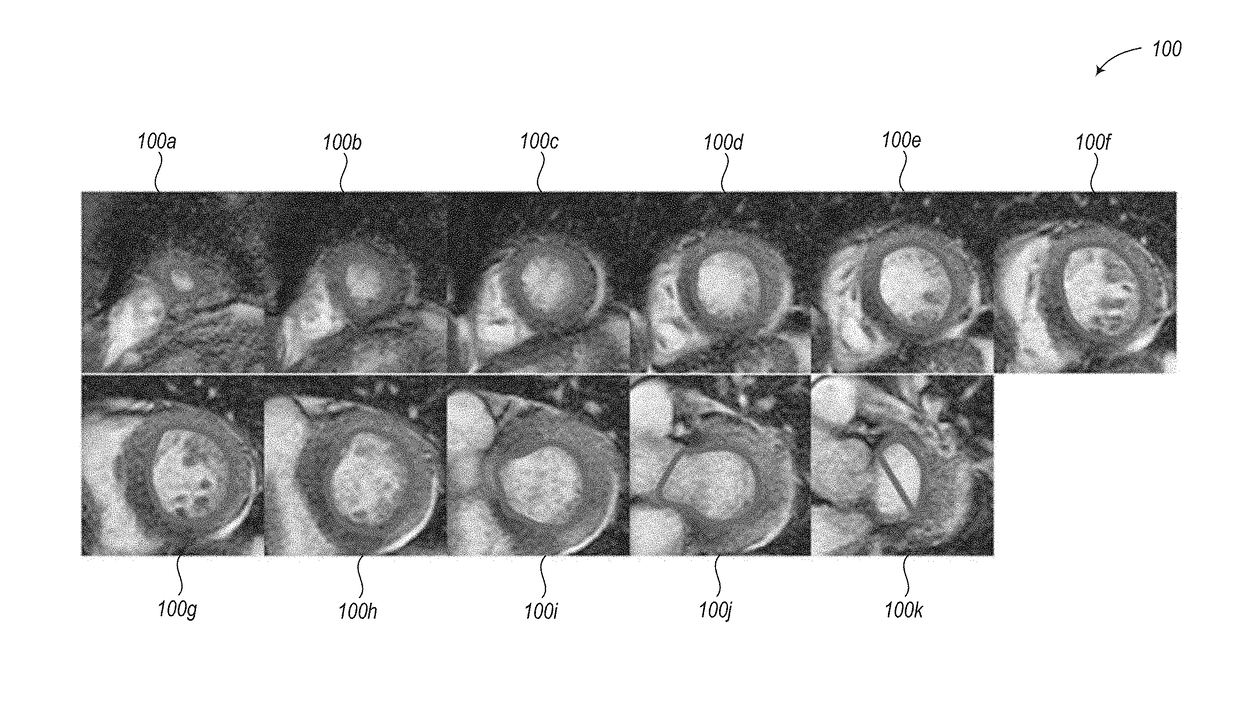
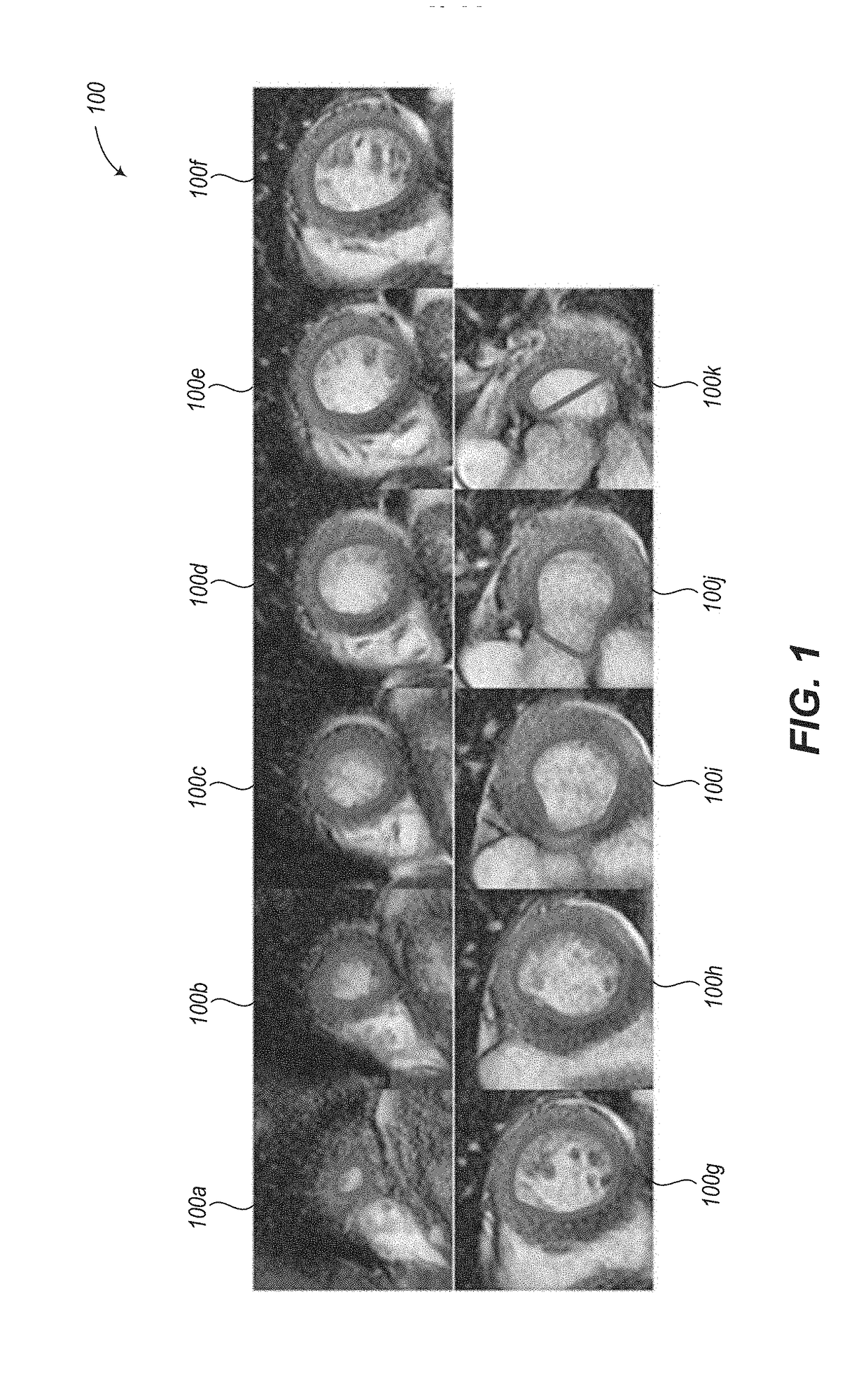
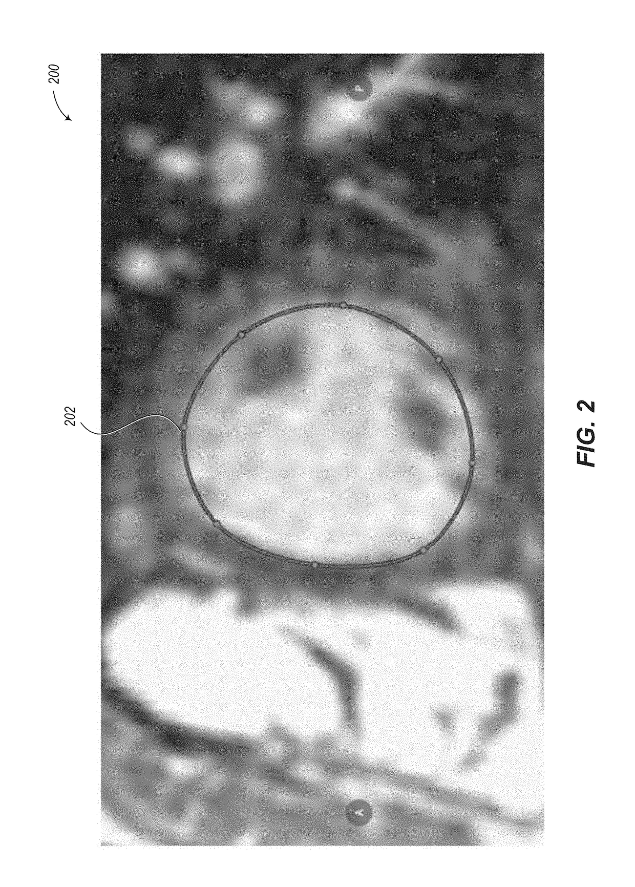

Image

  • Automated segmentation utilizing fully convolutional networks
  • Automated segmentation utilizing fully convolutional networks
  • Automated segmentation utilizing fully convolutional networks

Examples


Embodiment Construction

[0119]In the following description, certain specific details are set forth in order to provide a thorough understanding of various disclosed implementations. However, one skilled in the relevant art will recognize that implementations may be practiced without one or more of these specific details, or with other methods, components, materials, etc. In other instances, well-known structures associated with computer systems, server computers, and / or communications networks have not been shown or described in detail to avoid unnecessarily obscuring descriptions of the implementations.

[0120]Unless the context requires otherwise, throughout the specification and claims that follow, the word “comprising” is synonymous with “including,” and is inclusive or open-ended (i.e., does not exclude additional, unrecited elements or method acts).

[0121]Reference throughout this specification to “one implementation” or “an implementation” means that a particular feature, structure or characteristic de...


PUM


Abstract

Systems and methods for automated segmentation of anatomical structures (e.g., heart). Convolutional neural networks (CNNs) may be employed to autonomously segment parts of an anatomical structure represented by image data, such as 3D MRI data. The CNN utilizes two paths, a contracting path and an expanding path. In at least some implementations, the expanding path includes fewer convolution operations than the contracting path. Systems and methods also autonomously calculate an image intensity threshold that differentiates blood from papillary and trabeculae muscles in the interior of an endocardium contour, and autonomously apply the image intensity threshold to define a contour or mask that describes the boundary of the papillary and trabeculae muscles. Systems and methods also calculate contours or masks delineating the endocardium and epicardium using the trained CNN model, and anatomically localize pathologies or functional characteristics of the myocardial muscle using the calculated contours or masks.

Description

BACKGROUNDTechnical Field[0001]The present disclosure generally relates to automated segmentation of anatomical structures.Description of the Related Art[0002]Magnetic Resonance Imaging (MRI) is often used in cardiac imaging to assess patients with known or suspected cardiac pathologies. In particular, cardiac MRI may be used to quantify metrics related to heart failure and similar pathologies through its ability to accurately capture high-resolution cine images of the heart. These high-resolution images allow the volumes of relevant anatomical regions of the heart (such as the ventricles and muscle) to be measured, either manually, or with the help of semi- or fully-automated software.[0003]A cardiac MRI cine sequence consists of one or more spatial slices, each of which contains multiple time points (e.g., 20 time points) throughout a full cardiac cycle. Typically, some subset of the following views are captured as separate series: The short axis (SAX) view, which consists of a se...

Claims


Application Information

Patent Timeline
Patent Type & Authority Applications(United States)
IPC IPC(8): G06T7/00G06T7/149G06T7/136G06T7/143
CPCG06T7/0012G06T7/149G06T7/136G06T7/143G06T2207/10088G06T2207/20084G06T2207/20081G06T2207/30048G06T7/11G06N3/084G06N3/045G06T7/10G06N3/08G06T2207/30004
Inventor GOLDEN, DANIEL IRVINGLE, MATTHIEULIEMAN-SIFRY, JESSELAU, HOK KAN
Owner ARTERYS INC
Learn More