Radio-frequency electrical membrane breakdown for the treatment of high risk and recurrent prostate cancer, unresectable pancreatic cancer, tumors of the breast, melanoma or other skin malignancies, sarcoma, soft tissue tumors, ductal carcinoma, neoplasia, and intra and extra luminal abnormal tissue

a radiofrequency electrical membrane and breakdown technology, applied in the field of medical devices and treatment methods, can solve the problems of not being able to stimulate a specific immune response, both heat-based and cryoablation techniques, and not being able to spare normal structures in the treatment zone,

Active Publication Date: 2018-09-20
IMMUNSYS INC
View PDF11 Cites 12 Cited by
  • Summary
  • Abstract
  • Description
  • Claims
  • Application Information

AI Technical Summary

Benefits of technology

[0019]The present invention is an imaging, guidance, planning and treatment system integrated into a single unit or assembly of components, and a method far using same, that can be safely and effectively deployed to treat prostate cancer in all medical settings, including in a physician's office or in an outpatient setting. The system utilizes the novel process of Radio-Frequency Electrical Membrane Breakdown (“EMB” or “RFEMB”) to destroy the cellular membranes of unwanted or cancerous tissue without denaturing the intra-cellular contents of the cells comprising the tissue, thereby exposing tumor antigens and other intra-cellular components which can have an immunologic effect on local or distant cancerous tissue, with or without the addition of immunologic adjutant drugs.

Problems solved by technology

Because cellular proteins are denatured by the heat of thermal ablation techniques, they are not available to stimulate a specific immune response as they may be with cryoablation.
Both heat based and cryoablation techniques suffer from the drawback, that they have little or no ability to spare normal structures in the treatment zone and so can be contraindicated based on tumor location or lead to complications from collateral injury.
Above a critical transmembrane potential and with longer exposure times, poration becomes irreversible leading to eventual cell death due an influx of extracellular ions resulting in loss of homeostasis and subsequent apoptosis.
However, in all cases the mechanism of cellular destruction and death by IRE is apoptotic which requires considerable time to pass and is not visible pathologically in a time frame to be clinically useful in determining the efficacy of IRE treatment which is an important clinical drawback to the method.
In this context, irreversible electroporation leading to cell death was viewed as a failure in as much as the treated cells did not survive to realize the modification as intended.
However, the DC pulses used in currently available IRE methods and devices have characteristics that can limit their use or add risks for the patient because current methods and devices create severe muscle contraction during treatment.
This is a significant disadvantage because it requires that a patient be placed and supported under general anesthesia with neuromuscular blockade in order for the procedure to be carried out, and this carries with it additional substantial inherent patient risks and costs.
Moreover, since even relatively small muscular contractions can disrupt the proper placement of IRE electrodes, the efficacy of each additional pulse train used in a therapy regimen may be compromised without even being noticed during the treatment session.
In addition, because cells ablated by IRE methods undergo apoptotic death without membrane rupture their ability to induce a supplemental immune response as observed with cryoablation is impaired.
When used as the sole ablative tool in a treatment protocol, IRE's inability to induce a supplemental immune response is a substantial limitation to its therapeutic benefit for patients.
On the other hand, cryoablation is limited by significant clinical disadvantages arising from the extreme cold and its capacity to destroy nearby critical healthy structures.
therapeutic action and whole gland therapies, such as radical prostatectomy or radiation therapy, which are often associated with significant morbidities, particularly in the setting of high risk prostate cancer, and which often are not even clinically useful in cases of recurrent disease.
At the present time, all methods for carrying out focal therapy involve technologies that require full operating room capabilities, or imaging capabilities such as MRI, both of which are very expensive and in relatively limited supply.

Method used


Image

  • Radio-frequency electrical membrane breakdown for the treatment of high risk and recurrent prostate cancer, unresectable pancreatic cancer, tumors of the breast, melanoma or other skin malignancies, sarcoma, soft tissue tumors, ductal carcinoma, neoplasia, and intra and extra luminal abnormal tissue
  • Radio-frequency electrical membrane breakdown for the treatment of high risk and recurrent prostate cancer, unresectable pancreatic cancer, tumors of the breast, melanoma or other skin malignancies, sarcoma, soft tissue tumors, ductal carcinoma, neoplasia, and intra and extra luminal abnormal tissue
  • Radio-frequency electrical membrane breakdown for the treatment of high risk and recurrent prostate cancer, unresectable pancreatic cancer, tumors of the breast, melanoma or other skin malignancies, sarcoma, soft tissue tumors, ductal carcinoma, neoplasia, and intra and extra luminal abnormal tissue

Examples


Embodiment Construction

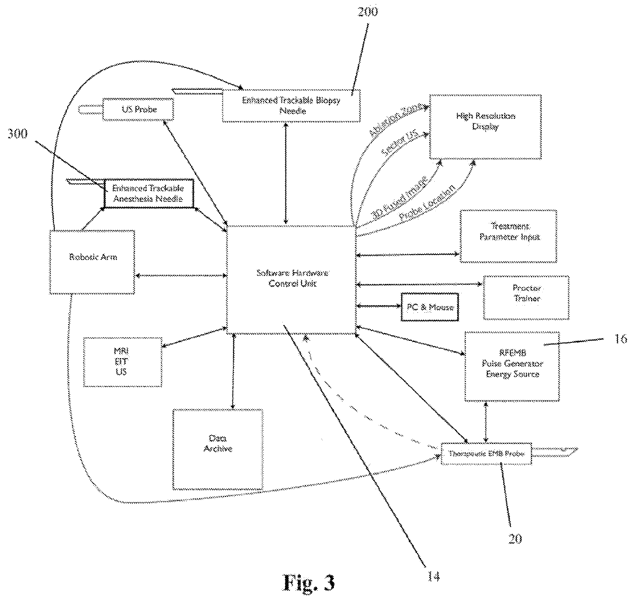
[0052]In general, the software-hardware controller unit (SHCU) operating the proprietary office based prostate cancer treatment system software according to the present invention facilitates the treatment of prostate cancer by directing the placement of EMB treatment probe(s) 20, biopsy needle(s) 200 and, optionally, anesthesia needle(s) 300, and by delivering electric pulses designed to cause EMB within the cancerous tissue to EMB treatment probe(s) 20, all while the entire process may be monitored in real time via one or more two- or three-dimensional imaging devices and via one or more biopsy samples taken at strategic locations to measure cell death. The system is such that the treatment may be performed by a physician under the guidance of the software, or may be performed completely automatically, from the process of imaging the treatment area to the process of placing one or more probes using robotic arms operatively connected to the SHCU to the process of delivering electric...


PUM


Abstract

An imaging, guidance, planning and treatment system integrated into a single unit or assembly of components, and a method for using same, that can be safely and effectively deployed to treat prostate cancer in all medical settings, including in a physician's office or in an outpatient setting. The system utilizes the novel process of Radio-Frequency Electrical Membrane Breakdown (“EMB” or “RFEMB”) to destroy the cellular membranes of unwanted or cancerous tissue without denaturing the intra-cellular contents of the cells comprising the tissue, thereby exposing tumor antigens and other intra-cellular components which can have an immunologic effect on local or distant cancerous tissue, with or without the addition of immunologic adjutant drugs. The system preferably comprises at least one EMB treatment probe 20, at least one trackable biopsy needle 200, at least one trackable anesthesia needle 300, and at least one controller unit for at least partially automating the treatment process.

Description

CROSS-REFERENCE TO RELATED APPLICATIONS[0001]The present invention is a continuation-in-part of U.S. patent application Ser. No. 14 / 451,333, filed Aug. 4, 2014.BACKGROUND OF THE INVENTION1. Field of the Invention[0002]The present invention relates generally to medical devices and treatment methods, and more particularly, to a device and method of treating prostate cancer by ablating cancerous tissue using applied electric fields.2. Description of the Background[0003]Cancer is not one single disease but rather a group of diseases with common characteristics that often result in sustained cell proliferation, reduced or delayed cell mortality, cooption of bodily angiogenesis and metabolic processes and evasion of bodily immune response which results in undesirable soft tissue growths called neoplasms or, more commonly, tumors. Removal or destruction of this aberrant tissue is a goal of many cancer treatment methods and modalities. Surgical tumor excision is one method of accomplishing ...

Claims


Application Information

Patent Timeline
Patent Type & Authority Applications(United States)
IPC IPC(8): A61B18/14A61B10/02A61B18/18
CPCA61B18/1477A61B10/0233A61B18/18A61B2018/00196A61B2018/00577A61B2018/00994A61B2018/1475A61B2034/2051A61B8/12A61B18/1492A61B2018/00547A61B2018/00982
Inventor ONIK, GARY M.MIESSAU, JAMES A.BOSTWICK, DAVID G.
Owner IMMUNSYS INC
Why Eureka
Try Eureka