Cartilage repair, preservation and growth by stimulation of bone-chondral interface and delivery system and methods therefor
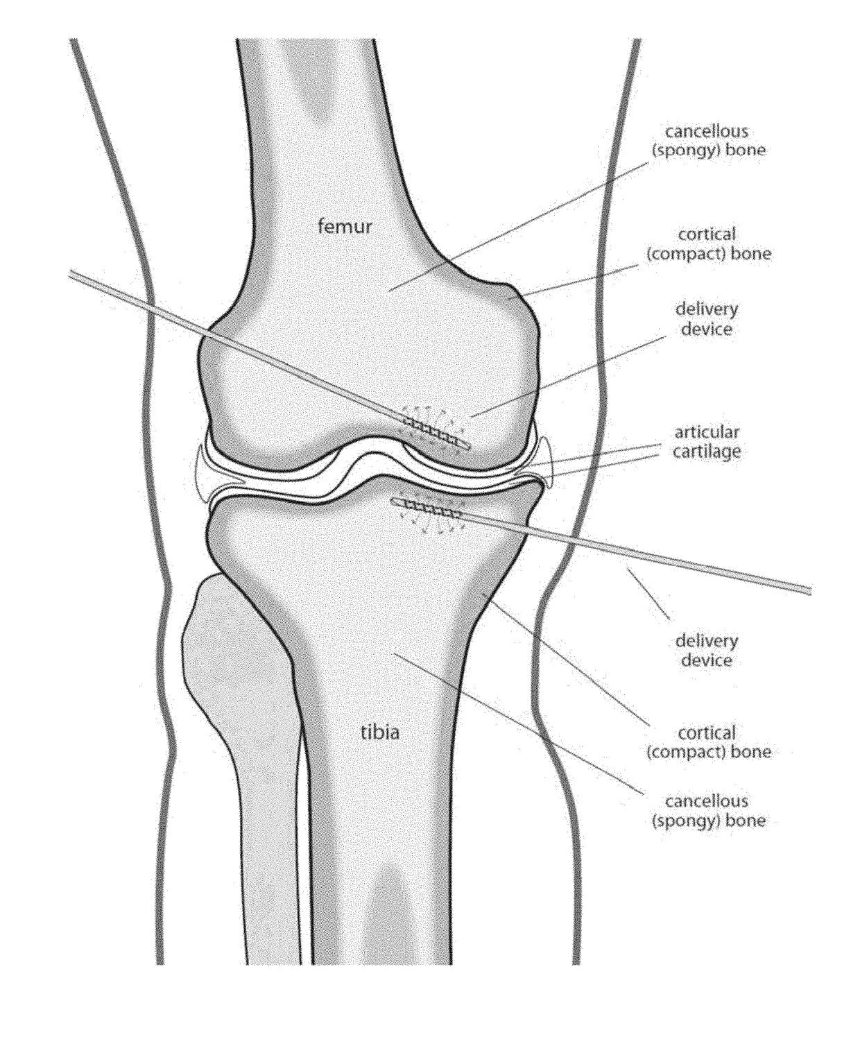
a cartilage and bone-chondral interface technology, applied in trocar, other medical devices, infusion needles, etc., can solve the problems of increased risk of coronary artery disease, adverse impact on cartilage, increased risk of hypertension, etc., to improve cartilage repair, preserve and grow cartilage, and reduce table time. , the effect of preserving cartilag
- Summary
- Abstract
- Description
- Claims
- Application Information
AI Technical Summary
Benefits of technology
Problems solved by technology
Method used
Image
Examples
example a
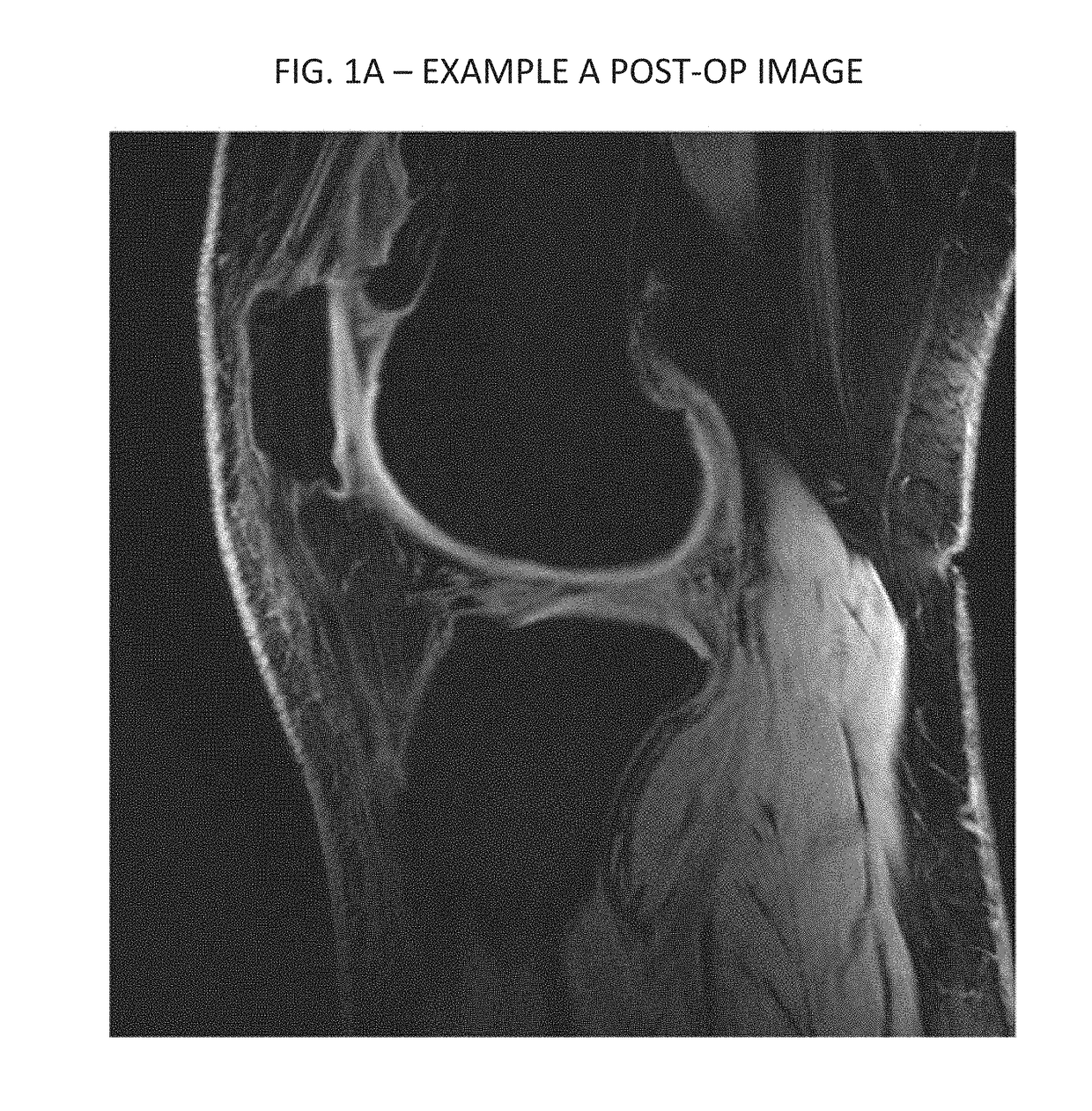
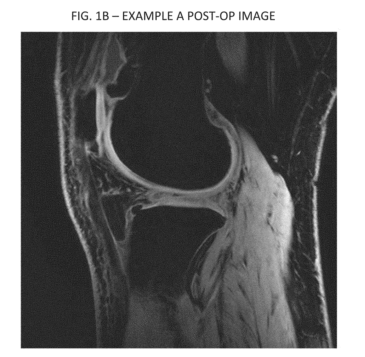
[0085]In an experimental trial of treatment methods according to an exemplary embodiment of the present invention, a 50 year old female with advanced degenerative osteoarthritic disease of the knee was treated. Prior to the treatment, a pre-operative MRI was done. Post treatment a three month follow-up MRI was performed on the patient and read by an independent radiologist. Provided hereto as Appendix A is the independent radiologist's report. As noted in the report, the post operative MRI showed an increase in cartilage matrix from 1.8 mm pre-treatment to 3.7 mm at the follow-up MRI. Thus, the inventive technique shows early promise for cartilage repair and possible regeneration.
example b
[0086]Imaging data from another experimental case are provided in FIGS. 1-4. As can be seen therein, FIGS. 1 and 2 are respectively axial and sagittal images of a patient's knee from a preoperative scan. FIG. 1 depicts an area of decreased blood flow due to an ischemia, as shown by the red arrow, also seen in FIG. 2 pointed to by the red and green arrows. Injections similarly performed according to the above described protocol, to the femoral and tibial compartments by drilling and injecting at the bone-cartilage interface, followed by injections into the knee joint. FIGS. 3 and 4 are corresponding axial and sagittal images form a MRI taken three months following the treatment. As can be seen, significant new cartilage has grown, and the ischemia has been essentially resolved, as shown in FIG. 3.
example c
[0087]FIG. 5 depicts side by side comparisons of sagittal images of a knee of a third patient. The left panel is an image form a preoperative scan, and the right panel a corresponding image from a post operative MRI. As shown in FIG. 5, the post operative MRI showed an increase in cartilage matrix from 1.60 mm to 1.87 mm at the follow-up MRI.
[0088]In exemplary embodiments of the present invention, the therapeutic protocol described above can similarly be used for osteoarthritis and avascular necrosis, as well as for treating meniscal and labral injuries in the joints.
[0089]Exemplary Variations of the Protocol
[0090]In exemplary embodiments of the present invention, variations on the above-described protocols can be used for other anatomical areas. Examples of these are next described.
[0091]Joints—for joints, the step of injecting GCSF for stimulating bone marrow may be skipped. The drilling / twisting alone of the delivery device (as described below) will stimulate bone marrow combined...
PUM
 Abstract
Description
Claims
Application Information
 - R&D
- Intellectual Property
- Life Sciences
- Materials
- Tech Scout
- Unparalleled Data Quality
- Higher Quality Content
- 60% Fewer Hallucinations
Browse by: Latest US Patents, China's latest patents, Technical Efficacy Thesaurus, Application Domain, Technology Topic, Popular Technical Reports.
© 2025 PatSnap. All rights reserved.Legal|Privacy policy|Modern Slavery Act Transparency Statement|Sitemap|About US| Contact US: help@patsnap.com


