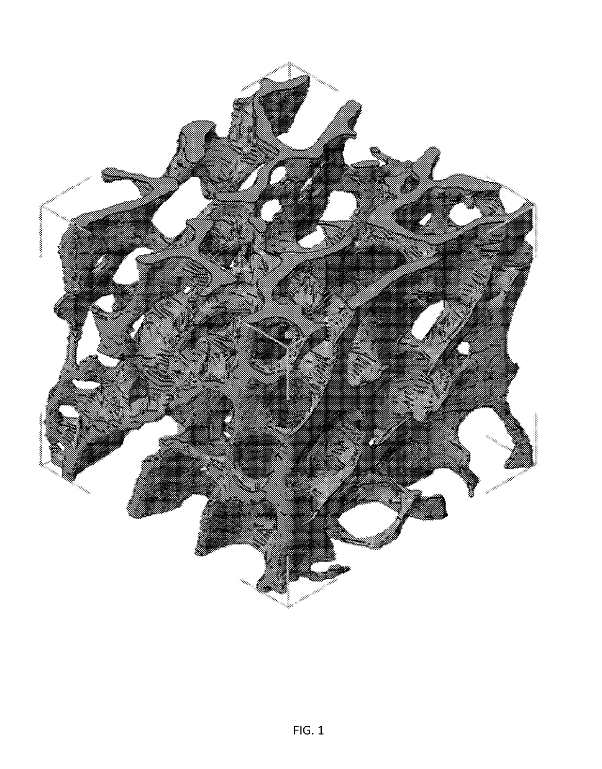
Three dimensional porous cartilage template
a cartilage template and three-dimensional technology, applied in the field of three-dimensional porous cartilage templates, can solve the problems of severe bone defects and fractures, generating an annual cost of $2.5 billion, and bone tissue is also susceptible to malignant growths and metastases from surrounding organs, so as to promote the repair of severe bone defects and promote the repair of bone defects
- Summary
- Abstract
- Description
- Claims
- Application Information
AI Technical Summary
Benefits of technology
Problems solved by technology
Method used
Image
Examples
example 1
ion of 3D Porous Cartilage Template Printing Using Human Mscs
[0141]Rationale.
[0142]The first step in developing a model of endochondral ossification is the generation of a porous cartilage template. While cartilage has been engineered for decades using human MSCs cultured on porous scaffolds, chondrocytes do not reside on porous structures in the body. Rather, they are encapsulated within dense ECM, even when this structure constitutes a macroscopically porous template like it does during endochondral ossification. For this reason, cartilage engineering is typically conducted by encapsulating the cells within a matrix that closely resembles the native ECM, such as a hydrogel. Hydrogels, 3D crosslinked polymer networks swollen with water, can be prepared from synthetic or naturally derived polymers. The stiffness and crosslinking density of the hydrogel matrix affects the development of cartilage tissue. However, the generation of a porous hydrogel structure in which chondrocytes are...
example 2
f Dynamic Matrix Composition on Chondrocyte Hypertrophy
[0148]Rationale.
[0149]The composition of the ECM is extremely important in cartilage development. Many investigators have explored the effects of incorporating various ECM components into hydrogels, including glycosaminoglycans (GAGs) and different types of collagen. However, in normal cartilage development, the content of the ECM varies dramatically over time. For example, MSCs undergoing chondrogenic differentiation produce the ECM component fibronectin for about 10 days, and then it is downregulated. The importance of temporal control over this biochemical cue in MSC chondrogenesis was demonstrated when fibronectin fragments were released from synthetic hydrogels via a light-activated degradation strategy according to the temporal profile observed in development. Chondrogenic differentiation of encapsulated MSCs was enhanced compared to hydrogels containing persistent levels of fibronectin. In order to examine the effects of ...
example 3
es Between Bone-Like Structure (Described Embodiments), a Lattice Structure (Control 1) and a Non-Porous Structure (Control 2)
[0153]A. Percent porosity and pose size of bone-like and lattice structures is calculated from CAD designs. Similar measurement values between the two structures removes porosity as a variable in experiments comparing structure.
[0154]B. The stress distribution of all three structures is evaluated using finite element analysis. The described embodiments have preferred stress distribution characteristics.
[0155]C. Chondrocyte pellet-laden gels are printed into all three structures and cultured for three days. Live / dead staining of all constructs is performed to assess cell viability. Glycosaminoglycans (GAGs) staining of all constructs after three days of culture is performed to evaluate cartilage tissue formation.
[0156]D. The culture described in (C) is extended to three weeks after extrusion of all structures. The constructs are stained for collagen and GAGs t...
PUM
| Property | Measurement | Unit |
|---|---|---|
| volume | aaaaa | aaaaa |
| volume | aaaaa | aaaaa |
| volume | aaaaa | aaaaa |
Abstract
Description
Claims
Application Information
 - R&D
- Intellectual Property
- Life Sciences
- Materials
- Tech Scout
- Unparalleled Data Quality
- Higher Quality Content
- 60% Fewer Hallucinations
Browse by: Latest US Patents, China's latest patents, Technical Efficacy Thesaurus, Application Domain, Technology Topic, Popular Technical Reports.
© 2025 PatSnap. All rights reserved.Legal|Privacy policy|Modern Slavery Act Transparency Statement|Sitemap|About US| Contact US: help@patsnap.com


