Methods of detecting cell-free DNA in biological samples

a cell-free dna and biological sample technology, applied in combinational chemistry, biochemistry apparatus and processes, library screening, etc., can solve the problems of high degraded fragments and (partially) single-stranded fragments of dna in circulation that remain undetected, detection of cultivable organisms, and complications due to viral infection of the urinary tra

Pending Publication Date: 2020-02-13
CORNELL UNIVERSITY
View PDF1 Cites 4 Cited by
  • Summary
  • Abstract
  • Description
  • Claims
  • Application Information

AI Technical Summary

Benefits of technology

[0018]In some embodiments, the method further comprises determining that bacteria causing the bacterial infection are actively replicating if the coverage of the bacterial genome is high and a higher coverage is observed at the bacterial replication origin relative to the rest of the bacterial genome; and determining that bacteria causing the bacterial infection are not replicating if the coverage of the bact

Problems solved by technology

These methods, although relevant to a wide range of applications, are not sensitive to the full diversity of circulating DNA; in particular shorter fragments, highly degraded fragments and (partially) single-stranded fragments of DNA in circulation remain undetected.
In addition, complications due to viral

Method used


Image

  • Methods of detecting cell-free DNA in biological samples
  • Methods of detecting cell-free DNA in biological samples
  • Methods of detecting cell-free DNA in biological samples

Examples


example 1

and Methods

Study Design and Sample Collection.

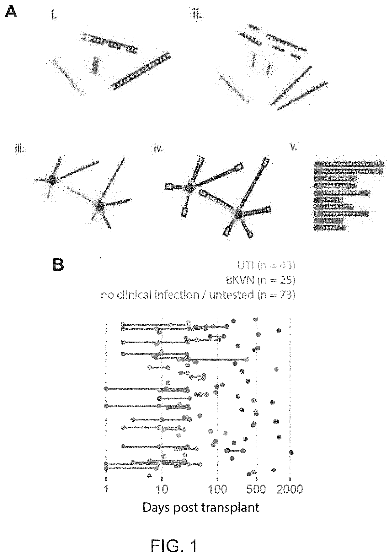
[0093]113 (1 mL) urine samples and 10 (4 mL) urine samples from a cohort of kidney transplant patient. Urine was stored at −80° C. and was only thawed when ready for extraction. Cell-free DNA was extracted from one (BK virus-associated nephropathy (BKVN), Urinary Tract Infection (UTI) and UTI control) or four mL (BKVN control only) of urine using Qiagen Circulating Nucleic Acid Kit according to the manufacturer's instructions. Following extraction, libraries were prepared from extracted DNA using a single-stranded adapter ligation (P. Burnham et al., Sci. Rep., vol. 6, 27859, 2016).

cfDNA Extraction from Urine.

[0094]Urine supernatants are thawed and centrifuged. cfDNA was extracted using the QIAamp circulating nucleic acid kit (Qiagen Valencia, Calif.). The concentration of total cfDNA in the sample was measured using a QuBit fluorometer. cfDNA extracts were stored at −20° C.

Negative Control.

[0095]To control for environmental and sample-t...

example 2

ell-Free DNA Extraction and Library Preparation

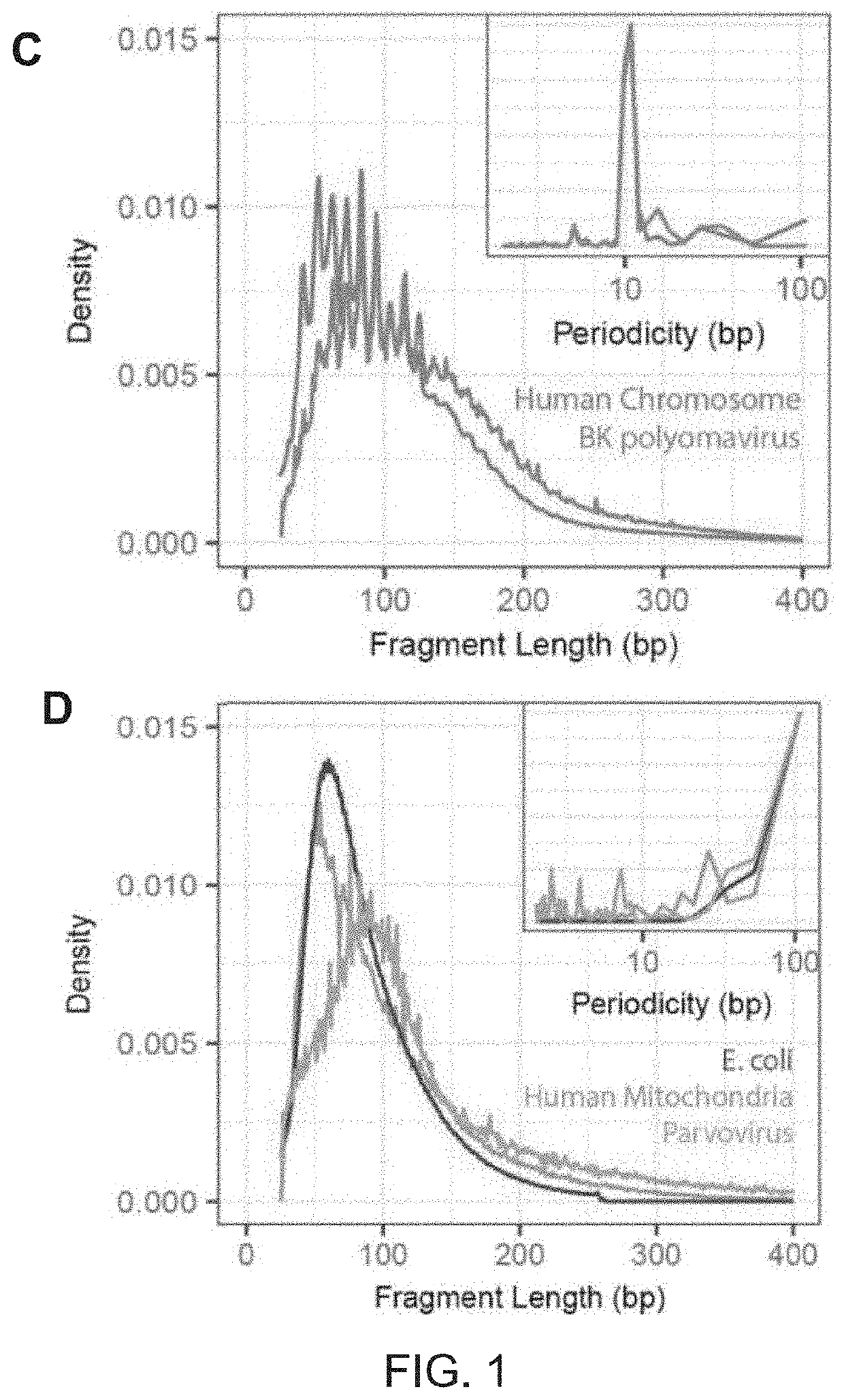
[0117]Urinary cell-free DNA is comprised of chromosomal, mitochondrial, and microbial cfDNA released from host cells and microbes in the urinary tract and of plasma-derived cfDNA that passes trans-renally from the circulation into urine. Urine can be collected non-invasively in large volumes, and therefore represents an attractive target for diagnostic assays. Compared to plasma DNA, relatively few studies have examined the properties and diagnostic potential of urinary cfDNA. The urinary environment degrades nucleic acids more rapidly than plasma resulting in fewer DNA fragments that are far shorter (J. Zhang et al., Clin. Chem., vol. 45, 1741-1746, 1999), consequently sequence analyses of urinary cfDNA have to date required relatively large volumes of urine (>10 ml) (Y.-H. Su et al., J. Mol. Diagn., vol. 6, no. 2, 101-7, 2004), (N. B. Y. Tsui, et al., PLoS One, vol. 7, no. 10, 1-7, 2012). Here, the inventors applied a single-stranded ...

example 3

Screening

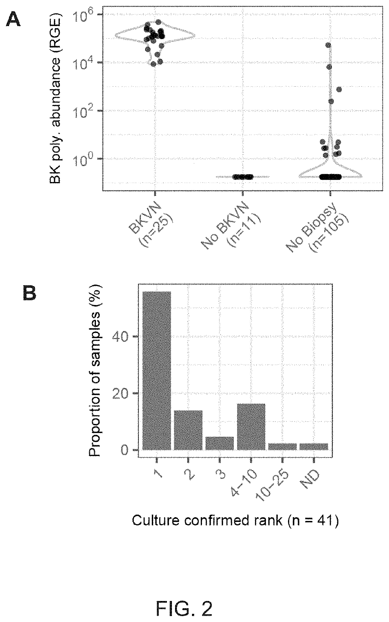
[0119]The presence of cfDNA was assessed from bacterial and viral pathogens reported in clinical diagnostic workups. Here, previously described bioinformatic approaches were used to quantify non-human cfDNA sequences in the datasets (I. De Vlaminck, et al., Proc. Natl. Acad. Sci., vol. 112, no. 43, 13336-13341, 2015). Briefly, human sequences were identified by alignment of the sequences to the human reference and removed. Remaining sequences were BLAST-ed against a custom database of microbial reference genomes. The relative genomic representation of different species was estimated using GRAMMy (L. C. Xia, et al., PLoS One, vol. 6, no. 12, e27992, 2011). In order to directly compare the measured microbial abundance across samples and species, the representation of microbial genome copies was computed, relative to the representation of host genomes in the datasets, and expressed this quantity as relative genome equivalents (RGE).

[0120]An extremely high load of BK Polyomavir...


PUM

PropertyMeasurementUnit
Lengthaaaaaaaaaa
Electrical resistanceaaaaaaaaaa
Fractionaaaaaaaaaa

Abstract

The present disclosure is directed to methods of detecting cell-free DNA (cfDNA) in biological samples and using it to quantify organ damage and identify pathogens. In some aspects, the biological samples are from patients who have undergone solid-organ transplantation. The disclosure is also directed to methods of detecting and analyzing methylation patterns in cell-free DNA from organ transplant patients to identify the presence of pathogens as well as quantify contributing tissue proportions as a measurement of the host response.

Description

CROSS REFERENCE TO RELATED APPLICATION[0001]This application claims the benefit of priority from U.S. Provisional Application No. 62 / 482,272, filed Apr. 6, 2017, the entire contents of which are incorporated herein by reference.STATEMENT REGARDING FEDERALLY SPONSORED RESEARCH OR DEVELOPMENT[0002]This invention was made with government support under Grant Nos. R21A1133331 and R21A1124237, awarded by the National Institutes of Health. The government has certain rights in the invention.INCORPORATION BY REFERENCE OF SEQUENCE LISTING[0003]The Sequence Listing in an ASCII text file, named as 34855.7811-02-PC.seq.updated_ST25.txt of 2 KB, created on Apr. 3, 2018, and submitted to the United States Patent and Trademark Office via EFS-Web, is incorporated herein by reference.BACKGROUND[0004]Cell-free DNA (cfDNA) exists in many shapes and forms, including fragments of the nuclear genome, the mitochondrial genome and microbial genomes. The predominant type of cfDNA is derived from the nuclear ...

Claims


Application Information

Patent Timeline
IPC IPC(8): C12Q1/6883C12Q1/689C12Q1/70C40B30/06
CPCC12Q1/701C12Q2600/156C12Q1/6883C40B30/06C12Q1/689C12Q2600/158C12Q2600/154C12Q1/6869
Inventor DE VLAMINCK, IWIJNLEE, JOHN RICHARDBURNHAM, PHILIP SMITHCHENG, ALEXANDRE PELLANSUTHANTHRIAN, MANIKKAMDADHANIA, DARSHANA
Owner CORNELL UNIVERSITY
Why Eureka
Try Eureka