Non-leaking or minimally-leaking choroidal or retinal revascularization

a choroidal or retinal revascularization, non-leaking or minimally-leaking technology, applied in the field of ocular diseases, can solve the problems of increasing non-leaking or minimally-leaking choroidal perfusion, and reducing the hypoxia of the outer retina and so as to reduce the hypoxia of the retina of the eye of the subject, the effect of reducing the void

Inactive Publication Date: 2020-02-27
RGT UNIV OF CALIFORNIA +2
View PDF0 Cites 0 Cited by
  • Summary
  • Abstract
  • Description
  • Claims
  • Application Information

AI Technical Summary

Benefits of technology

[0040]In some embodiments, non-leaking or minimally-leaking choroidal perfusion in the subject is increased after the formulation is administered to the subject. Non-leaking or minimally-leaking choroidal revascularization in the subject can be promoted after the formulation is administered to the subject. Macular flow voids can be reduced, hypoxia in the outer retina and retinal pigment epithelium (RPE) of the eye of the subject can be reduced, and / or ischemia in the outer retina and RPE of the eye of the subject can be reduced after the formulation is administered to the subject. Non-leaking or minimally-leaking choroidal perfusion in the subject can be increased after the formulation is administered to the subject.
[0041]In some embodiments, the label indicates the formulation is for treating a disease selected from a group comprising dry age-related macular degeneration (AMD) and / or geographic atrophy (GA), or a combination thereof. In some embodiments, the label indicates the formulation is for treating a disease selected from a group comprising wet age-related macular degeneration (AMD), choroidal neovascularization (CNV), polypoidal choroidal vasculopathy, degenerative (pathologic) myopia, or a combination thereof. In some embodiments, the label indicates the formation is for treating a disease selected from a group comprising diabetic macular edema, macular edema from retinal vein occlusion, diabetic retinopathy, retinal vein occlusion, retinopathy of prematurity, retinal neovascularization, optic nerve neovascularization, familial exudative vitreoretinopathy, sickle cell disease, or a combination thereof. Non-leaking or minimally-leaking retinal perfusion in the subject can increase after the formulation is administered to the subject. Non-leaking or minimally-leaking retinal revascularization in the subject can be promoted after the formulation is administered to the subject. Hypoxia in the retina of the eye of the subject can be reduced, and / or ischemia in the retina of the eye of the subject can be reduced after the formulation is administered to the subject. Non-leaking or minimally-leaking retinal perfusion in the subject can increase after the formulation is administered to the subject.

Problems solved by technology

Loss of normal macular tissue in wet AMD also causes irreversible visual loss.
In some embodiments, the method results in increasing non-leaking or minimally-leaking choroidal perfusion in the subject.
In some embodiments, the method results in promoting non-leaking or minimally-leaking choroidal revascularization in the subject.
In some embodiments, the method results in increasing non-leaking or minimally-leaking choroidal perfusion in the subject.
In some embodiments, the method results in promoting non-leaking or minimally-leaking choroidal revascularization in the subject.
In some embodiments, the method results in increasing non-leaking or minimally-leaking choroidal perfusion in the subject.
In some embodiments, the method results in promoting non-leaking or minimally-leaking choroidal revascularization in the subject.
In some embodiments, the method results in increasing choroidal perfusion in the subject.
The method can thereby result in reversing, halting, or slowing the progression of the disease.
In some embodiments, the method results in increasing non-leaking or minimally-leaking retinal perfusion in the subject.
In some embodiments, the method results in promoting non-leaking or minimally-leaking retinal revascularization in the subject.
In some embodiments, the method results in increasing non-leaking or minimally-leaking retinal perfusion in the subject.
In some embodiments, the method results in promoting non-leaking or minimally-leaking retinal revascularization in the subject.
In some embodiments, the method results in increasing non-leaking or minimally-leaking retinal perfusion in the subject.
In some embodiments, the method results in promoting non-leaking or minimally-leaking retinal revascularization in the subject.
In some embodiments, the method thereby results in increasing non-leaking or minimally-leaking retinal perfusion in the subject.
The method can thereby result in halting or slowing the progression of the disease.
The method can thereby result in reversing, halting, or slowing the progression of the disease.
The subject can have received a radiation treatment for a disease that is an intraocular tumor a head tumor, a neck tumor, or a combination thereof, resulting in delayed onset of the disease.
The composition, after being administered to the subject, can result in reversing, halting, or slowing of the progression of the disease in the subject.
The composition, after being administered to the subject, can result in reversing, halting, or slowing the progression of the disease in the subject.
The composition, after being administered to the subject, can result in halting or slowing of the progression of the disease in the subject.
The composition, after being administered to the subject, can result in reversing, halting, or slowing of the progression of the disease in the subject.
The subject may have received a radiation treatment for a disease that is an intraocular tumor a head tumor, a neck tumor, or a combination thereof, resulting in delayed onset of the disease.

Method used


Image

  • Non-leaking or minimally-leaking choroidal or retinal revascularization
  • Non-leaking or minimally-leaking choroidal or retinal revascularization
  • Non-leaking or minimally-leaking choroidal or retinal revascularization

Examples


example 1

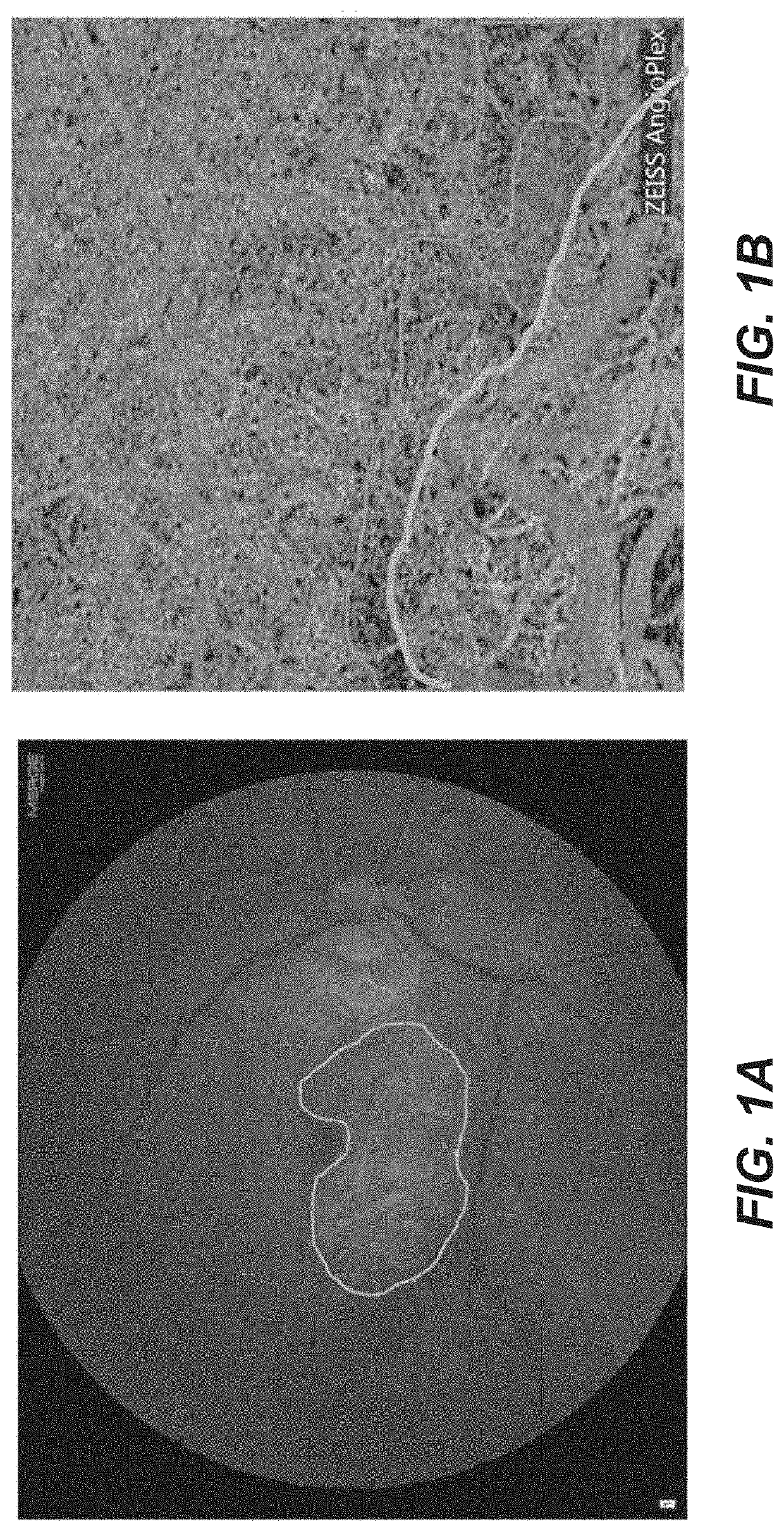
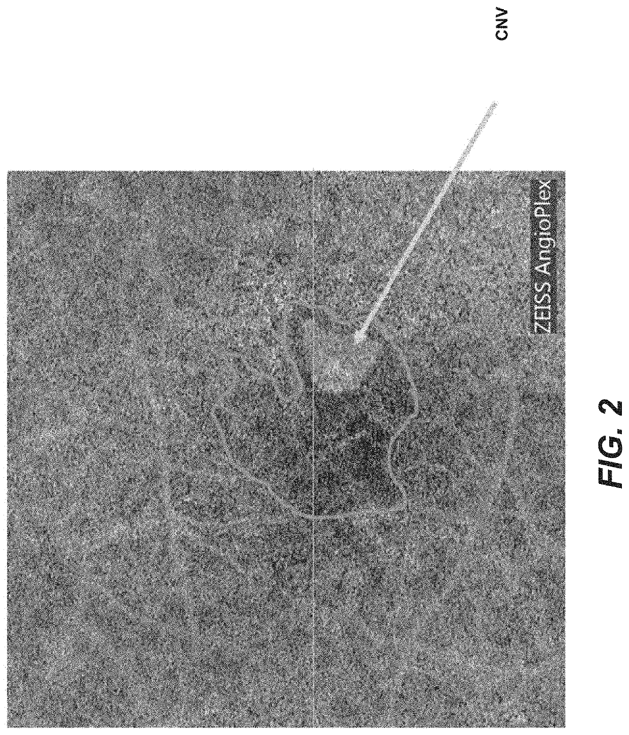
Non-Leaking Choroidal Neovascularization Protects Overlying Tissue from Developing Geographic Atrophy

[0122]There is clinical support for the hypothesis that a mature neovascular network underneath the fovea can support normal foveal architecture and prevent development of geographic atrophy. OCT angiography has shown that patients with type 1 CNV (sub-RPE) underneath the fovea develop GA eccentric to the retina overlying the neovascularization, suggesting that the CNV maintains the viability of the overlying macular tissue.

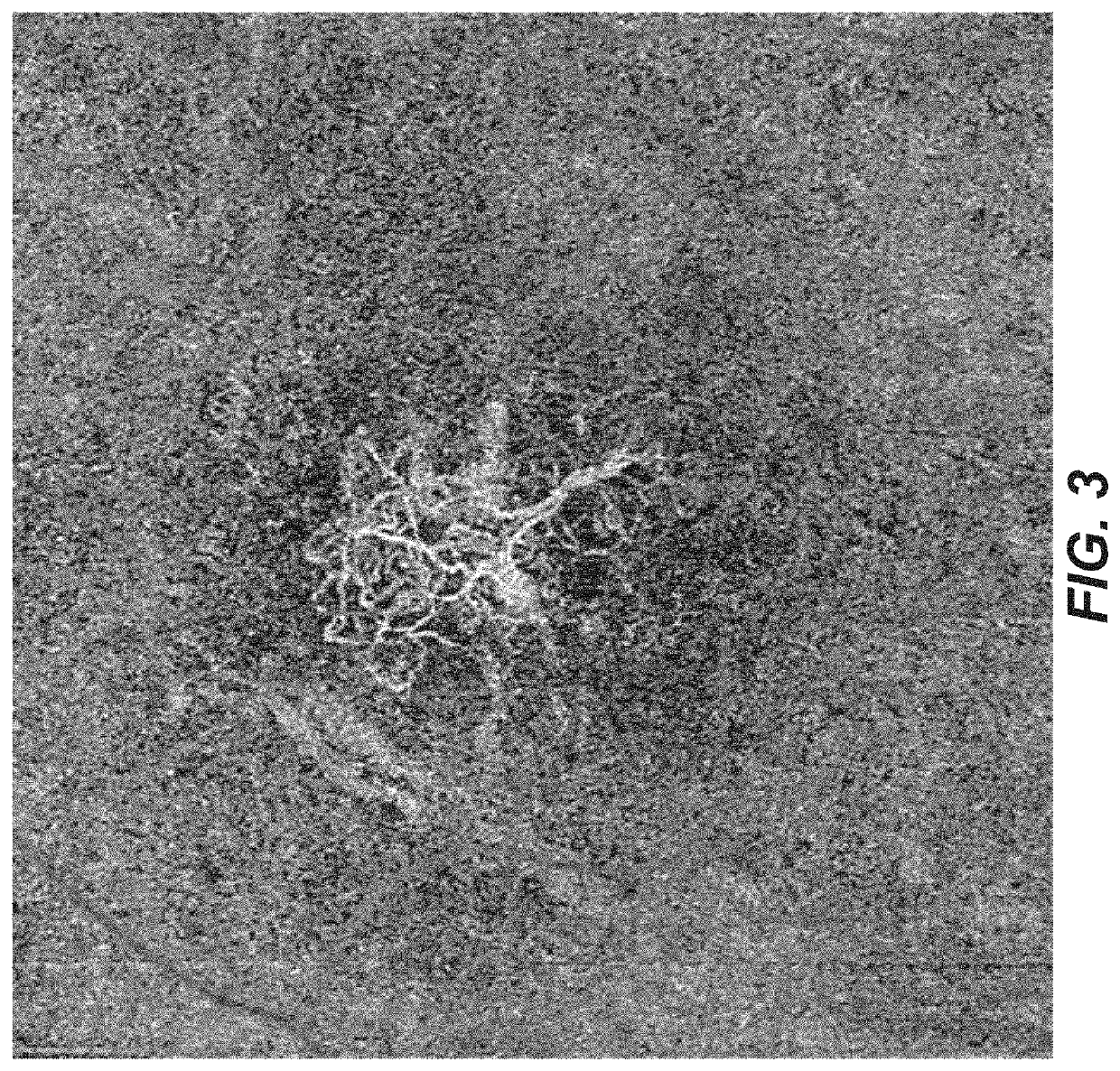
[0123]An example of this is shown below in an 88 year-old subject with disciform scar OS. The subject had received numerous injections in OD until he got post-injection endophthalmitis one year previous to the OCT angiography study shown in FIG. 3. The OCT and OCT angiogram reveal subfoveal CNV that is non-leaking (Let's show a picture of the OCT, too; don't' have it). Then, he did not want to have further injections, He has had no exudation nor progression of atr...

example 2

AMD with Progressively Expanding Extrafoveal Geographic Atrophy (GA)

[0124]A patient with AMD, good central visual acuity (≥20 / 40) and early GA that spares fixation is considered for treatment with pro-angiogenic therapy. OCTA examination reveals macular flow voids indicating reduced choriocapillaris perfusion. Pro-angiogenic compound(s) is / are administered intravitreally. Alternatively, pro-angiogenic therapy can be administered by a suprachoroidal route or injected into the subretinal space in the macular region. Following pro-angiogenic therapy, the patient is followed by sequential ocular examination, including visual acuity assessment, fundus auto-fluorescence (FAF), OCT and OCTA examinations. On follow up, GA progression is halted or slowed as demonstrated by serial FAF and OCT examination. OCTA demonstrates reduction in macular flow voids, including at the margin of the existing GA lesion(s). If during follow-up there is progressive choriocapillaris attrition with progression ...

example 3

AMD with CNV

[0125]OCTA in patients with CNV shows increased flow voids (choriocapillaris perfusion defects) surrounding the neovascular membrane. Resultant hypoxia results in VEGF secretion by RPE with subsequent CNV development. After treatment with anti-VEGF agents, CNV usually shows reduction in permeability. To minimize exudation and visual loss, repeated anti-VEGF injections are required. As an alternative, angiogenic factors that promote non-leaking choroidal neovascularization, reduce ischemia and resultant need for frequent VEGF injections. Pro-angiogenic choroidal revascularization therapy reconstitutes choriocapillaris and eliminates the ischemic drive for VEGF secretion. A patient with wet AMD is initially treated with an anti-VEGF agent that stops leakage from choroidal neovascularization. At the same time or shortly thereafter, a pro-angiogenic factor is administered intravitreally or subretinally, or in the suprachoroidal space, which promotes non-leaking choriocapilla...


PUM

PropertyMeasurementUnit
optical coherence tomographyaaaaaaaaaa
optical coherence tomography angiographyaaaaaaaaaa
compositionaaaaaaaaaa

Abstract

Disclosed herein include methods, kits, formulations, and compositions for increasing choroidal or retinal perfusion or promoting non-leaking or minimally-leaking choroidal or retinal revascularization in a subject in need thereof. An effective amount of an angiogenesis factor (e.g., a pro-angiogenic factor and/or a vascular maturation factor) can be administered to the subject.

Description

[0001]The present application claims priority under 35 U.S.C. § 119(e) to U.S. Application No. 62 / 720,441, filed Aug. 21, 2018. The content of the related application is incorporated herein by reference in its entirety.BACKGROUNDField[0002]The present disclosure relates generally to the field of treating ocular diseases, for example by increasing non-leaking or minimally leaking choroidal or retinal revascularization.Description of the Related Art[0003]Age-related macular degeneration (AMD) is the leading cause of central visual loss in the developed world. The “dry” form of the disease is characterized by yellow deposits (drusen), which accumulate underneath the retinal pigment epithelium (RPE). In some cases of dry AMD, there is progressive atrophy of the RPE, overlying photoreceptors, and subjacent choriocapillaris. This “geographic atrophy” (GA) often extends progressively around the macular center (fovea), until it finally involves the fovea and causes irreversible central visu...

Claims


Application Information

Patent Timeline
Patent Type & Authority Applications(United States)
IPC IPC(8): A61K38/18A61K9/00A61P27/02A61B3/10A61B3/12
CPCA61K38/1866A61P27/02A61B3/1241A61B3/102A61K9/0048A61K38/1891A61K38/204A61K39/3955A61K2300/00A61K2039/505A61K2039/545
Inventor GRUBBS, ROBERT H.SCHWARTZ, DANIEL M.
Owner RGT UNIV OF CALIFORNIA
Why Eureka
Try Eureka