A method for identification of low grade cervical cytology cases likely to progress to high grade/cancer

a cytology case and high-grade technology, applied in the direction of material excitation, optical radiation measurement, instruments, etc., can solve the problems of no current system, no current system, no clinical diagnosis, etc., and achieve high sensitivity and specificity. , the effect of time-consuming

Active Publication Date: 2020-04-16
TECHCAL UNIV DUBLIN +1
View PDF0 Cites 2 Cited by
  • Summary
  • Abstract
  • Description
  • Claims
  • Application Information

AI Technical Summary

Benefits of technology

[0047]The present invention provides a method of identifying and appropriately managing the treatment of women presenting with low grade cytological abnormalities, thus providing a cost-effective solution to distinguish between women with persistent low grade abnormalities at risk of disease progression and those likely to regress. The method of the present invention may be used to distinguish between transient and persistent HPV infection or to predict disease progression, in particular, to make a prognosis regarding the likelihood of low-grade squamous intraepithelial lesions (LSIL) progressing to high-grade squamous intraepithelial lesions (HSIL) or regressing to negative. An advantage of the present invention is that Raman spectroscopy alone may be used to provide a prognosis. This replaces the need to separately carry out cytology screening and test for the presence of HPV DNA and HPV mRNA. Thus, the present invention provides a method of using Raman spectroscopy for discriminating between LSIL cases likely to progress to HSIL or cancer and those likely to regress such that patients having low grade samples can avoid undergoing invasive colposcopy. This represents an important unmet clinical need as a high proportion of women with LSIL who are at a relatively low risk of developing cancer undergo unnecessary colposcopy follow up and in many instances also undergo unnecessary treatment.
[0048]Typically therefore the method does not include a separate step of cytology screening or screening for the presence of HPV DNA and / or HPV mRNA.
[0049]The step of analysing the Raman spectrum to determine whether the Raman spectrum falls within one or more predefined classes of cells may comprise using a classification model. A suitable classification model may be built using a database of reference Raman spectra. The reference Raman spectra may comprise Raman spectra for one or more predefined classes of cells. In particular, the reference Raman spectra may comprise Raman spectra for one, several or all of the following: (i) negative samples, (ii) biological samples comprising low-grade squamous intraepithelial lesions (LSIL) that are positive for HPV mRNA and positive for HPV DNA and therefore likely to progress to high-grade squamous intraepithelial lesions (HSIL); (iii) biological samples comprising LSIL that are negative for HPV mRNA and positive for HPV DNA and therefore unlikely to progress to HSIL and are likely to regress to negative; (iv) biological samples comprising LSIL that are negative for HPV mRNA and negative for HPV DNA and therefore unlikely to progress to HSIL and are likely to regress to negative; and (v) biological samples comprising HSIL. The database of reference Raman spectra may be compiled by obtaining Raman spectra for biological samples and subjecting biological samples to HPV DNA testing (e.g. Cobas) and HPV mRNA testing (e.g. APTIMA). Cells comprising LSIL that are likely to progress to HSIL are positive for HPV mRNA and positive for HPV DNA and cells comprising LSIL that are likely to regress to negative are negative for HPV mRNA and either positive or negative for HPV DNA. Specifically, the presence of HPV E6 / E7 mRNA indicates an active transforming HPV infection suggesting that the LSIL cases are more likely to progress to HSIL.
[0050]Raman Spectroscopy and HPV signatures are thus used to give a spectral signature which can identify whether the HPV infection is transient (and therefore likely to revert to normal) or persistent / integrated (and therefore likely to progress to cancer). This avoids the need for further testing for low grade cytology samples in cases where the HPV infection is transient. High sensitivity and specificity was achieved for the classification of LSIL cases with either an episomal HPV infection (i.e. not integrated into the host cell DNA) or a transforming HPV infection (i.e. integrated into the host cell DNA). The inventors have therefore shown that Raman spectroscopy can distinguish between women with LSIL cytology who are at risk of disease progression and those who are likely to regress.
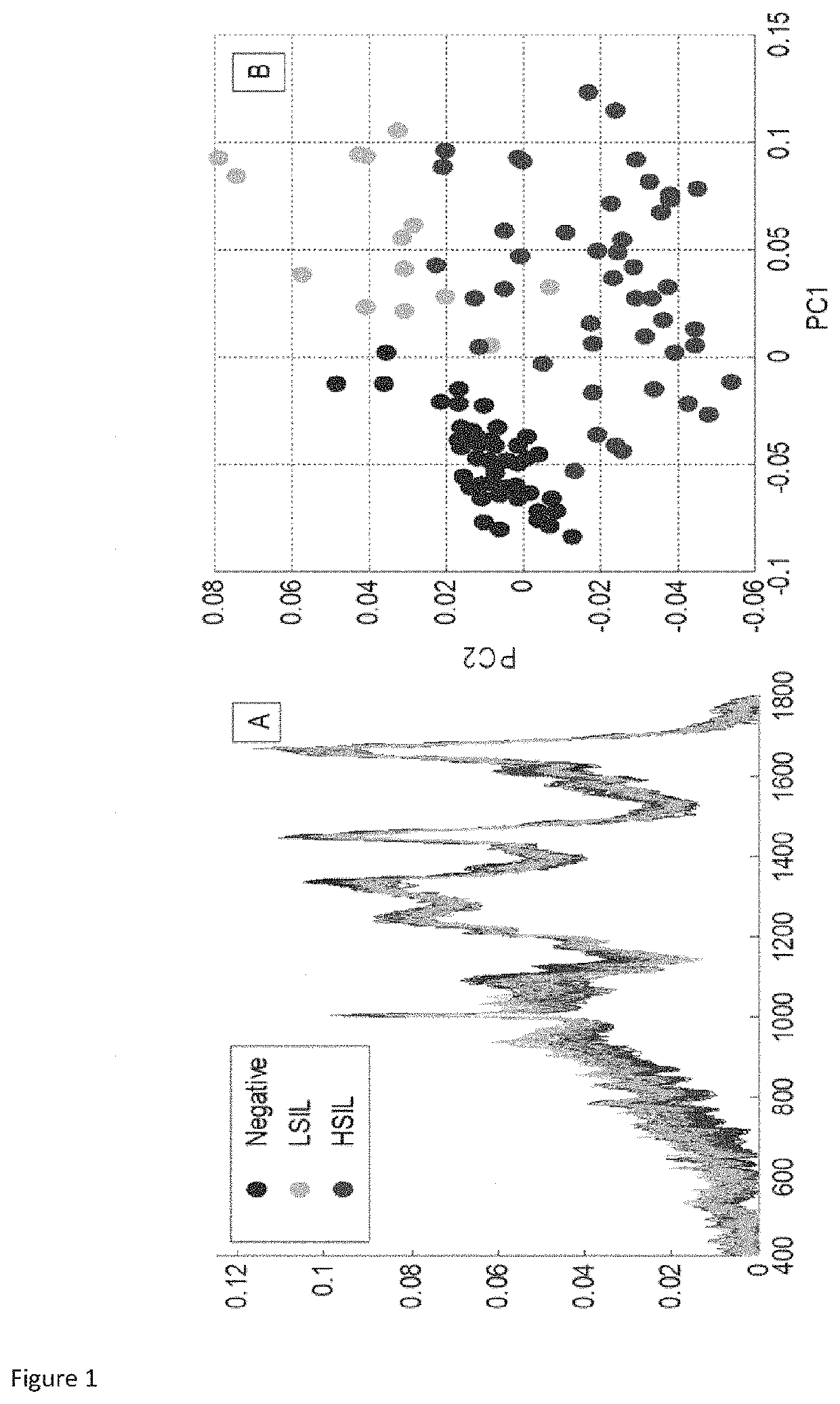
[0051]The classification model may comprise multivariate statistical analysis, such as Partial Least Squares Discriminant Analysis (PLS-DA), principal component analysis, linear discriminant analysis, support vector machines and random forest methods. As the visual difference between the Raman spectra may be very subtle, multivariate analysis, such as PLS-DA, may be used to determine the spectral differences.
[0052]The biological sample may be obtained during a smear test or a Pap smear. The biological sample or the Pap smear may be processed using a liquid-based cytology test or conventional cytology. The liquid-based cytology test may be ThinPrep® or SurePath®.

Problems solved by technology

These screening programmes are common in developed countries, greatly reducing the mortality rates due to cervical cancer, but are not yet implemented in developing countries due to lack of infrastructure and funding.
LSIL may regress, but HSIL is unlikely to do so and may progress to invasive disease.
None of the currently available systems, however, FocalPoint® GS Imaging system (BD) or the ThinPrep® Imaging system (Hologic), provides fully automated screening without human intervention at some stage.
The final decision still lies with the cytologist, resulting in the same subjectivity problem as in manual screening.
However, these tests are expensive, time-consuming and provide no information on cervical cytopathology.
Over the last decade, prophylactic HPV vaccines have been developed, but, despite the introduction of these vaccines, there is still a need for regular cervical screening, as the vaccines do not protect against all oncogenic HPV types.
Additionally, some women may not benefit from the vaccines if there is a pre-existing HPV infection or they do not receive the complete number of doses.
This leads to an increased risk of cervical cancer in the future so these women need to be monitored more closely.
However, the disclosed methods are based on histochemical staining or in situ hybridisation or other molecular biology methods and do not disclose the use of Raman spectroscopy in a method of distinguishing between transient HPV infection and persistent HPV infection, nor do the known methods use Raman spectroscopy to predict disease progression from low grade lesions to high grade lesions.
This is a major benefit as finding the rare abnormal cells on the unstained slide preparations is very difficult and time consuming.

Method used


Image

  • A method for identification of low grade cervical cytology cases likely to progress to high grade/cancer
  • A method for identification of low grade cervical cytology cases likely to progress to high grade/cancer
  • A method for identification of low grade cervical cytology cases likely to progress to high grade/cancer

Examples


example 1

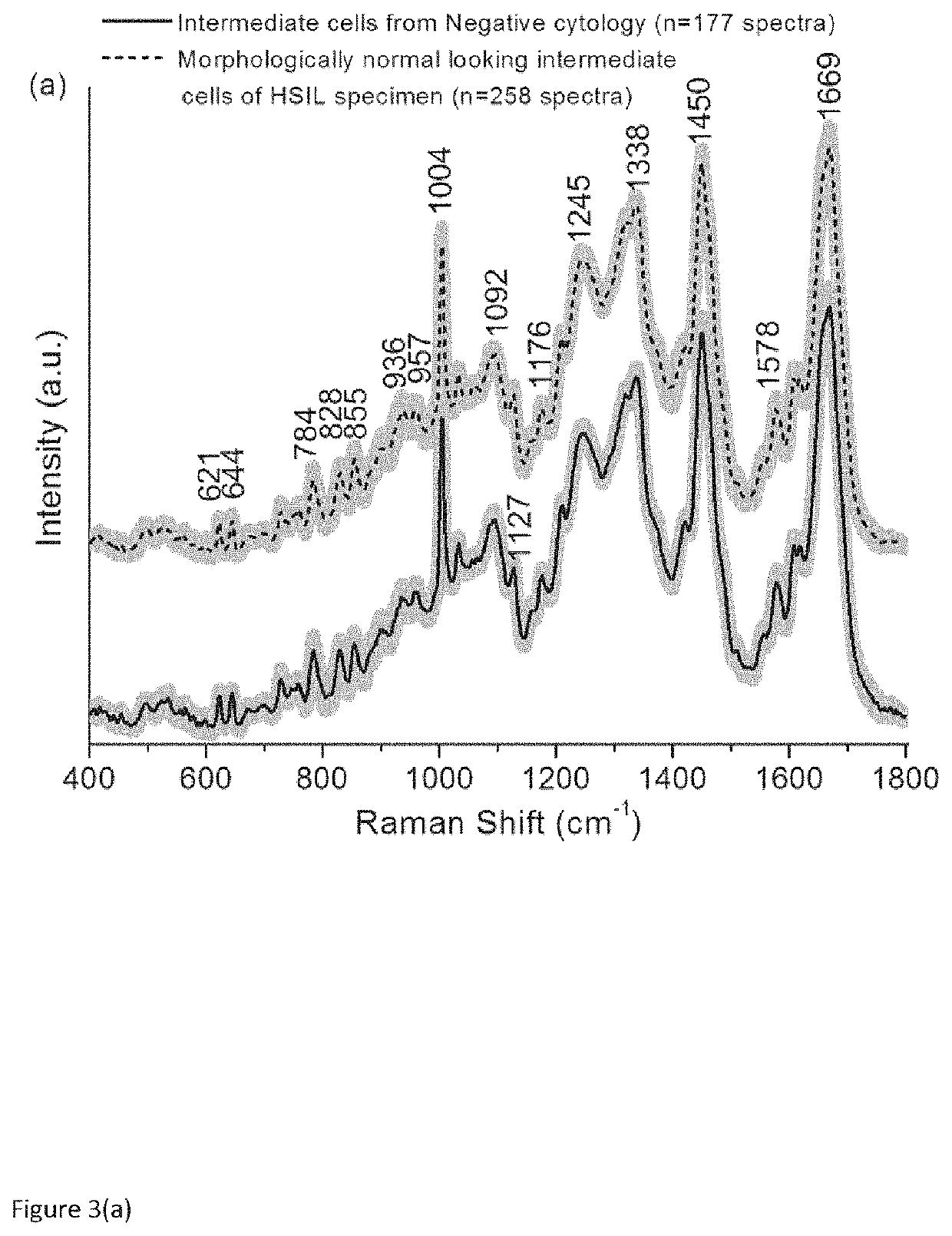
Use of Superficial or Intermediate Epithelial Cells for Discrimination of Negative and HSIL Cytology Cases

[0076]Materials and Methods

[0077]Sample Collection and Processing

[0078]True negative cervical liquid based cytology samples were obtained from the cytology laboratory, Coombe Women and Infants University Hospital (CWIUH), Dublin, Ireland. HSIL cytology specimens were collected during the routine Pap smear from the Colposcopy clinic, CWIUH, Dublin, Ireland. The collected smears were processed via the ThinPrep® method. This study was approved by the Research Ethics Committee CWIUH. The cells were collected from the cervix using a cyto brush and then rinsed in the specimen vial containing PreservCyt transport medium (ThinPrep® Pap Test; Cytyc Corporation, Boxborough, Mass.). The labelled ThinPrep® sample vial was sent to the cytology laboratory equipped with a ThinPrep® processor. All samples were prepared using a ThinPrep® 2000 processor (Hologic Inc., Marlborough, Mass. 01752). T...

example 2

LSIL-HPV

[0087]Materials and Methods

[0088]Sample Collection

[0089]This current study was approved by the Research Ethics Committee at the Coombe Women and Infants University Hospital (CWIUH), Dublin. A total of 39 cervical liquid based cytology samples (15 true negative (TN) specimens, 12 LSIL specimens and 12 HSIL specimens) were collected for this Raman study. True negative cytology samples were obtained from the cytology laboratory, CWIUH, Dublin, Ireland. The LSIL and HSIL cytology specimens were collected from the Colposcopy clinic, CWIUH, Dublin, Ireland. The collected smears were processed via ThinPrep® method. For ThinPrep®, an adequate sampling of cells was collected from the ectocervix of true negative, LSIL and HSIL patients using the cytobrush. The cytobrush was rinsed in the vial containing PreservCyt transport medium (ThinPrep® Pap Test; Cytyc Corporation, Boxborough, Mass.). The vial was named with the patient name and ID and then sent to the cytology laboratory equippe...


PUM

PropertyMeasurementUnit
diameteraaaaaaaaaa
poweraaaaaaaaaa
Raman spectrumaaaaaaaaaa

Abstract

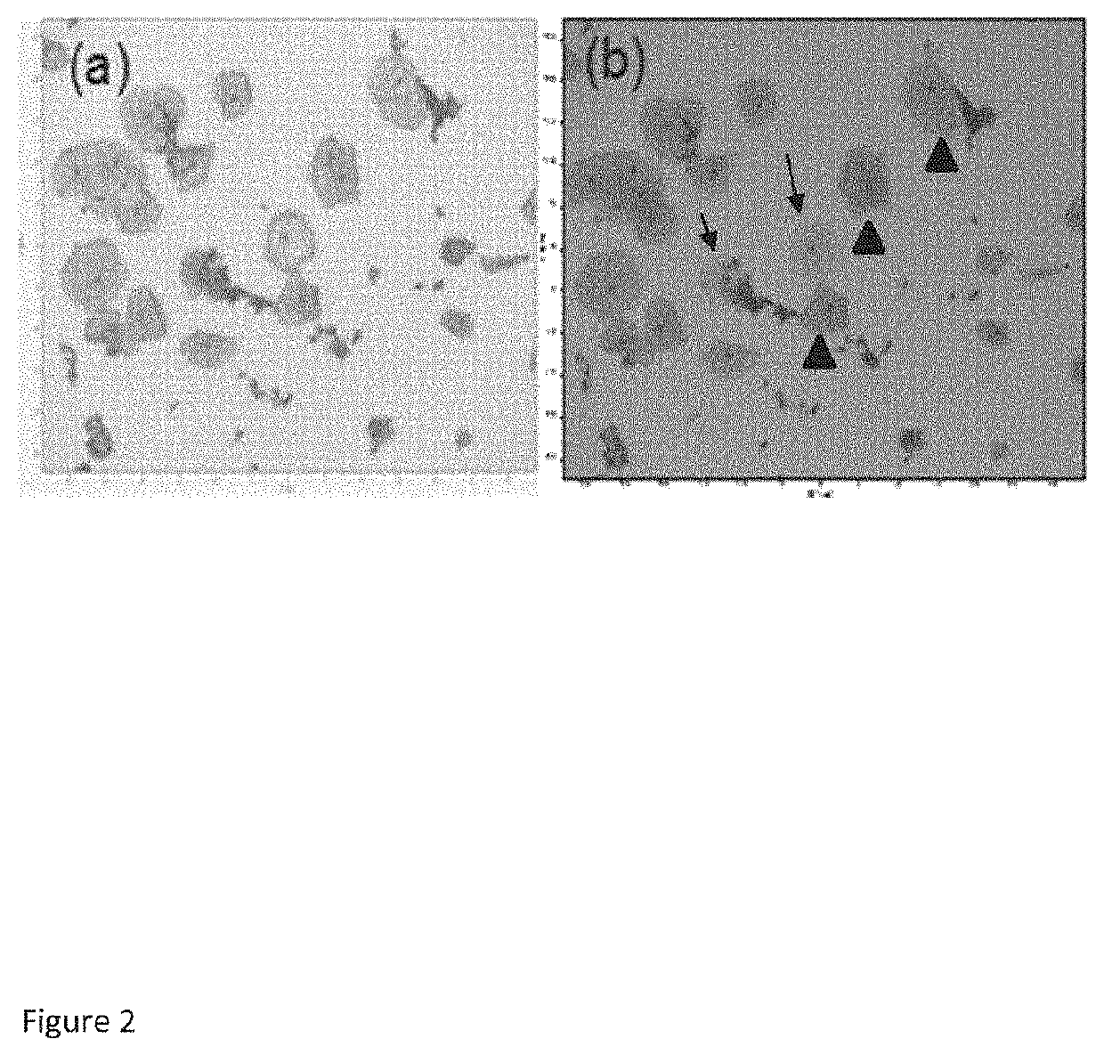
The present invention provides a method of using Raman spectroscopy for identification of low grade cervical cytology cases likely to progress to high grade / cancer. The Applicant has found that high quality Raman spectra can be successfully acquired from morphologically normal appearing cells from negative, LSIL and HSIL Thinprep® specimens and different grades of cervical pre-cancer can be separated with good sensitivities and specificities. Raman spectroscopy can further identify different categories of the LSIL cases i.e., whether they are likely to regress to negative or progress to HSIL cytology.

Description

FIELD[0001]The present application relates to a method for identification of low grade cervical cytology cases likely to progress to high grade / cancer.BACKGROUND OF THE INVENTION[0002]1. Cervical Cancer[0003]Cervical cancer is the fourth most common cancer in women worldwide, accounting for an estimated 528,000 new cases and 266,000 deaths in 2012. The mortality associated with cervical cancer can be reduced if this disease is detected at the early stages of development or at the pre-malignant stage (cervical intraepithelial neoplasia, CIN). Unlike most other types of cancer, cervical cancer affects mainly younger women, with about 60% of cases occurring in women under 50 years of age. Persistent infection with high risk human papillomavirus (HPV) (e.g., HPV types 16, 18) is accepted as the major cause for the development of cervical pre-cancer and cancer. Other risk factors include smoking, immunosuppression, long term use of oral contraceptives and socioeconomic status.[0004]Cervi...

Claims


Application Information

Patent Timeline
Patent Type & Authority Applications(United States)
IPC IPC(8): G01N21/65G01N33/543G01N33/574
CPCG01N33/54373G01N33/57411G01N21/65G01J3/44
Inventor DURAIPANDIAN, SHIYAMALALYNG, FIONATRAYNOR, DAMIENO'LEARY, JOHNMARTIN, CARAKEARNEY, PADRAIG
Owner TECHCAL UNIV DUBLIN
Why Eureka
Try Eureka