Specific cell fractionating and capturing methods

a specific cell and cell technology, applied in the field of specific cell fractionation and capturing methods, can solve the problems of difficult fractionation and capture of cancer cells, limited type of cancer cells that can be captured, etc., and achieve the effect of effectively capturing specific cells, effective fractionation of specific cells, and reducing adhesion or attachment of blood cells

Active Publication Date: 2020-07-30
SUMITOMO RUBBER IND LTD +1
View PDF13 Cites 0 Cited by
  • Summary
  • Abstract
  • Description
  • Claims
  • Application Information

AI Technical Summary

Benefits of technology

[0033]One aspect of the present invention relates to a method for fractionating specific cells present in blood or biological fluid. The method includes fractionating the blood or biological fluid by centrifugation to collect the specific cells in the blood or biological fluid. Further, the centrifugation is carried out using a container having a low protein adsorbing layer at least partially formed on the inner surface thereof. Such a method can effectively fractionate specific cells (e.g., many types of cancer cells, including cancer cells not expressing EpCAM). Thus, it is possible to sufficiently fractionate specific cells such as cancer cells from blood or biological fluid.
[0034]Another aspect of the present invention relates to a method for capturing specific cells present in blood or biological fluid. The method includes fractionating the blood or biological fluid by centrifugation to collect the specific cells in the blood or biological fluid, and then capturing the specific cells present in the collected liquid onto a hydrophilic polymer layer. Further, the centrifugation is carried out using a container having a low protein adsorbing layer at least partially formed on the inner surface thereof. Such a method can effectively capture specific cells (e.g., many types of cancer cells, including cancer cells not expressing EpCAM) and can also reduce adhesion or attachment of blood cells including red blood cells, white blood cells, and platelets. Thus, it is possible to selectively capture specific cells such as cancer cells from blood or biological fluid.

Problems solved by technology

However, a problem exists in that since the number of circulating tumor cells is very small (several to hundreds of cells / 1 mL of blood), such cancer cells are difficult to fractionate and capture.
This technique, which involves an antigen-antibody reaction (capture by EpCAM antibody), can only capture cancer cells expressing EpCAM, and the type of cancer cells that can be captured is limited (see, for example, Patent Literature 1).

Method used


Image

  • Specific cell fractionating and capturing methods
  • Specific cell fractionating and capturing methods
  • Specific cell fractionating and capturing methods

Examples


examples

[0118]The present invention is specifically described with reference to, but not limited to, examples below.

[0119]Specific cell-fractionating and -capturing methods were performed as described below. The results of Example a and Comparative Example a (specific cell-fractionating methods) are shown in Table 1; the results of Examples A to C and Comparative Example A (specific cell-capturing methods) are shown in Table 2; and the results of Examples 1 to 4 and Comparative Examples 1-1 to 1-3 and 2 to 5 (specific cell-fractionating or -capturing methods) are shown in Table 3.

Specific Cell-Fractionating Method

Example A

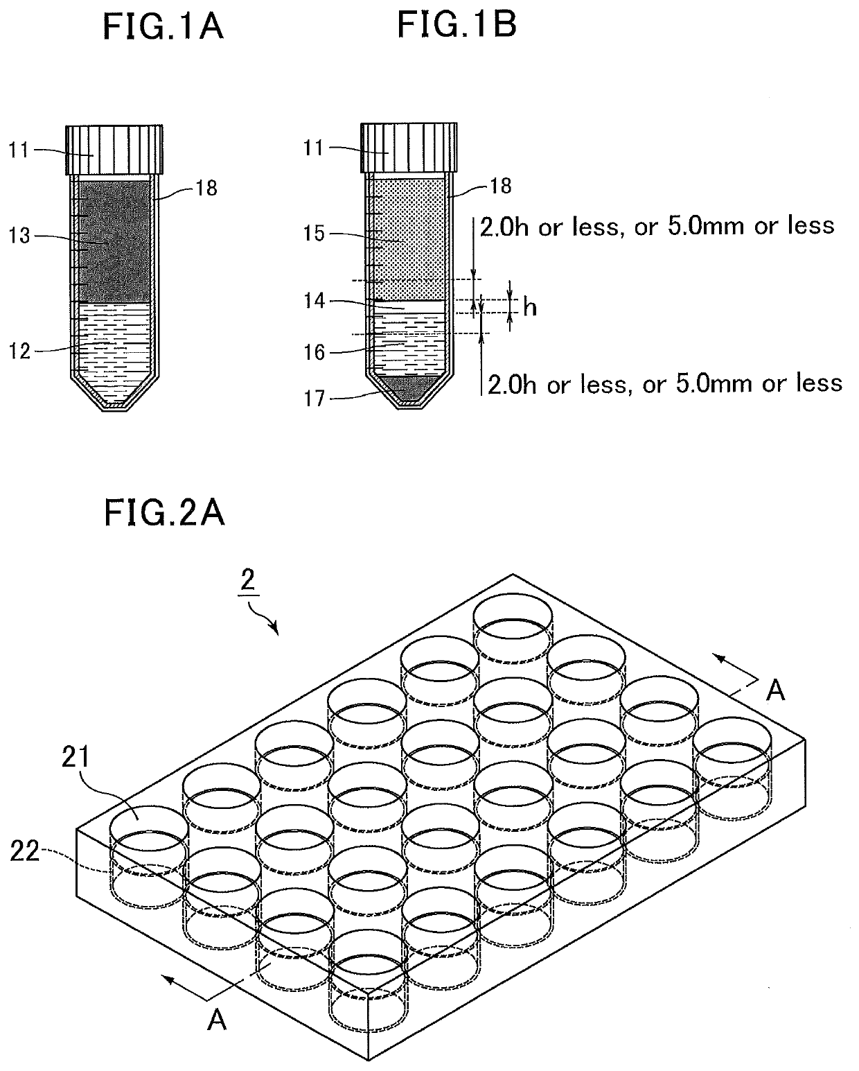
[0120]Stained human colon adenocarcinoma (HT-29) cells were suspended in whole blood at a concentration of 500,000 cells per mL of blood to prepare spiked blood. The spiked blood was diluted with an equal volume of a phosphate buffer solution to prepare a spiked blood dilution. Next, a separation liquid (Lymphoprep, density=1.077±0.001 g / mL) was placed in a 15 mL centrifug...

example a

[0132]Using azobisisobutyronitrile (AIBN), 2-methoxyethyl acrylate was thermally polymerized at 80° C. for six hours to produce poly(2-methoxyethyl acrylate) (molecular weight: Mn=about 15,000, Mw=about 50,000). Then, a 0.25% solution of the poly(2-methoxyethyl acrylate) in methanol was prepared.

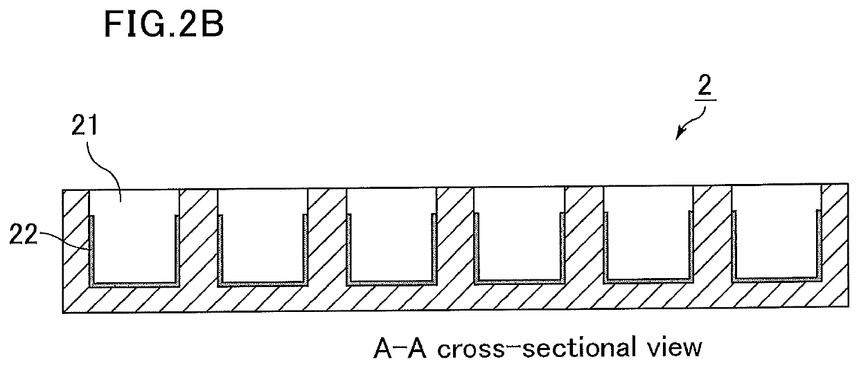
[0133]The poly(2-methoxyethyl acrylate) solution (0.25% by mass) was injected into a glass chamber slide and dried to form a hydrophilic polymer layer.

[0134]Further, fibronectin was adsorbed onto the part coated with poly(2-methoxyethyl acrylate) (hydrophilic polymer layer). Specifically, a 200 μg / mL solution of fibronectin in a phosphate buffered saline (PBS) solution was prepared, injected, and left at 40° C. for one hour, followed by washing it with a PBS solution to prepare a medical analysis device.

[0135]Stained human colon adenocarcinoma (HT-29) cells were suspended in whole blood at a concentration of 100 cells per mL of blood to prepare spiked blood. The spiked blood was diluted with...

example b

[0136]The cell capture ratio (%) was determined as in Example A, except that fibronectin was not adsorbed onto the hydrophilic polymer layer formed by injecting the poly(2-methoxyethyl acrylate) solution (0.25% by mass) into the glass chamber slide and drying it.


PUM

PropertyMeasurementUnit
thicknessaaaaaaaaaa
contact angleaaaaaaaaaa
densityaaaaaaaaaa

Abstract

Provided are specific cell-fractionating and -capturing methods which can fractionate and capture, respectively, specific cells (e.g., many types of cancer cells, including cancer cells not expressing EpCAM, or peripheral blood stem cells). Included is a method for fractionating specific cells present in blood or biological fluid, the method including fractionating the blood or biological fluid by centrifugation to collect the specific cells in the blood or biological fluid, the centrifugation being carried out using a container having a low protein adsorbing layer at least partially formed on the inner surface thereof.

Description

TECHNICAL FIELD[0001]The present invention relates to a method for fractionating (separating) specific cells in blood or biological fluid (e.g., cancer cells or stem cells present in blood or biological fluid), and a method for capturing the cells.BACKGROUND ART[0002]When cancer cells are formed, they are known to appear in due course in blood or biological fluid. Such cancer cells in blood are called “circulating tumor cells (CTCs)”. Thus, it is expected that the circulating tumor cells may be analyzed, e.g., to evaluate the cancer-treating effect, predict prognosis life expectancy, predict the effect of anticancer drugs before administration, or examine treatment methods based on genetic analysis of cancer cells.[0003]However, a problem exists in that since the number of circulating tumor cells is very small (several to hundreds of cells / 1 mL of blood), such cancer cells are difficult to fractionate and capture.[0004]For example, the CellSearch System is known as a technique for c...

Claims


Application Information

Patent Timeline
Patent Type & Authority Applications(United States)
IPC IPC(8): B01D21/26G01N1/28B01J20/26
CPCG01N1/28B01J20/261B01D21/262G01N33/5002G01N33/5094G01N33/491
Inventor MINAGAWA, YASUHISATANAKA, MASARUEMURA, HARUKASUTO, KAZUKI
Owner SUMITOMO RUBBER IND LTD
Why Eureka
Try Eureka