Automatic identification and segmentation of target regions in pet imaging using dynamic protocol and modeling

Inactive Publication Date: 2020-08-20
UNIV OF VIRGINIA ALUMNI PATENTS FOUND
View PDF0 Cites 2 Cited by
  • Summary
  • Abstract
  • Description
  • Claims
  • Application Information

AI Technical Summary

Benefits of technology

This patent describes a method for segmenting tumors from healthy tissue in PET images using a trained ANN network. The method first filters the images using a time activity curve approach to improve signal-to-noise ratio. The trained ANN network is then used to segment the tumor regions in other PET images from the same or different patients. The method has been tested on breast tumor and background and has shown good performance with the BP algorithm. The technical effect is the development of a reliable and efficient tool for tumor segmentation that can be used in clinics.

Problems solved by technology

Still, some breast cancers are not found by mammogram because radiologists can make negative diagnosis either because of the large number of mammograms to be analyzed or when coping with the inconclusive cases when inspecting noise contaminated images.

Method used


Image

  • Automatic identification and segmentation of target regions in pet imaging using dynamic protocol and modeling
  • Automatic identification and segmentation of target regions in pet imaging using dynamic protocol and modeling
  • Automatic identification and segmentation of target regions in pet imaging using dynamic protocol and modeling

Examples


example 1

[0105]A continuous dynamic positron emission tomography (PET) assembly for imaging a target region of a subject. The assembly may comprise: a radioactive tracer isotope injector configured to administer a radioactive isotope into the subject; a scintillator crystal configured to absorb ionizing radiation from the subject and emit scintillator light, wherein said scintillator crystal undertakes the absorption substantially at the same time of the start of administering the radioactive isotope; a photo detector in communication with said scintillator crystal, wherein the photodetector is configured to detect the emitted scintillation light and provide electrical signals as output; a signal digitizing circuitry converting the output electrical signals into digital data; and a processor configured to receive the digital data and implement a model to convert the digital data into a three dimensional, tomographic image reconstruction.

example 2

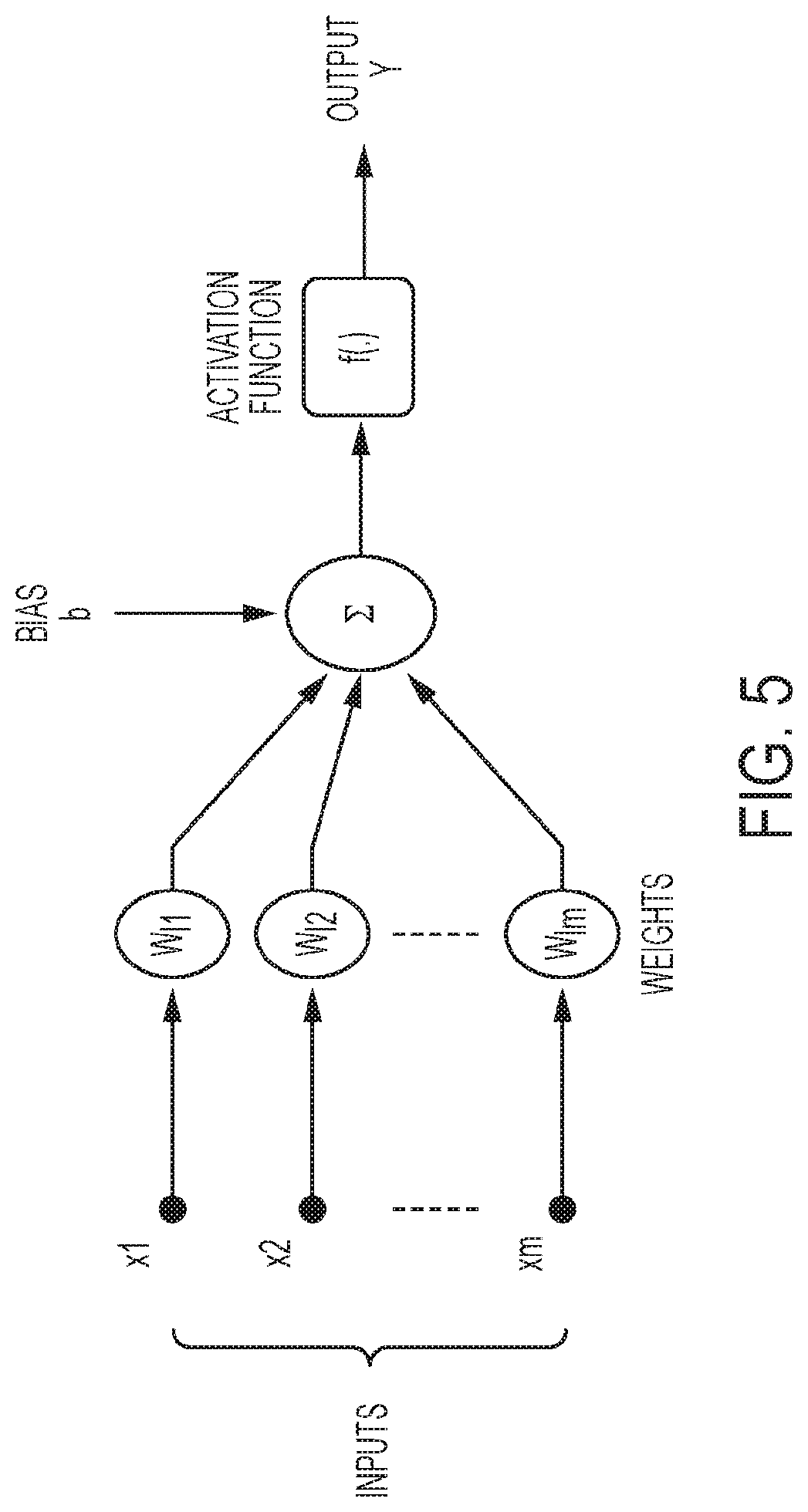
[0106]The PET assembly of example 1, wherein said model comprises an artificial Neural Network (ANN).

example 3

[0107]The PET assembly of example 1 (as well as subject matter in whole or in part of example 2), wherein undertaking the absorption, by the scintillator crystal, substantially at same time of the start of administering the radioactive isotope includes at least one of the following ranges of timing:

[0108]about 1 minute to about 60 minutes after the start of administering;

[0109]about 5 minutes to about 30 minutes after the start of administering;

[0110]about 1 minute to about 25 minutes after the start of administering;

[0111]about 10 to about 30 minutes after the start of administering;

[0112]about 15 to about 20 minutes after the start of administering;

[0113]about 25 minutes after the start of administering;

[0114]about 30 to about 60 minutes after the start of administering; or

[0115]about 0 to 30 minutes after the start of administering;

[0116]about 25 to 30 minutes after the start of administering;

[0117]about 1 minute to about 10 minutes after the start of administering; or about 60 m...


PUM


Abstract

A continuous dynamic positron emission tomography (PET) assembly for imaging a target region of a subject. The assembly includes a radioactive tracer isotope injector configured to administer a radioactive isotope into the subject and a scintillator crystal configured to absorb ionizing radiation from the subject and emit scintillator light. The scintillator crystal undertakes the absorption substantially at the same time of the start of administering the radioactive isotope. The assembly also includes a photo detector in communication with the scintillator crystal, wherein the photodetector is configured to detect the emitted scintillation light as input and provide electrical signals as output. The assembly further includes a signal digitizing circuitry converting the output electrical signals into digital data. Moreover, the assembly includes a processor configured to receive the digital data and implement a model to convert the digital data into a three dimensional, tomographic image reconstruction.

Description

RELATED APPLICATIONS[0001]The present application claims benefit of priority under 35 U.S.C § 119 (e) from U.S. Provisional Application Ser. No. 62 / 269,628, filed Dec. 18, 2015, entitled “Dynamic TOFPET Breast Imager with Full Breast View and Related Methods Thereof”, U.S. Provisional Application Ser. No. 62 / 362,914, filed Jul. 15, 2016, entitled “Automatic Identification and Segmentation of Breast Tumors in PET Imaging Using Dynamic Protocol and Artificial Neural Network (ANN)”, and U.S. Provisional Application Ser. No. 62 / 435,645, filed Dec. 16, 2016, entitled “Dynamic TOFPET Breast Imager with Full Breast View and Related Methods Thereof,” the disclosures of which are hereby incorporated by reference herein in their entirety.FIELD OF THE INVENTION[0002]The present invention relates generally to the field of automatic identification and segmentation of target region images in positron emission tomography (PET) imaging, whereby the imaging commences substantially at the same time o...

Claims


Application Information

Patent Timeline
IPC IPC(8): A61B6/03A61B6/00G01T1/29
CPCA61B6/4216A61B5/055G01T1/2985A61B6/037A61B5/0066A61B6/032A61B6/502A61B8/481A61M5/007G01T1/164G01T1/166G01T1/202G01T1/2023
Inventor LI, YINLINKUNDU, BIJOYMAJEWSKI, STANISLAW
Owner UNIV OF VIRGINIA ALUMNI PATENTS FOUND
Why Eureka
Try Eureka