Analysis method for extracellular vesicles, using size exclusion chromatography, and use for same
- Summary
- Abstract
- Description
- Claims
- Application Information
AI Technical Summary
Benefits of technology
Problems solved by technology
Method used
Image
Examples
example 1
ion and Analysis of Reference Extracellular Vesicles
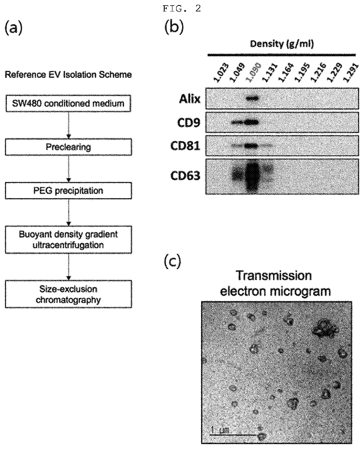
[0067]The colorectal cancer cell line SW480 culture medium was centrifuged at 500×g for 10 min and 2,000×g for 20 min to remove precipitates. To primarily purify and precipitate extracellular vesicles present in the supernatant, the supernatant was subjected to addition of a polyethylene glycol solution (8.4% polyethylene glycol 6000, 250 mM NaCl, 20 mM HEPES, pH 7.4), stored in a refrigerator for 16 h, and then centrifuged at 12,000×g for 30 min to harvest the precipitated extracellular vesicles, which were then dissolved in HEPES-buffered saline (20 mM HEPES, 150 mM NaCl, pH 7.4).
[0068]To secondarily purify extracellular vesicles by using density and buoyancy, the sample was subjected to 30%-20%-5% OptiPrep buoyant density gradient ultracentrifugation at 200,000×g for 2 h. After the ultracentrifugation, regions having an equivalent density (1.08-1.12 g / ml) to extracellular vesicles were harvested. To tertiarily purify the purifie...
example 2
of Reference Extracellular Vesicles Using Pump-Type Size-Exclusion Chromatography and Fluorescently Labeled Antibodies
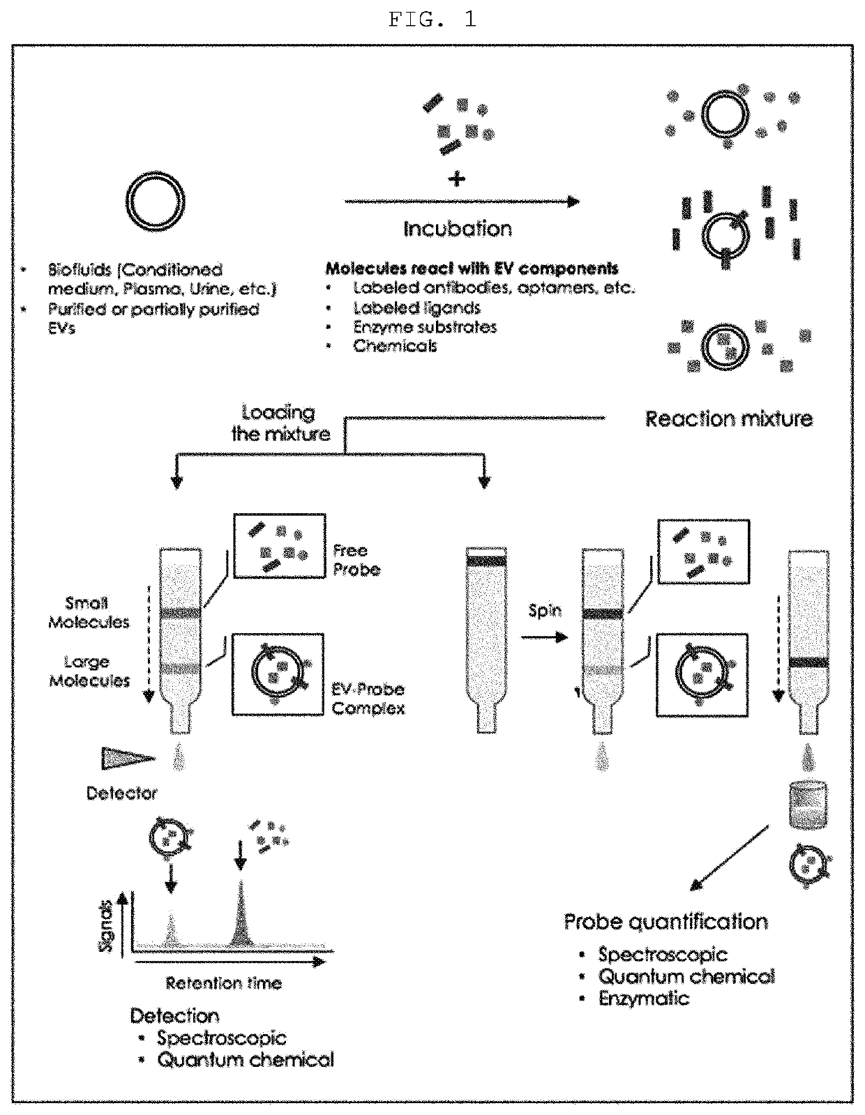
[0070]To establish that the components of extracellular vesicles can be quantitatively analyzed by using the size-specific fractionation ability of size-exclusion chromatography and quantifying various kinds of probes recognizing the components of extracellular vesicles, the following tests were carried out.
[0071]2-1. Fluorescently Labeled Mouse Antibody (Normal Mouse IgG)
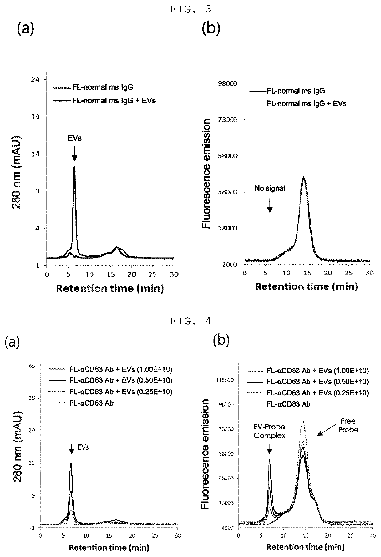
[0072]The purified reference extracellular vesicles were mixed with a fluorescently labeled mouse antibody (normal mouse IgG), followed by incubation at 37° C. for 30 min, and then the mixture was injected into a column packed with Sephacryl S500 and developed using the HPLC system, and the 280 nm absorption chromatogram (FIG. 3A) and the fluorescence chromatogram (FIG. 3B) were analyzed.
[0073]The absorption chromatogram results confirmed that the reference extracellular vesicles were detected at 6....
example 3
of Extracellular Vesicles in Cell Cultures Using Pump-Type Size-Exclusion Chromatography and Fluorescently Labeled Antibodies
[0088]3-1. Fluorescently Labeled Anti-C63 Antibody
[0089]To analyze the amount or components of extracellular vesicles in the cell culture using the analysis method of the present invention, SW480 colorectal cancer cells were cultured in RPMI media for 24 h to harvest the cell culture. A group of RPMI medium without cell culture mixed with fluorescently labeled anti-CD63 antibody and a group of a colorectal cancer cell culture mixed with fluorescently labeled anti-CD63 antibody were incubated at 37° C. for 30 min, and injected into TSK6000 HPLC column, followed by development using the HPLC system, and a fluorescence chromatogram was analyzed.
[0090]As a result, as shown in FIG. 9, only the fluorescence band of the free antibody detected at 22 min was observed in the group of RPMI medium mixed with fluorescently labeled anti-CD63 antibody, but additional fluores...
PUM
| Property | Measurement | Unit |
|---|---|---|
| Wavelength | aaaaa | aaaaa |
| Wavelength | aaaaa | aaaaa |
| Fluorescence | aaaaa | aaaaa |
Abstract
Description
Claims
Application Information
 - R&D
- Intellectual Property
- Life Sciences
- Materials
- Tech Scout
- Unparalleled Data Quality
- Higher Quality Content
- 60% Fewer Hallucinations
Browse by: Latest US Patents, China's latest patents, Technical Efficacy Thesaurus, Application Domain, Technology Topic, Popular Technical Reports.
© 2025 PatSnap. All rights reserved.Legal|Privacy policy|Modern Slavery Act Transparency Statement|Sitemap|About US| Contact US: help@patsnap.com


