Deformable registration for multimodal images

a multi-modal image and registration technology, applied in image enhancement, measurement using nmr, instruments, etc., can solve the problems of increasing the risk of invasive access or treatment of healthy tissue regions, clinicians may not be able to completely access or treat pathological tissue, and achieve the effect of facilitating surgical operations

Active Publication Date: 2021-02-11
GENERAL ELECTRIC CO
View PDF4 Cites 4 Cited by
  • Summary
  • Abstract
  • Description
  • Claims
  • Application Information

AI Technical Summary

Benefits of technology

The invention is an image registration system that uses pre-operative magnetic resonance (MR) images and ultrasound images to guide surgical operations. The system receives intra-operative ultrasound images during the operation and aligns them with the pre-operative images using a spatial transformation that accounts for non-linear deformations. The transformed MR images provide guidance for the surgery. The technical effect of the invention is improved accuracy and precision in surgical procedures.

Problems solved by technology

However, due to the limited patient space in the magnet bore, requirement of MRI compatible surgical instruments and low imaging frame rate, it is difficult to integrate real-time MRI into surgical operation workflows.
Thus, real-time MR images are typically not available in the surgical operation phase, which increases the risks of invasive access or treatment of healthy tissue regions that were not targeted in the planning phase.
In addition, clinicians may fail to completely access or treat the pathological tissue due to the lack of real-time tracing of pathological tissue boundaries.
However, ultrasound imaging provides poorer soft tissue contrast, which may lead to inaccurate localization of the tissue of interest (e.g., pathological tissue, such as tumors) and / or the boundaries of such tissue.
These limitations may in turn limit the ability to locate or delineate pathology or target lesions.
However, in this example, during the subsequent brain surgical operation, there are deformations of the tissues inside the skull due to the fluid and pressure loss associated with the dura opening, or due to the tumor resection.
However, in such combined or multi-modal imaging context, linking real-time ultrasound imaging with pre-acquired MR images (acquired at a different time) may be time-consuming, computationally intensive, and may have inaccuracies that adversely affects the precision and outcome of the surgical operation.
In addition, image contrast and spatial resolution in MRI and ultrasound imaging are different and these differences exacerbate the difficulty in aligning or registering MR and ultrasound images.

Method used


Image

  • Deformable registration for multimodal images
  • Deformable registration for multimodal images
  • Deformable registration for multimodal images

Examples


Embodiment Construction

[0019]One or more specific embodiments will be described below. In an effort to provide a concise description of these embodiments, not all features of an actual implementation are described in the specification. It should be appreciated that in the development of any such actual implementation, as in any engineering or design project, numerous implementation-specific decisions must be made to achieve the developers' specific goals, such as compliance with system-related and business-related constraints, which may vary from one implementation to another. Moreover, it should be appreciated that such a development effort might be complex and time consuming, but would nevertheless be a routine undertaking of design, fabrication, and manufacture for those of ordinary skill having the benefit of this disclosure.

[0020]When introducing elements of various embodiments of the present disclosure, the articles “a,”“an,”“the,” and “said” are intended to mean that there are one or more of the el...


PUM


Abstract

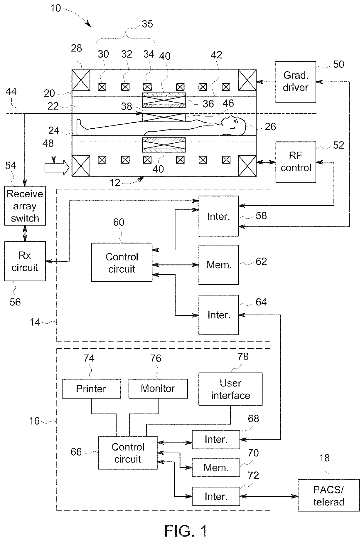
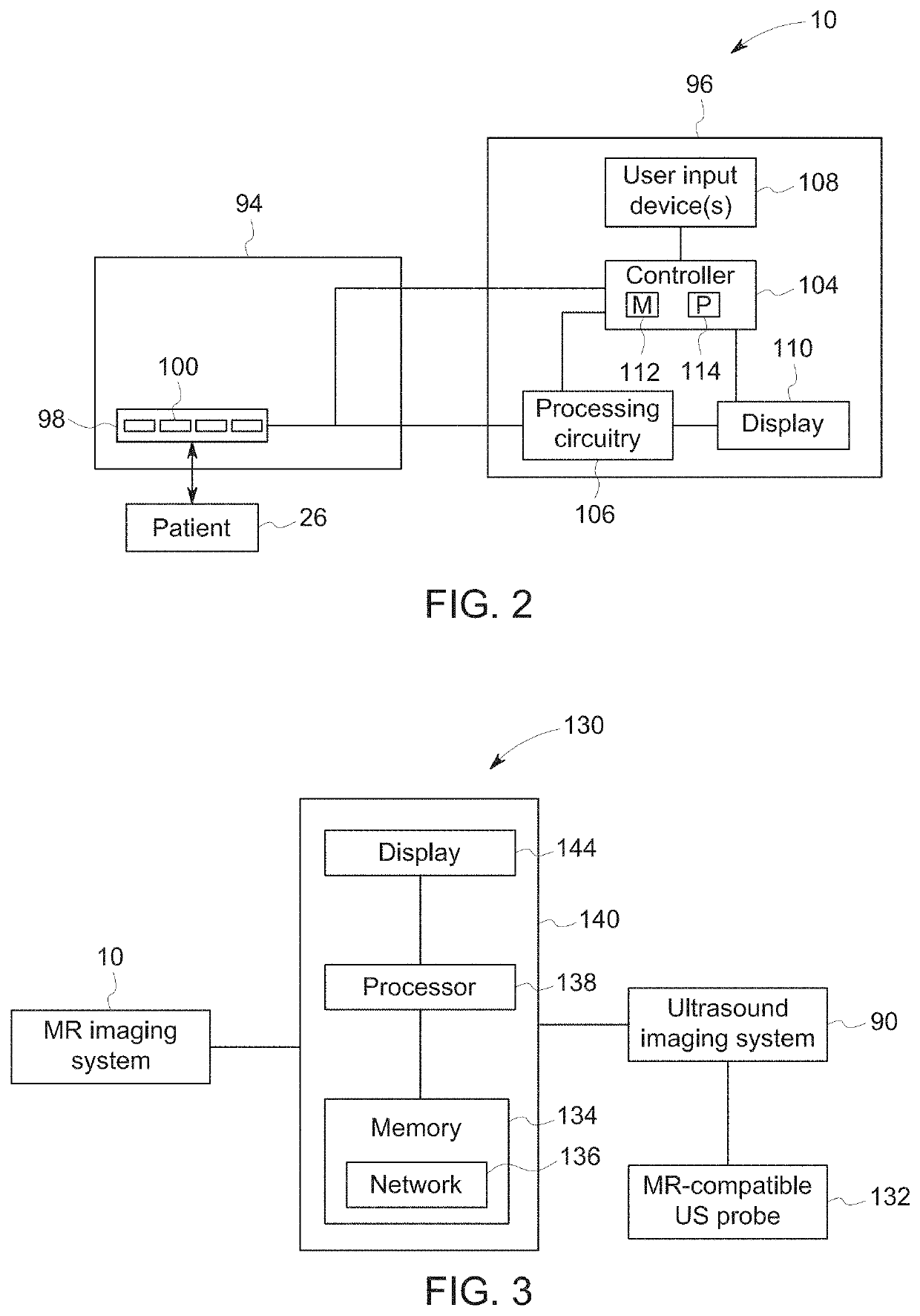
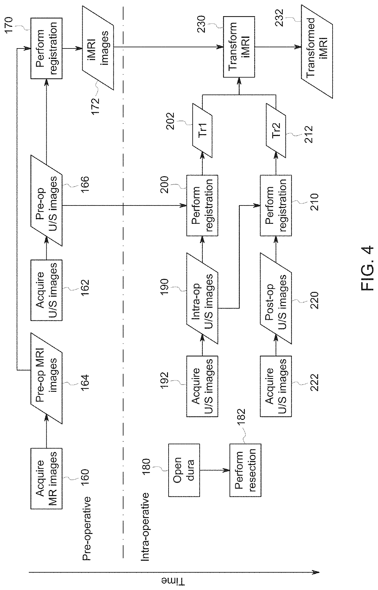
The subject matter discussed herein relates to the automatic, real-time registration of pre-operative magnetic resonance imaging (MRI) data to intra-operative ultrasound (US) data (e.g., reconstructed images or unreconstructed data), such as to facilitate surgical guidance or other interventional procedures. In one such example, brain structures (or other suitable anatomic features or structures) are automatically segmented in pre-operative and intra-operative ultrasound data. Thereafter, anatomic structure (e.g., brain structure) guided registration is applied between pre-operative and intra-operative ultrasound data to account for non-linear deformation of the imaged anatomic structure. MR images that are pre-registered to pre-operative ultrasound images are then given the same nonlinear spatial transformation to align the MR images with intra-operative ultrasound images to provide surgical guidance.

Description

BACKGROUND[0001]The subject matter disclosed herein relates to automatic multimodal image registration that utilizes structure segmentation to facilitate guidance and which may be used for real-time tracking of moving objects.[0002]Non-invasive imaging technologies allow images of the internal structures or features of a patient / object to be obtained without performing an invasive procedure on the patient / object. In particular, such non-invasive imaging technologies rely on various physical principles (such as the differential transmission of X-rays through a target volume, the reflection of acoustic waves within the volume, the paramagnetic properties of different tissues and materials within the volume, the breakdown of targeted radionuclides within the body, and so forth) to acquire data and to construct images or otherwise represent the observed internal features of the patient / object.[0003]By way of example, magnetic resonance imaging (MRI) is a useful imaging modality in the c...

Claims


Application Information

Patent Timeline
IPC IPC(8): G06T3/00G06T11/00G01R33/56A61B8/08
CPCG06T3/0068G06T11/006G06T2210/41A61B8/5207G01R33/56G06T7/33G06T7/0012G16H50/30G06T2207/10088G06T2207/10136G06T2207/30008G01R33/5608G01R33/4814G06T7/0016G06T2207/30016G06T2207/20084A61B8/5261A61B8/4416A61B8/565G06T3/14
Inventor GHOSE, SOUMYAMITRA, JHIMLIMILLS, DAVID MARTINSMITH, LOWELL SCOTTYEO, DESMOND TECK BENGFOO, THOMAS KWOK-FAH
Owner GENERAL ELECTRIC CO
Why Eureka
Try Eureka