In situ cell analysis in cell culture system

a cell culture system and in situ cell technology, applied in the field of in situ cell analysis, can solve the problems of inability to monitor cells without, lack of suitable in-situ controls,

Active Publication Date: 2021-03-11
LIFETAQ ANALYTICS GMBH
View PDF0 Cites 0 Cited by
  • Summary
  • Abstract
  • Description
  • Claims
  • Application Information

AI Technical Summary

Benefits of technology

[0020]It is the object of the present invention to provide improved detecting methods which permit the possibility of determining a molecule in its qualitative environment. In particular, a specific molecule is to be detected in cell culture systems.

Problems solved by technology

In this context, there is currently a lack of suitable in-situ controls that can measure intermediate products in a cell culture system without influencing the cells.
Measurement by means of a flow cytometer meets the current requirements for sensitivity and specificity, but suffers from the necessity to isolate the analytes (cells) from the culture and to discard them after the analysis.
However, this method has the disadvantage that the fluorescent dyes have to be removed by a purification process which changes the culture conditions and / or subsequent further analyses.
It is not possible to monitor cells without interfering with the growth conditions.

Method used


Image

  • In situ cell analysis in cell culture system
  • In situ cell analysis in cell culture system
  • In situ cell analysis in cell culture system

Examples


example 1

Structure of Analyte Probes

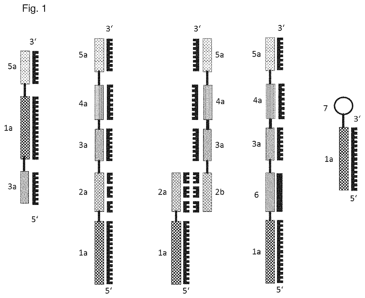
[0159]As shown in FIG. 1, analyte probes can consist of the following parts:

[0160]1. from the left—An aptamer (1a) as a single strand may contain one or two identification elements as identification sequences (3a, 5a).

[0161]2. from the left—Shown here as a single strand. These identification sequences can be attached 5′ or 3′ at the aptamer. A connecting element (2a) (RNA or DNA) can be provided between aptamer and identification sequence. The connecting element can be single-stranded (2. from the left, 2a) or double-stranded (3. from the left, 2a and 2b).

[0162]3. from the left—2b shows a strand of the connecting element that is complementary to strand 2a. In the double-stranded embodiment, the identification element and the detection element can be present on different strands or on the same strands. Double strandedness allows the use of double strand-specific restriction enzymes to cleave the connecting element. Furthermore, a qPCR identification sequenc...

example 2

Process Flow

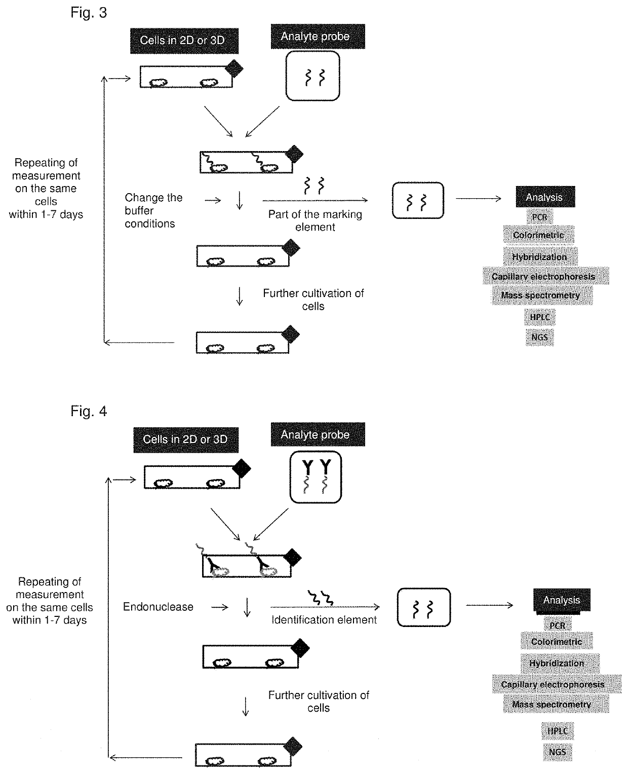
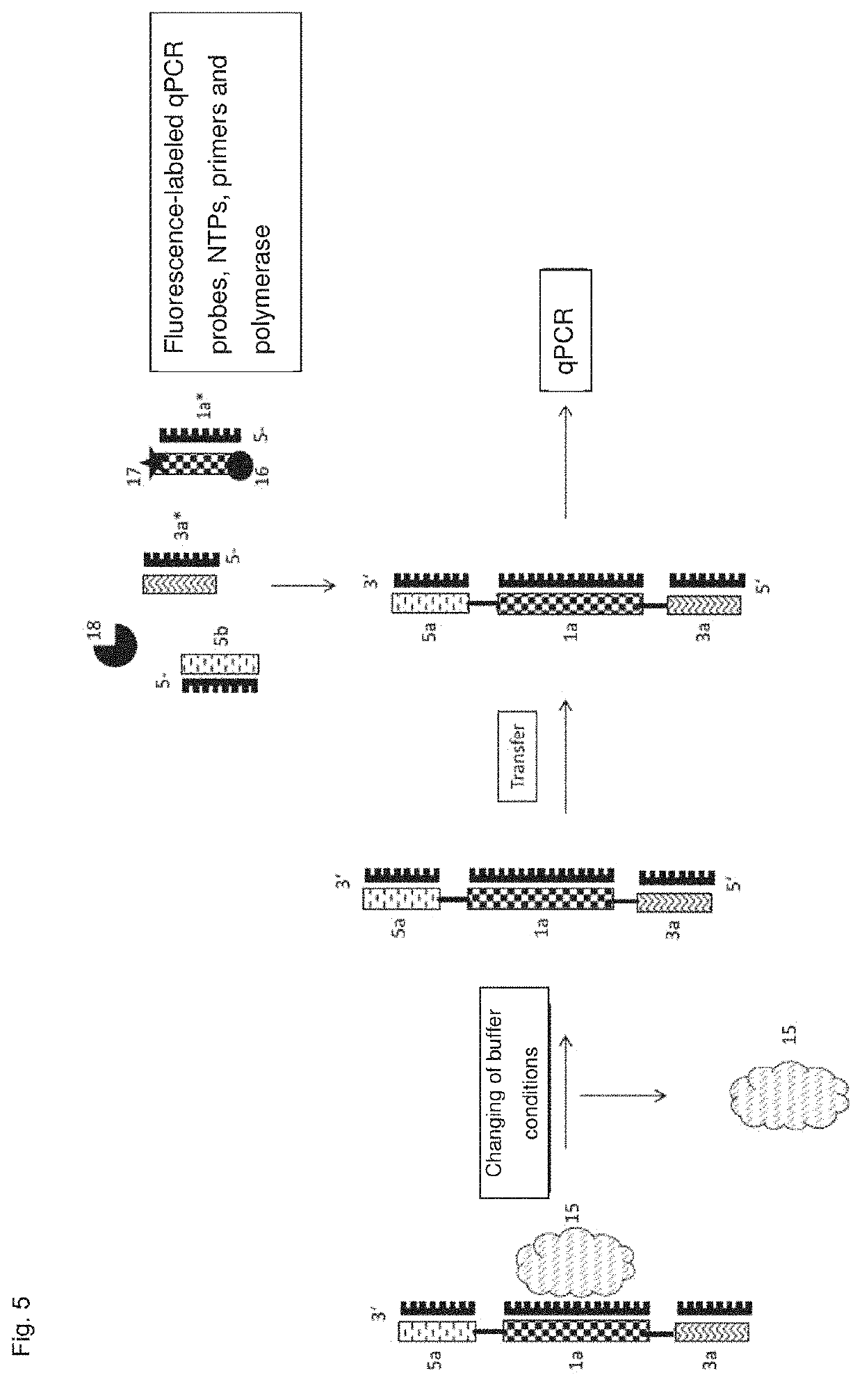
[0171]FIGS. 3 and 4 show schematically the processes of methods according to the invention. From top to bottom, from left to right, following the arrows: Cells are cultivated in 2D or 3D. Analyte probes (in FIG. 3 with antibody detection element; in FIG. 4 as aptamer) bind to the cells via the detection element. The identification element is released—in FIG. 3 by changing the buffer conditions and thus interruption of the binding to the cells, in FIG. 4 by an endonuclease. The cells can then be cultured further (bottom left). The identification element is now separated from the cells and transferred to another container and submitted for analysis, e.g. via PCR and determination of the amplificates, colorimetrically, by hybridization with a labeling probe, capillary electrophoresis, mass spectrometric determination, HPLC, NGS. FIG. 5 shows a process flow for use of an analyte probe with an aptamer 1a, which binds a cell surface molecule (15) as the detection element. The ...

example 3

Aptamer Development

[0173]For the selection of appropriate aptamers as the detection element a DNA aptamer library (Trilink Biotechnologies) was chosen, consisting of a randomized 40 base sequence and flanking defined primers. The first two SELEX rounds were performed directly against adipocyte-derived mesenchymal stem cells (adMSCs / Thermofischer), followed by 10× SELEX rounds against His-tagged CD105 protein (Sino-Biological). Before the first round against cells, the aptamer library was amplified by means of PCR (Biomers For Primer / biotRevPrimer, Solis BioDyne Hot-Start FIRE-pol DNA polymerase) and then incubated with streptavidin beads (Promega). After incubation, the double strand was denatured for 5 min using 0.1 M NaOH, transferred to a new Eppendorf tube and brought to pH 7 with 0.1 M HCl.

[0174]For the first SELEX round, adMSCs (P3) were washed in a T-75 flask (Biogreiner) with 5 ml of Alpha-Mem (SigmaAldrich) without FCS. The aptamer library was diluted in 3 ml of Alpha-Mem w...


PUM

PropertyMeasurementUnit
timeaaaaaaaaaa
timeaaaaaaaaaa
timeaaaaaaaaaa

Abstract

The present invention includes an in-situ method, comprising
    • a) determining a molecule selected from the group consisting of cell surface molecules and extracellular matrix molecules in a two- or three-dimensional cell culture system comprising living cells and cell culture medium, comprising the steps of
      • i) providing an analyte probe consisting of a detection element, which binds the molecule, and one or more identification elements;
      • ii) binding of the analyte probe to the molecule in the cell culture system, wherein the growth ability of the contained living cells is not substantially impaired by this step;
      • iii) optionally removing unbound analyte probes;
      • iv) releasing the analyte probe;
      • v) transferring the analyte probe into a container which differs from the cell culture system;
      • vi) detecting the identification element(s); and
    • b) continuing the cell cultivation in the cell culture system.

Description

FIELD OF THE INVENTION[0001]The present invention relates to the field of in-situ analysis of the cell surface, cell surface components or unbound cell components in a cell culture system.BACKGROUND OF THE INVENTION[0002]The standard method for characterizing animal cells is flow cytometry. Here, cells or beads can be measured in a thin channel with the aid of lasers and detectors. The cells are mixed with fluorescence-labeled antibodies which bind and label specific molecules on the surface of the cells. The cells can then be analyzed in the flow cytometer based on the light scattering and the resulting fluorescence signal.[0003]The monitoring of cells during the cultivation process is of particular interest in the context of cell-therapeutic products. To ensure and improve the safety and efficacy of these products constant testing the starting material, intermediates and the final product is required. The quality control system is of crucial importance here as it ensures the consi...

Claims


Application Information

Patent Timeline
Patent Type & Authority Applications(United States)
IPC IPC(8): C12Q1/6841C12N15/115C12Q1/6804
CPCC12Q1/6841C12N15/115C12Q2525/205C12Q2525/161C12Q1/6804C12Q1/02C12Q1/68G01N33/53G01N33/537G01N33/5375G01N33/54306
Inventor TASCHNER, MANFREDLORBER, VOLKER
Owner LIFETAQ ANALYTICS GMBH
Why Eureka
Try Eureka