
Mass spectrometric determination of particular tissue states

a mass spectrometric and tissue state technology, applied in mass spectrometers, instruments, particle separator tubes details, etc., can solve the problems of insufficient certainty in the detection of known markers for tissue states and tissue degenerations from mass spectral individual pixels, and achieve the effect of improving spectral quality and significantly increasing the probability of being able to identify known markers for tissue states and tissue degenerations with certainty

Active Publication Date: 2021-03-11
BRUKER DALTONIK GMBH & CO KG
View PDF0 Cites 0 Cited by
  • Summary
  • Abstract
  • Description
  • Claims
  • Application Information

AI Technical Summary

Benefits of technology

The invention provides a way to analyze tissue without needing any prior information. It can determine areas of the tissue that have similar mass spectra and then combine these to improve the accuracy of identifying known markers for tissue states and degenerations. This method can work on a large or small scale, and can help to identify specific markers that are associated with certain tissue conditions.

Problems solved by technology

The detection of known markers for tissue states and tissue degenerations from mass spectra of individual pixels is often not possible with sufficient certainty because the quality of the spectra is not high enough.

Method used


Image

  • Mass spectrometric determination of particular tissue states
  • Mass spectrometric determination of particular tissue states
  • Mass spectrometric determination of particular tissue states

Examples


Embodiment Construction

[0027]While the invention has been illustrated and explained with reference to a number of embodiments, those skilled in the art will recognize that various changes in form and detail may be made to it without departing from the scope of the technical teaching defined in the attached patent claims.

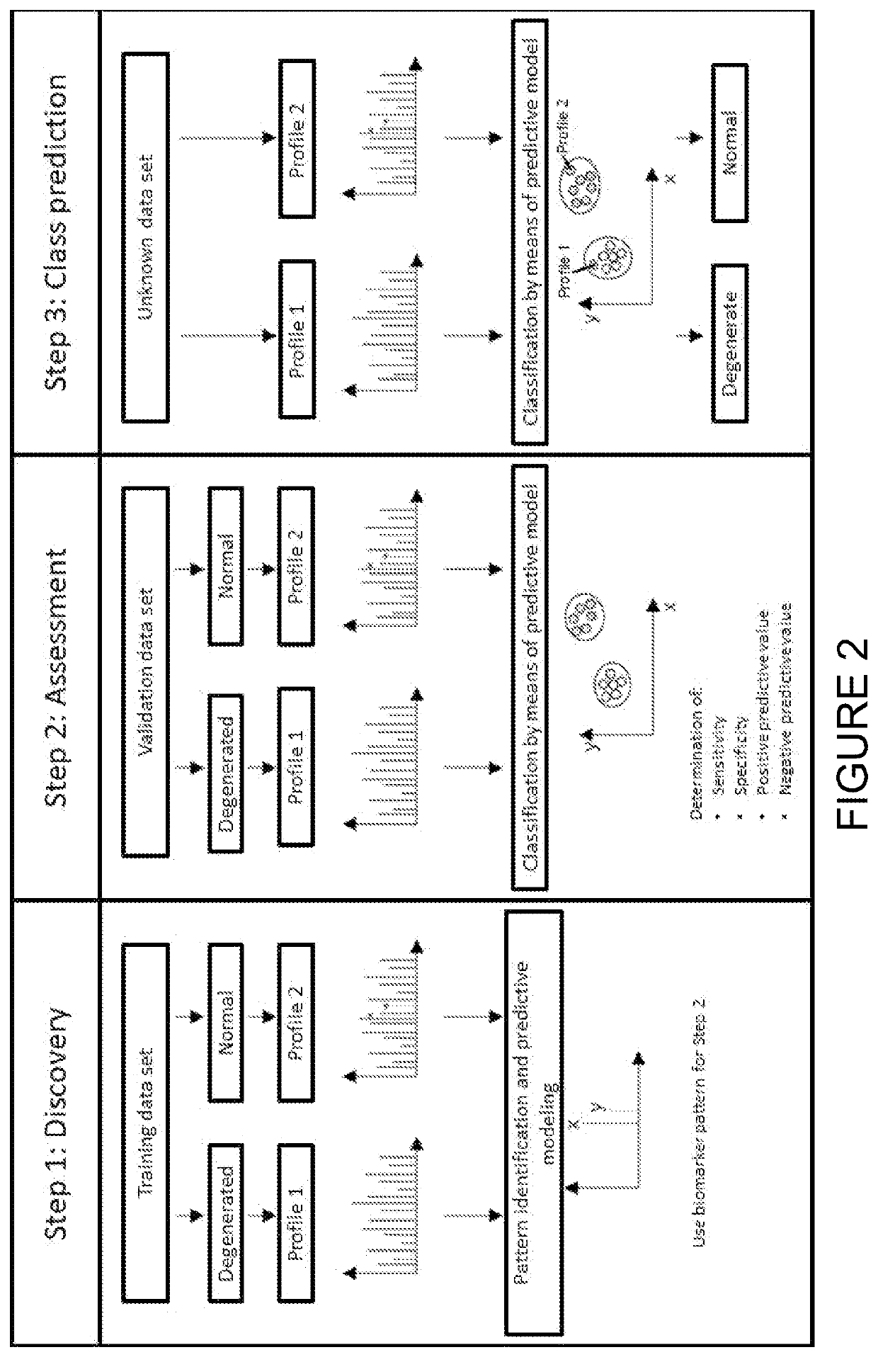
[0028]It is again pointed out here that the terms “particular tissue state” and “tissue degeneration” should be understood here as states of areas of a tissue section in terms of a stress, a pathological change, an infection, a change brought about by the effect of a xenobiotic, a change brought about by genetic engineering, a change caused by mutations, a particular metabolic phenotype or other change compared to a normal state of this tissue. For the particular tissue state, a marker should be known (for example, determined by the method shown in FIG. 2) which forms a certain, characteristic intensity pattern of mass signals in a mass spectrum of the tissue. The marker can comprise a sin...


PUM


Abstract

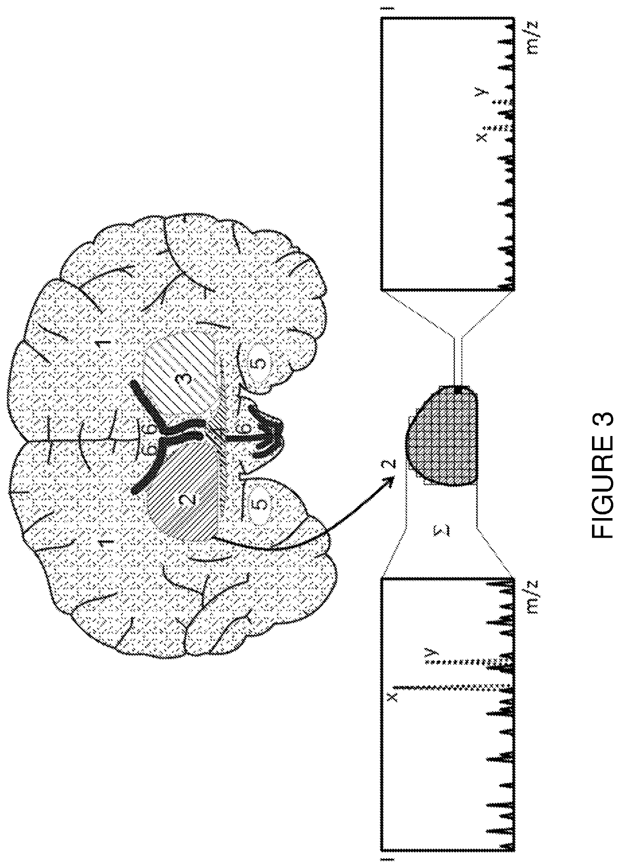
The invention relates to the identification and visualization of the spatial distribution of particular tissue states in histological tissue sections from mass spectrometric signals acquired with spatial resolution. The invention proposes a method by means of which regions of the tissue with similar mass spectra are found automatically, and it is further proposed that mass spectra of these regions are summed in order to improve the spectral quality to such an extent that known markers for tissue degenerations can be identified with increased certainty. Regions of similar mass spectra can be interconnected on a large scale, but can also be isolated from each other on a small scale.

Description

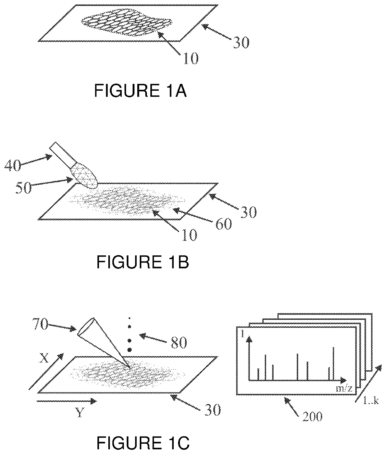
BACKGROUND OF THE INVENTIONField of the Invention[0001]This invention relates to the determination and visualization of the spatial distribution of particular tissue states, for example oncological tissue degenerations, in histological tissue sections from spatially resolved mass spectrometric signals.Description of the Related Art[0002]Spatially resolved mass spectra of a thin tissue section may be obtained by imaging mass spectrometric analysis (i.e., “Imaging Mass Spectrometry”, or “IMS”). As shown in FIG. 1A, a thin tissue section (10) is produced, for example using a microtome, from a deep frozen piece of tissue of an organ of interest from a human, animal or even plant individual, and is put onto a specimen slide (30). When a MALDI method is used for ionization (MALDI=ionization by matrix-assisted laser desorption), a matrix (60) which absorbs laser energy is subsequently applied to the surface of the specimen. This can be done by the intermittent spraying of a matrix solution...

Claims


Application Information

Patent Timeline
Patent Type & Authority Applications(United States)
IPC IPC(8): H01J49/00H01J49/04G01N1/28
CPCH01J49/0004H01J49/0418H01J49/40H01J49/0036G01N1/2813G01N27/64G01N33/6848G01N21/25G01N33/4833H01J49/164
Inventor DEININGER, SÖREN-OLIVERSCHÜRENBERG, MARTIN
Owner BRUKER DALTONIK GMBH & CO KG
Learn More