Method and system for assessing vessel obstruction based on machine learning

a machine learning and vessel technology, applied in the field of methods and systems to assess the severity of vessel obstruction, can solve the problems of reduced oxygen supply to the myocardium, ischemia and chest pain, and physiological significance of atherosclerotic lesions

Pending Publication Date: 2021-10-28
PIE MEDICAL IMAGING
View PDF0 Cites 5 Cited by
  • Summary
  • Abstract
  • Description
  • Claims
  • Application Information

AI Technical Summary

Benefits of technology

[0047]In accordance with aspects herein, the one or more processors can be configured to perform classifying operation by utilizing secondary information to perform the classification. The secondary information can include one or more of the following parameters: coronary tree anatomy, demographic information of the patient, coronary artery calcification, coronary plaque, spectral multi-energy or photon counting, ECG parameters, cardiac biomarkers, adipose tissue surrounding or within the heart, shape of myocardium, or the like.
[0048]In accordance with aspects herein, the one or more processors can be configured to perform the analyzing operation by extracting, for each of the data segments, a feature vector that comprises multiple factors that are measured or extracted from the corresponding data segment, wherein the multiple factors describe or characterize a nature of the corresponding region.
[0049]In accordance with aspects herein, the one or more processors can be configured to extract a feature vector comprising a series of factors, where each of the factors has a value representing an amount of variation in a characteristic of interest over multiple clusters.
[0050]In accordance with aspects herein, the one or more processors can be configured to extract a feature vector comprising a series of factors, where each of the factors represents an intensity of a characteristic of interest over multiple segments of the myocardium.
[0051]In accordance with aspects herein, the one or more processors can be configured to extract a feature vector comprising a series of factors, where a subset of the factors in the series represent intensity within segments, and where another subset of the factors in the series represent values indicative of myocardium volume, minimum myocardium thickness and / or maximum myocardium thickness.

Problems solved by technology

CAD generally refers to conditions that involve narrowed or blocked blood vessels that can lead to reduced or absent blood supply to the sections distal to the stenosis resulting in reduced oxygen supply to the myocardium, resulting in, for instance, ischemia and chest pain (angina).
Although objectivity, reproducibility, and accuracy in assessment of lesion severity has improved by means of quantitative coronary analysis tools (QCA), the physiological significance of atherosclerotic lesions, which is the most important prognostic factor in patients with coronary artery disease, cannot be appreciated by X-ray angiography.
Overestimation of the severity of the stenosis can cause a treatment which in hindsight would not have been necessary and therefore exposing the patient to risks that are not necessary.
Underestimation of the severity of the stenosis, however, could induce risks because the patient is left untreated while the stenosis is in reality severe and actually impedes flow to the myocardium.
The technique is associated with the additional cost of a pressure wire which can only be used once.
Furthermore, measuring FFR requires invasive catheterization with the associated cost and procedure time.
Also, in order to induce (maximum) hyperemia, additional drug infusion (adenosine or papaverine) is required, which is an extra burden for the patient.
Coronary computed tomography (CT) angiography (CCTA) is a non-invasive imaging modality for the anatomic assessment of coronary arteries but does not assess the functional significance of coronary lesions.
This potential for false positive results has raised concerns that widespread use of CCTA may lead to clinically unnecessary coronary revascularization procedures.
This lack of specificity of CCTA is one of the main limitations of CCTA in determining the hemodynamic significance of CAD (Meijboom et al, “Comprehensive assessment of coronary artery stenoses: computed tomography coronary angiography versus conventional coronary angiography and correlation with fractional ow reserve in patients with stable angina,” Journal of the American College of Cardiology 52 (8) (2008) 636-643).
As a result, CCTA may lead to unnecessary interventions on the patient, which may pose added risks to patients and may result in unnecessary health care costs.
This process is computationally complex and time-consuming and may require several hours.
There are several limitations for physiologic assessment of coronary stenosis by FFRCT besides its long computation time as mentioned above.
However, due to their small size these collateral vessels are not commonly visible on CCTA, and further steps are needed to determine the presence of collateral flow.
Fifth, because FFRCT requires accurate anatomic models, numerous artifacts on CCTA may affect FFRCT calculation, such as blooming artefacts caused by large arterial calcifications and stents.
In addition, motion, lower SNR, and mis-registration may compromise its accuracy.
Moreover, the method requires further clinical validation as it was only validated on a small set of CCTA scans.
Taking into account that a mild coronary obstruction has an average diameter between 1.5-2.5 mm and that spatial resolution of CCTA is in the range of 0.25 mm isotropic, obtaining accurate 3D morphology by means of segmentation is a very challenging task.
Although this approach is promising as it merges the anatomic information of CCTA with functional analysis, it requires an additional CT scan which inevitably leads to higher radiation dose and longer examination time and the need for injection of pharmacological stress agents.

Method used


Image

  • Method and system for assessing vessel obstruction based on machine learning
  • Method and system for assessing vessel obstruction based on machine learning
  • Method and system for assessing vessel obstruction based on machine learning

Examples


Embodiment Construction

[0073]The term “unseen”, as used throughout, refers to “non-training” items. For example, an unseen image is not a training image, an unseen feature is not a training feature. Instead, the unseen features, images, geometries, and other unseen items refer to aspects of a patient or object of interest that is being analyzed during the prediction phase of operation.

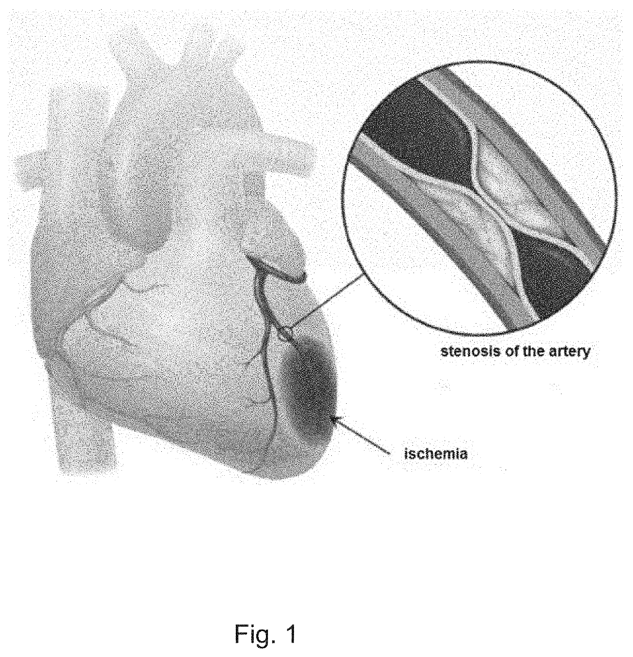
[0074]The present application relates to a method and system for machine learning to assess the hemodynamic functional severity of one or more vessel obstructions of a target organ based on contrast enhanced volumetric image dataset. In a preferred embodiment, the target organ represents the myocardium and the vessels the coronary arteries. A functionally significant stenosis is a hemodynamically significant obstruction of a vessel, and with respect to coronary arteries it defines the likelihood that coronary artery obstruction(s) impedes oxygen delivery to the heart muscle and causes anginal symptoms. Fractional flow reserv...


PUM


Abstract

Methods and systems are provided for assessing the presence of functionally significant stenosis in one or more coronary arteries, further known as a severity of vessel obstruction. The methods and systems can implement a prediction phase that comprises segmenting at least a portion of a contrast enhanced volume image data set into data segments corresponding to wall regions of the target organ, and analyzing the data segments to extract features that are indicative of an amount of perfusion experiences by wall regions of the target organ. The methods and systems can obtain a feature-perfusion classification (FPC) model derived from a training set of perfused organs, classify the data segments based on the features extracted and based on the FPC model, and provide, as an output, a prediction indicative of a severity of vessel obstruction based on the classification of the features.

Description

CROSS-REFERENCE TO RELATED APPLICATION(S)[0001]The present application is a continuation-in-part of U.S. application Ser. No. 16 / 508,996, filed on Jul. 11, 2019, which is a continuation of U.S. application Ser. No. 16 / 241,165, filed on Jan. 7, 2019, now U.S. Pat. No. 10,395,366, which is a continuation of U.S. application Ser. No. 15 / 933,854, filed on Mar. 23, 2018, now U.S. Pat. No. 10,176,575, which claims priority from U.S. Provisional App. No. 62 / 476,382, filed on Mar. 24, 2017, all of which are herein incorporated by reference in their entireties.BACKGROUND1. Field[0002]The present application relates generally to methods and systems to assess a severity of vessel obstruction, and methods and systems to form feature-perfusion classification (FPC) models that classify training features in connection with assessing a severity of vessel obstruction.2. State of the Art[0003]Coronary artery disease (CAD) is one of the leading causes of death worldwide. CAD generally refers to condit...

Claims


Application Information

Patent Timeline
Patent Type & Authority Applications(United States)
IPC IPC(8): G06T7/00A61B6/00G06K9/00G16H50/50G16H30/40G06T7/10G06K9/62G06N3/08G06V10/764
CPCG06T7/0012A61B6/507G06K9/00214G16H50/50G16H30/40G06T2207/30104G06K9/6269G06N3/088G06K2209/05G06T2207/20081G06T7/10A61B6/504A61B6/032G06V10/443G06V2201/031G06T2207/10081G06T2207/20084G16H50/20G06N20/10G06V10/763G06V10/764G06N3/045G06F18/23213G06V20/653G06V2201/03G06F18/2411
Inventor ISGUM, IVANAZREIK, MAJDLEINER, TIMABEN, JEAN-PAUL
Owner PIE MEDICAL IMAGING
Why Eureka
Try Eureka