
Dual-modality nanoprobe targeting glioblastoma and preparation method thereof

a nanoprobe and glioblastoma technology, applied in the field of biomedical materials, can solve the problems of low content of drugs or nanoparticles entering the brain, low application efficiency, and difficult to achieve the above-mentioned purpose of imaging diagnostic methods commonly used in clinics, and achieve enhanced imaging function, clear anatomical structure information of brain tumors, and clear image results.

Inactive Publication Date: 2021-11-04
WEST CHINA HOSPITAL SICHUAN UNIV
View PDF0 Cites 1 Cited by
  • Summary
  • Abstract
  • Description
  • Claims
  • Application Information

AI Technical Summary

Benefits of technology

The present patent provides a dual-modality nanoprobe that targets glioblastoma, which is a type of brain tumor. The nanoprobe combines the benefits of magnetic resonance and fluorescence imaging, allowing for clearer anatomical structure information of brain tumors and the ability to specifically identify target sites. The nanoprobe also has better in vivo stability, making it more effective for targeting glioblastoma. Overall, the patent provides a solution for improving the accuracy and effectiveness of brain tumor imaging.

Problems solved by technology

However, the imaging diagnostic methods commonly used in clinic are difficult to achieve the above purpose.
However, due to the limited injection volume of this method, the content of drugs or nanoparticles entering the brain is low, thus leading low application efficiency.
In addition, the BBB structure can be reversibly destroyed by physical methods such as focused ultrasound or magnetic field heating to achieve the purpose of drug delivery, but this method also allows other harmful substances to enter the brain.
On the other hand, in the prior art, enhanced magnetic resonance imaging utilizing gadolinium chelate T1 contrast agent is a commonly used clinical method for diagnosing glioblastoma, but Gd-based contrast agent still has many defects in the detection.
Besides the leakage of blood vessels outside the tumor will have a great impact on enhanced imaging of T1, it is difficult for enhanced imaging of T1 itself to accurately understand postoperative reaction, especially in evaluating false positive progress after radiotherapy or false positive reaction during anti-angiogenesis therapy.
However, due to the high replacement rate of cerebrospinal fluid, the close connection between brain endothelial cells and the low endocytosis efficiency of brain endothelial cells, more than 98% of small molecules and 100% of large molecules cannot enter the brain parenchyma, which seriously hinders the brain aggregation of nanoprobes and the brain delivery of nanodrugs.
At the same time, the single imaging mode has obvious limitations, such as MRI is limited by low sensitivity, and optical imaging is affected by low spatial resolution and lack of tissue permeability.

Method used


Image

  • Dual-modality nanoprobe targeting glioblastoma and preparation method thereof
  • Dual-modality nanoprobe targeting glioblastoma and preparation method thereof
  • Dual-modality nanoprobe targeting glioblastoma and preparation method thereof

Examples


example 1

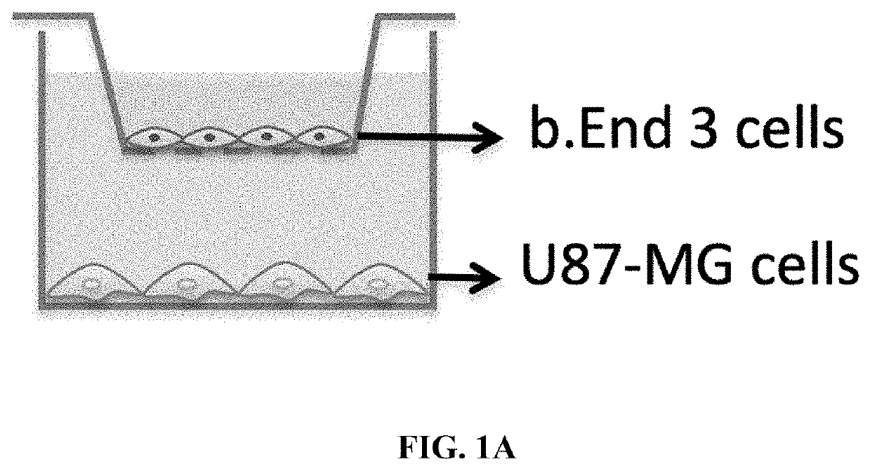
[0072]This Example illustrates the construction and evaluation of BBB model in vitro.

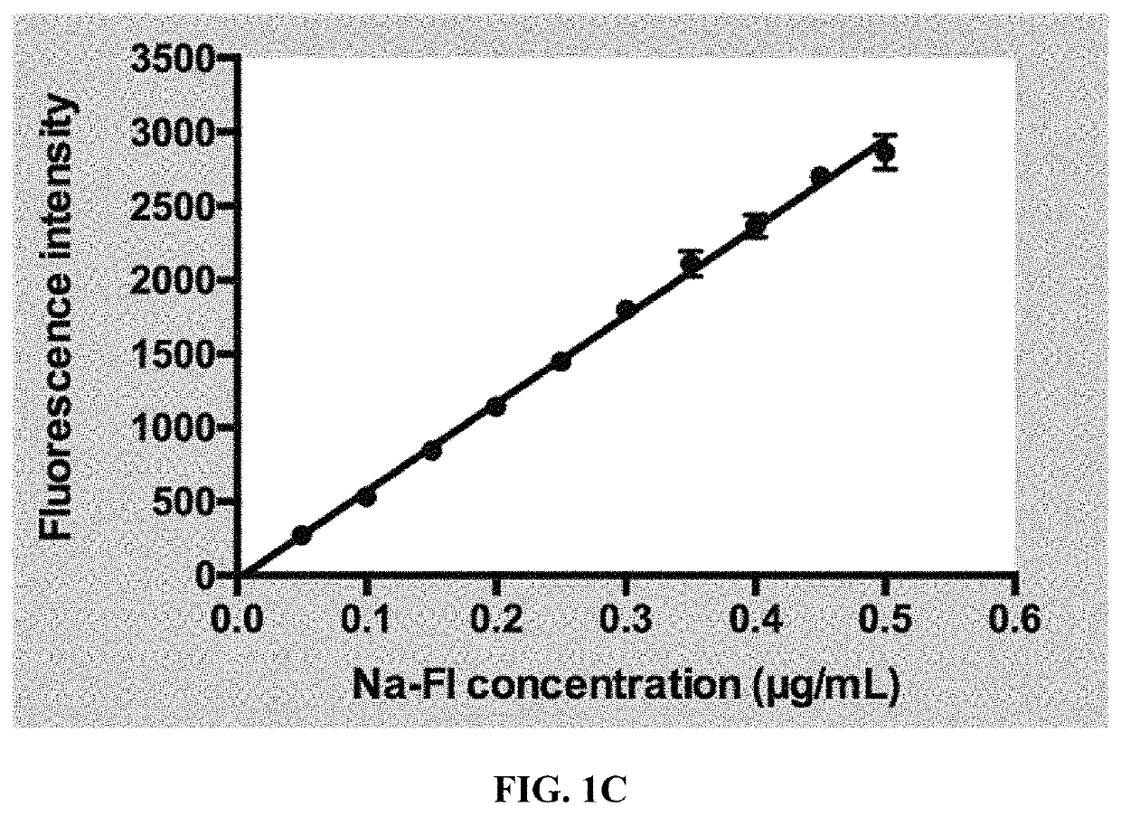
[0073]Experimental method: an in vitro blood-brain barrier model was established to evaluate the permeability of dual-modality nanoprobes. U87-MG cells and bEnd.3 cells were cultured in high glucose DMEM containing 10% fetal bovine serum and 1% penicillin / streptomycin at 37° C. and 5% CO2. A thin layer of collagen solution was evenly coated on the inner side of Transwell poly microporous lipid membrane, the membrane was placed on an ultra-clean table for about 30 min, and it was used after naturally dried. bEnd.3 cells were inoculated into the upper chamber of a 24-well Transwell at a density of 5×10⁴ cells / well. Two days later, U87-MG cells were inoculated into the lower chamber of the upper chamber at a density of 1×10⁵ cells / well, and the two kind of cells were continued to co-culture for one week.

[0074]Firstly, the BBB model was tested and evaluated by leak test. The upper and lower chambers of ...

example 2

[0081]In this Example, the performance of dual-modality nanoprobe penetrating in vitro BBB model was evaluated.

[0082]Experimental method: after successfully constructing BBB model in vitro by the method of Example 1, the upper and lower chambers of Transwell were gently cleaned twice with PBS, and then PBS, Cy7-PEG-DSPE-SPIONs and two kinds of peptide / Cy7-PEG-DSPE-SPIONs solutions (the two kinds of peptide / Cy7-PEG-DSPE-SPIONs solutions were ANG / Cy7-PEG-DSPE-SPIONs and DANG / Cy7-PEG-DSPE-SPIONs; similarly, the two peptide / Cy7-PEG-DSPE-SPIONs solutions mentioned in the following examples will also be ANG / Cy7-PEG-DSPE-SPIONs and DANG / Cy7-PEG-DSPE-SPIONs, without repeated in details)were added into the upper chamber, and the concentration of each group is 10.0 μg / mL, with a total of 200 μL; 900 μL PBS was added into the lower chamber, and incubated for 60 min, 100 μL of buffer solution in the lower chamber was added into PET black 96-well plate, the fluorescence intensity of the lower ch...

example 3

[0084]In this Example, the uptake of different probes by bEnd.3 cells and U87-MG cells is quantitatively analyzed.

[0085]Experimental method: cell plating: U87-MG cells and bEnd.3 cells were cultured with high glucose DMEM containing 10% fetal bovine serum and 1% penicillin / streptomycin at 37° C. and 5% CO2. When the cells grew well, the two kinds of cells were spread in 12-well plates with a density of 1×10⁵ cells per well.

[0086]Flow cytometry: after the cells adhered to the wall, two probe solutions (Peptides / Cy7-PEG-DSPE-SPIONs) with a concentration of 50 μg / mL (Fe3O4 concentration) were added, the cells cultured at 37° C. and 5% CO2 respectively for 1 h, 2 h and 4 h, the cells were collected for flow cytometry at the set time points, and the detection channel was set as APC-A750, the data results were processed and analyzed by flow cytometry software Flowjo.

[0087]Experimental results: two kinds of targeting polypeptide modified probes (Peptides / Cy7-PEG-DSPE-SPIONs) are incubated ...


PUM

PropertyMeasurementUnit
temperatureaaaaaaaaaa
constant temperatureaaaaaaaaaa

Abstract

A dual-modality nanoprobe targeting glioblastoma and preparation method thereof are described. The dual-modality nanoprobe of the present disclosure comprises DSPE-PEG(2000)-Amine, superparamagnetic iron oxide nanoparticles (SPIONs), Cy7-NHS molecule, targeting polypeptide and / or trans-mirror structure (i.e., enantiomer) thereof. The dual-modality nanoprobe can integrate the advantages of magnetic resonance and fluorescence imaging and thus provide clearer anatomical structure information of brain tumors; in the imaging process, the dual-modality nanoprobe can not only provide clearer image results, but also can specifically identify target sites. Also, the dual-modality nanoprobe of the present disclosure also has good in vivo stability.

Description

CROSS REFERENCE TO RELATED APPLICATION[0001]This application claims the priority of Chinese Patent Application No. 202010375037.8, entitled “Dual-Modality Nanoprobe Targeting Glioblastoma And Preparation Method Thereof” filed with China National Intellectual Property Administration on May 3, 2020, which is incorporated herein by reference in its entirety.TECHNICAL FIELD[0002]The disclosure relates to the technical field of biomedical materials, in particular to a dual-modality nanoprobe targeting glioblastoma and preparation method thereof.BACKGROUND ART[0003]Due to the location specificity, heterogeneity and invasiveness of glioblastoma, it is necessary to obtain high spatial resolution and high sensitivity images before surgery to guide further surgery. However, the imaging diagnostic methods commonly used in clinic are difficult to achieve the above purpose.[0004]On the one hand, penetration of blood-brain barrier (BBB) is considered as the main target of nano-drug or probe to tr...

Claims


Application Information

Patent Timeline
Patent Type & Authority Applications(United States)
IPC IPC(8): A61K49/18A61K49/00
CPCA61K49/1866A61K49/0002B82Y25/00A61K49/0056A61K49/0054A61K49/186B82Y30/00B82Y40/00B82Y20/00A61K49/0032B82Y5/00B82Y15/00H01F1/0054
Inventor WU, MINGONG, QIYONGLV, SUZENG, FANXINXIE, RUOXI
Owner WEST CHINA HOSPITAL SICHUAN UNIV
Learn More