Method and system for imaging

Pending Publication Date: 2022-06-16
4DMEDICAL LTD
View PDF0 Cites 1 Cited by
  • Summary
  • Abstract
  • Description
  • Claims
  • Application Information

AI Technical Summary

Benefits of technology

The present invention provides a method for detecting irregularities in the pulmonary vasculature through the use of an Hessian filter. The method can segment the vasculature data from a 3D image without the need for contrast agents, which is less expensive and safer for patients. The automated analysis of the probability data and scale data can quickly and accurately assess whether a patient has vascular irregularities, such as pulmonary emphysema. The method can also use existing contrast-free patient images, avoiding additional collection of images and reducing costs.

Problems solved by technology

However, the vasculature and changes to the vasculature (such as PE) are difficult to clearly visualise in and typically require the use of contrast agents such as iodine to enhance contrast and hence visibility.
The obstruction of the blood flow through the lung vasculature, and the resultant pressure on the right ventricle of the heart, lead to the signs and symptoms of PE, such as difficulty in breathing, chest pain, heart palpitations, or possibly patient collapse.
However, adverse medical conditions can be caused by administration of contrast agents.
a. Complete filling defects where contrast does not fill the vessel due to the existence of a large embolus. Image intensities downstream of the embolus are darker as there is no contrast in this part of the vessel,
b. Partial filling defects where little contrast passes through and there are annular regions of high image intensity around the embolus,
c. And other defects such as: wedge-shaped areas of hyperattenuation represent regions of lung infarction that are associated with loss of blood supply to that region of the lung; and contrast material flowing through thickened but smaller arteries that have re-opened (recanalization).
The investigation found that non-contrast CT is not suitable for detecting central pulmonary embolism.

Method used


Image

  • Method and system for imaging
  • Method and system for imaging
  • Method and system for imaging

Examples


Embodiment Construction

[0105]The method of the present invention provides a means for obtaining vessel calibre measures from contrast free computed tomography (CT) images. The laboratory based imaging system discussed herein yielded images with sufficient resolution to resolve smaller blood vessels in vivo. The imaging system and method of the present invention presents an alternative angiography technique for small animal studies, and human scanning, without the need for contrast agents, and theoretically could allow repeat imaging in the same animals over time as disease processes progress or in response to treatments.

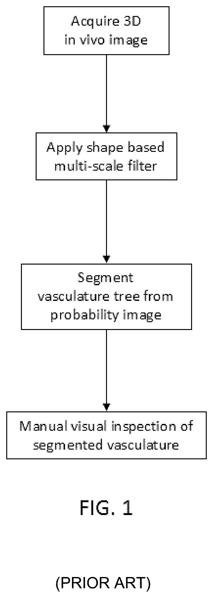
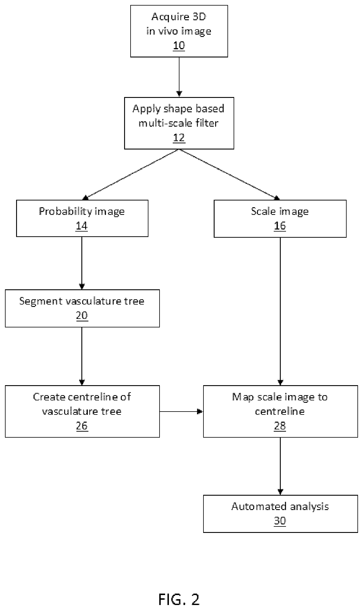
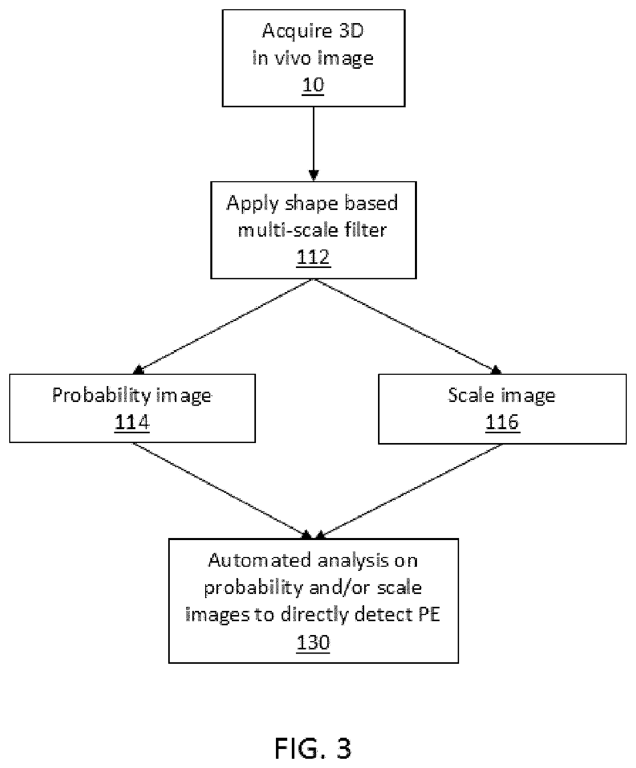
[0106]FIGS. 1 to 3 are flow charts illustrating method steps for prior art (FIG. 1), and method steps for embodiments of the invention (FIG. 2 and FIG. 3). FIG. 1 illustrates typical method steps of the prior art in which a multi-scale filter is applied to images without contrast agent in order to segment the vasculature for manual visual inspection by a user. By contrast, FIG. 2 depicts a...


PUM


Abstract

The present invention relates to the field of medical imaging in the absence of contrast agents. In one form, the invention relates to the field of imaging vessels, particularly blood vessels such as the pulmonary vasculature and is suitable for use as a technique for detecting pulmonary embolism (PE), such as acute PE. Embodiments of the present invention provide improved image processing techniques having the capability to extract and use image data to overcome the need for contrast agents to distinguish between different types of tissue. Furthermore, it has also been realised that the image data accessed by the improved image processing can be used to identify irregularities in vessels.

Description

CROSS-REFERENCE TO RELATED APPLICATION(S)[0001]This application is a divisional application of U.S. patent application Ser. No. 16 / 081,854, filed Aug. 31, 2018, for “Method and System for Imaging,” which is a U.S. National Phase of International Patent Application Serial No. PCT / AU2017 / 000054 entitled “Method and System for Imaging,” filed on Feb. 24, 2017. International Patent Application Serial No. PCT / AU2017 / 000054 claims priority to Australian Patent Application No. 2016900354, filed on Feb. 3, 2016, and Australian Patent Application 2016900817, filed on Mar. 4, 2016. The entire contents of each of the above-cited applications are hereby incorporated by reference in their entirety for all purposes.FIELD OF INVENTION[0002]The present invention relates to the field of medical imaging in the absence of contrast agents.[0003]In one form, the invention relates to the field of imaging vessels, particularly blood vessels such as the pulmonary vasculature.[0004]In one particular aspect ...

Claims


Application Information

Patent Timeline
IPC IPC(8): A61B6/00G06T7/143G06T7/187G06T7/62A61B5/08A61B5/02G06T7/35G06T7/00G06T7/11A61B6/03
CPCA61B6/5217A61B5/026G06T7/187G06T7/62A61B5/08A61B5/02007G06T7/35G06T7/0012G06T7/11A61B6/508A61B6/541A61B6/032A61B6/481A61B6/504A61B6/507G06T7/0016G06T2207/10081G06T2207/20016G06T2207/20076G06T2207/30061G06T2207/30172G06T7/143G06T2207/10076G06T2207/10088G06T2207/10136G06T2207/30104A61B5/7285A61B5/113A61B2576/02G06T2207/30101G06T2207/20024
Inventor FOURAS, ANDREASSAMARAGE, CHAMINDA RAJEEV
Owner 4DMEDICAL LTD
Why Eureka
Try Eureka