Urinary exosome biomarker for diagnosing antibody-mediated rejection after kidney transplantation or predicting prognosis of patient after kidney transplantation

a technology of antibody-mediated rejection and urinary exosomes, which is applied in the field of urinary exosome biomarkers for diagnosing antibody-mediated rejection after kidney transplantation or predicting the prognosis of patient after kidney transplantation, can solve the problem of no studies to discover biomarkers, and achieve accurate and rapid diagnosis, accurately and quickly identify antibody-mediated rejection, and increase expression

Pending Publication Date: 2022-08-11
UNIV OF ULSAN FOUND FOR IND COOPERATION +1
View PDF0 Cites 0 Cited by
  • Summary
  • Abstract
  • Description
  • Claims
  • Application Information

AI Technical Summary

Benefits of technology

[0026]Since the biomarkers provided in the present invention are specifically and remarkably increased in expression in patients exhibiting antibody-mediated rejection after kidney transplantation, it is possible to accurately and quickly diagnose whether kidney transplantation is rejected and the type of rejection through a composition, a kit or a detection method including a material for measuring the expression level of the biomarker protein of the present invention or a gene encoding the same.
[0027]In addition, since the biomarkers of the present invention are specifically expressed in patients with antibody-mediated rejection, differentially from patients with other types of transplantation rejection or BK virus nephropathy, which is common after transplantation, it is possible to accurately and quickly identify antibody-mediated rejection, and it is possible to provide information required to determine a treatment policy thereafter.

Problems solved by technology

However, there are no studies to discover biomarkers that can differentiate antibody-mediated rejection from T cell-mediated rejection, and furthermore, to discover biomarkers that can differentiate and diagnose BK virus nephropathy and transplantation rejection, which are complications after kidney transplantation, and to utilize the same in the selection of treatment direction and the judgment of treatment progress after kidney transplantation.

Method used


Image

  • Urinary exosome biomarker for diagnosing antibody-mediated rejection after kidney transplantation or predicting prognosis of patient after kidney transplantation
  • Urinary exosome biomarker for diagnosing antibody-mediated rejection after kidney transplantation or predicting prognosis of patient after kidney transplantation
  • Urinary exosome biomarker for diagnosing antibody-mediated rejection after kidney transplantation or predicting prognosis of patient after kidney transplantation

Examples


example 1

[0092]Selection of Patients and Preparation of Urine Samples

[0093]In order to discover biomarkers for diagnosing antibody-mediated rejection in kidney transplantation patients, mine samples were collected 2 to 3 hours before the biopsy from a total of 60 patients, including patients and donors who underwent an indication biopsy within 5 years of kidney transplantation. In addition, urine samples were collected from living kidney donors immediately before kidney donation surgery. The patients were classified into 5 groups according to the pathological diagnosis of biopsy: 12 antibody-mediated rejection (ABMR) groups, 8 T cell-mediated rejection (ABMR) groups, 5 BK virus nephropathy (BKVN) groups, 11 groups without major abnormalities (NOMOA) and 24 donor groups (DONOR). Urine from patients with mixed allopathic lesions was excluded. This study was conducted with the approval of the Medical Institution Evaluation Committee at Asan Medical Center, and written consent was obtained from ...

example 2

[0094]Processing of Urine Samples and Separation of Exosomes

[0095]By slightly modifying the methods of Pisitkim et al. (2004) and Alvarez et al. (2012), exosomes were separated from the prepared urine samples by step-wise ultracentrifugation. Specifically, 30 mL or more of urine samples was collected from each experiment participant, and 400 μL of a protease inhibitor mixture [50 μM 4-(2-animoethyl) benzenesulfonyl fluoride hydrochloride (AEBSF-HCl, Sigma Aldrich), 2 μM leupeptin-hemisulfate (Sigma Aldrich) and 3.3 mM sodium azide (Sigma Aldrich)] was added to each urine sample. In order to remove urinary sediments including whole cells, large membrane particles and other debris, the urine samples were centrifuged at 4,000 rpm and 4° C. for 15 minutes, and stored at −80° C. until the exosomes were extracted and used in the experiment.

[0096]Afterwards, the frozen urine samples were thawed by 15 mL each, vortexed for 1 minute and then centrifuged at 17,000×g for 15 minutes at room tem...

example 3

[0097]LC-MS Analysis and Protein Identification

[0098]Sample preparation for proteomic analysis was subjected to freeze drying, protein solubilization and digestion. The resulting peptide mixture was desalted, dried using C18 reverse phase chromatography, and then analyzed by high resolution mass spectrometry combined with nano-flow liquid chromatography.

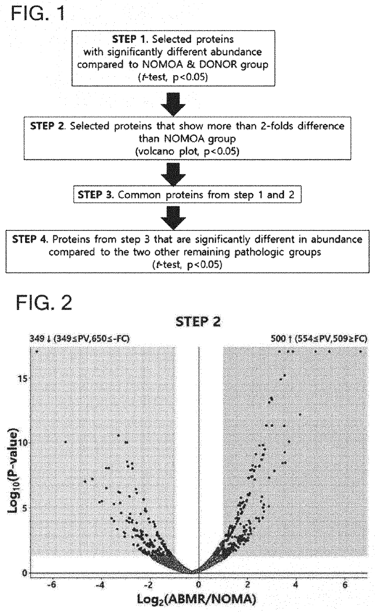
[0099]Sequence database analysis and label free quantitation (LFQ) analysis were applied using Proteome Discoverer 2.2 (Thermo Fisher Scientific) to search for biomarker candidate substances for diagnosing or predicting kidney transplantation rejection. From this, 1,820 urine exosome proteins were identified. Gene ontology assignments, molecular function and Kegg pathway analysis were performed using the DAVID bioinformation database.


PUM

PropertyMeasurementUnit
concentrationaaaaaaaaaa
drug resistanceaaaaaaaaaa
lengthaaaaaaaaaa

Abstract

The present invention relates to a biomarker for the non-invasive diagnosis of kidney transplantation rejection and use thereof, and particularly to: a biomarker composition and kit for diagnosing antibody-mediated rejection after kidney transplantation or predicting the prognosis of a patient after kidney transplantation, comprising any one or more proteins selected from the group consisting of LBP and CST3, or gene(s) encoding for the same; a method for providing information required for diagnosing antibody-mediated rejection after kidney transplantation or predicting the prognosis of a patient after kidney transplantation, by using the marker composition; a method for providing information required for determining a therapy for rejection after kidney transplantation; a method for the diagnosis and treatment of antibody-mediated rejection after kidney transplantation; and a method for screening for a therapeutic agent for antibody-mediated rejection after kidney transplantation.

Description

TECHNICAL FIELD[0001]The present invention relates to a biomarker associated with antibody-mediated rejection (ABMR) after kidney transplantation, a composition and a kit for diagnosing antibody-mediated rejection or predicting a prognosis for rejection after kidney transplantation, and a method for providing information for diagnosing antibody-mediated rejection or predicting a prognosis of rejection after kidney transplantation using the same.BACKGROUND ART[0002]The field of organ transplantation is slowly becoming a part of precision medicine. When the current standard immunosuppressive therapy is used, acute rejection still occurs in 15 to 20% of patients who have received kidney transplantation, and until now, if there is an increase in serum creatinine concentration, new proteinuria expression or the like, it is diagnosed through tissue biopsy. Since the renal condition at the time when there is an increase in serum creatinine concentration or new proteinuria expression is a c...

Claims


Application Information

Patent Timeline
Patent Type & Authority Applications(United States)
IPC IPC(8): G01N33/68C12Q1/68
CPCG01N33/68G01N2333/435G01N2800/245C12Q1/68C12Q1/6883C12Q2600/158G01N33/6893C12Q2600/118C12Q2600/136G01N2333/4716G01N2333/8121G01N2333/8139G01N2333/918
Inventor SHIN, SUNG
Owner UNIV OF ULSAN FOUND FOR IND COOPERATION
Why Eureka
Try Eureka