Non-invasive localization of a light-emitting conjugate in a mammal

a conjugate and non-invasive technology, applied in the field of non-invasive methods, can solve the problems of limited monitoring of the progression of infectious diseases, and achieve the effect of accurate determination of the concentration of the selected substan

Inactive Publication Date: 2001-04-17
LELAND STANFORD JUNIOR UNIV OF THE BOARD OF TRUSTEES THE
View PDF13 Cites 300 Cited by
  • Summary
  • Abstract
  • Description
  • Claims
  • Application Information

AI Technical Summary

Benefits of technology

In yet another embodiment, the invention includes a method of measuring the concentration of selected substances, such as dissolved oxygen or calcium, at specific sites in an organism. The method includes entities, such as cells, containing a concentration sensor--a light-generating molecule whose ability to generate light is dependent on the concentration of the selected substance. The entity containing the light-generating molecule is administered such that it adopts a substantially uniform distribution in the animal or in a specific tissue or organ system (e.g., spleen). The organism is imaged, and the intensity and localization of light emission is correlated to the concentration and location of the selected substance. Alternatively, the entity contains a second marker, such as a molecule capable of generating light at a wavelength other than the concentration sensor. The second marker is used to normalize for any non-uniformities in the distribution of the entity in the host, and thus permit a more accurate determination of the concentration of the selected substance.

Problems solved by technology

The ability to monitor the progression of infectious diseases is limited by the current ex vivo methods of detecting and quantifying infectious agents in tissues.

Method used


Image

  • Non-invasive localization of a light-emitting conjugate in a mammal
  • Non-invasive localization of a light-emitting conjugate in a mammal

Examples


example 1

B. Example 1

Transformation of Salmonella with pCGLS1 lux Plasmid

Salmonella strains SL1344, BJ66 and LB5000 were transformed with pCGLS1, a pUC!8-based vector encoding the lux operon from Xenorhabdus luminescens (Frackman, et al., 1993).
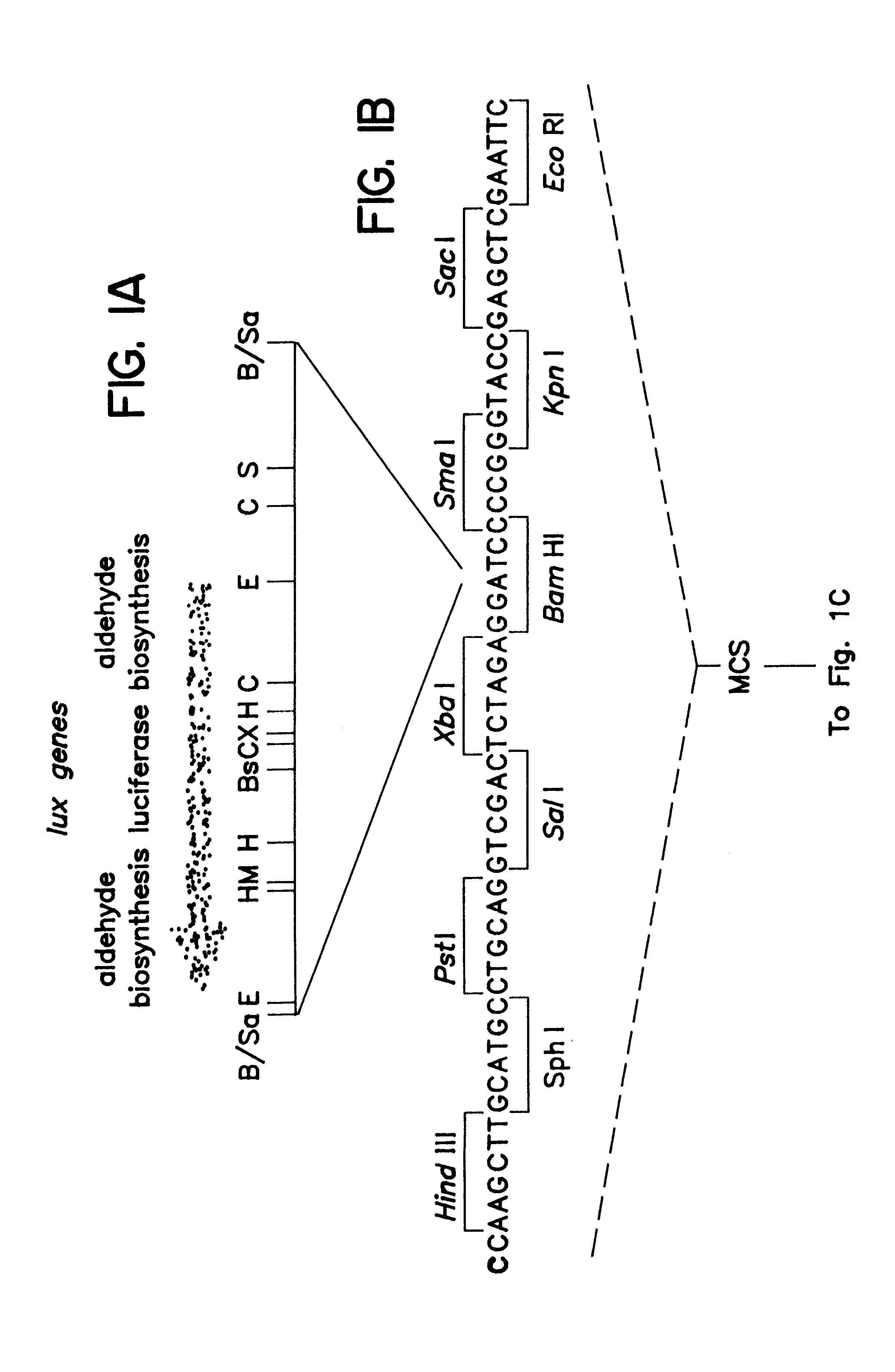
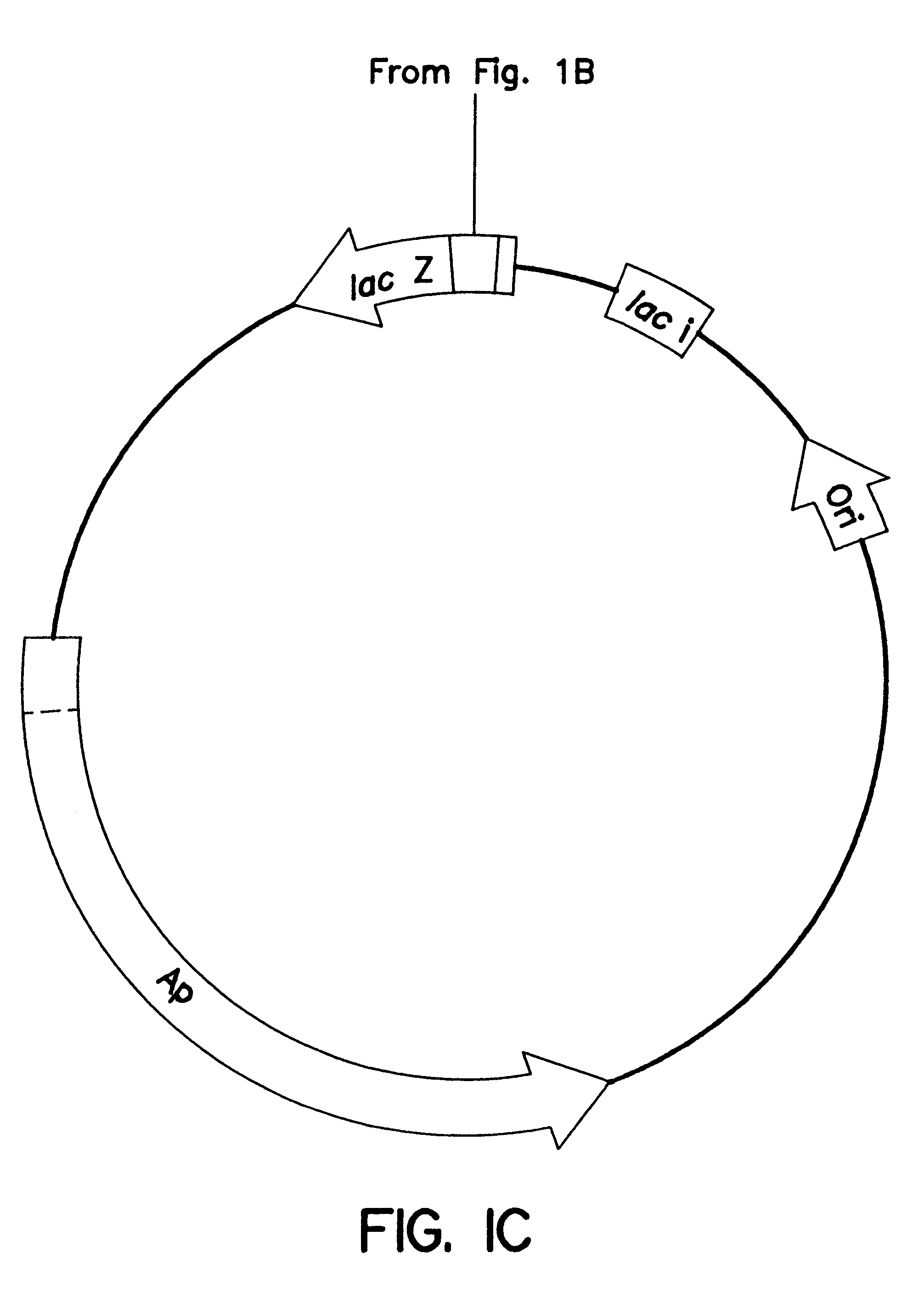
1. pCGLS1 Plasmid

A schematic of the pCGLS1 plasmid is shown in FIGS. 1A, 1B and 1C. The plasmid was constructed by cloning an .about.11 kb region encoding the lux genes from the soil bacterium Xenorhabdus luminescens (FIG. 1A; Frackman, et al., 1990) into the Bam HI site (FIG. 1B) of pUC18 (FIG. 1C; Clontech, Palo Alto, Calif.). The construction of the vector is described by Frackman, et al., (1990).

Restriction enzyme sites in FIG. 1A are represented as follows: Bs, Bst EII; C, Cla I; E, Eco RI; H, Hind III; M, Mlu I; S, Sca I; X, Xba I; B / Sa, Bam HI and Sau 3A junction. A sequence included in the multiple cloning site (MCS) is provided in FIG. 1B, with the Bam HI site indicated in bold type.

A graphical representation of a pUC18 vector with no insert ...

example 2

C. Example 2

Invasive Potential Of Normal And Transformed Salmonella

The invasive potential of six strains of Salmonella (SL1344lux, LB5000lux, BJ66lux, SL1344, LB5000 and BJ66) was determined using two types of bacterial adherence and entry assays. Colony-forming units (CFU) assays were performed essentially as previously described (Finlay and Falkow, 1989, Mol. Microbiol. 3:1833-1841) with modifications (Lee, et al., 1990, PNAS 87:4304-4308). Bioluminescence assays were performed essentially like the CFU assays, except that the number of cells was quantitated using bioluminescence, as opposed to CFUs.

Briefly, HEp-2 cells and primary murine peritoneal macrophages were seeded into 24-well tissue culture dishes at 1.times.10.sup.5 cells per well in RPMI (Gibco / BRL, Grand Island, N.Y.) supplemented with 20 mM glutamine (Gibco / BRL) and 5 fetal calf serum (Hyclone, Logan, Utah). Twenty four hours (HEp-2) or seven days (macrophages) after cell seeding, bacteria from static cultures (see "M...

example 3

D. Example 3

In vitro Luminescence of Transformed Salmonella

10 .mu.l of four 10-fold serial dilutions (ranging from 10.sup.6 cells to 10.sup.3 cells per ml) of LB5000lux Salmonella were placed in four 100 .mu.l glass capillary tubes (Clay-Adams div. of Becton Dickinson, Parsippany, N.J.). The bacterial suspensions formed columns of fluid in the tubes, with pockets of air at both ends. One end of each tube was sealed with critoseal (Clay-Adams). The medium in which dilutions were made was saturated with O.sub.2 through exposure to air.

The tubes were wrapped with clear plastic wrap and luminescence was determined by imaging for 30 seconds as described above. An exemplary image is shown in FIG. 3A. Four tubes are pictured. They contained (from top to bottom) 10.sup.6, 10.sup.5, 10.sup.4 and 10.sup.3 Salmonella cells / ml (10.sup.4, 10.sup.3, 10.sup.2 and 10 cells / tube). Luminescence could be detected in suspensions containing as few as 10.sup.4 cells / ml (100 cells). The luminescence is co...


PUM

PropertyMeasurementUnit
wavelengthaaaaaaaaaa
wavelengthsaaaaaaaaaa

Abstract

Methods and compositions for detecting and localizing light originating from a mammal are disclosed. Also disclosed are methods for tracking light emission to selected regions, as well as for tracking entities within the mammal. In addition, animal models for disease states are disclosed, as are methods for localizing and tracking the progression of disease or a pathogen within the animal, and for screening putative therapeutic compounds effective to inhibit the disease or pathogen.

Description

I. FIELD OF THE INVENTIONThe present invention relates to noninvasive methods and compositions for detecting, localizing and tracking light-emitting entities and biological events in a mammalian subject.II. BACKGROUND OF THE INVENTIONThe ability to monitor the progression of infectious diseases is limited by the current ex vivo methods of detecting and quantifying infectious agents in tissues. The replication of an infectious agent in a host often involves primary, secondary and tertiary sites of replication. The sites of replication and the course that an infectious agent follows through these sites is determined by the route of inoculation, factors encoded by the host as well as determinants of the infecting agent.Experience may offer, in some cases, an estimate of probable sites of replication and the progress of an infection. It is more often the case, however, that the sites of infection, and the pace of the disease are either not known or can only roughly be estimated. Moreove...

Claims


Application Information

Patent Timeline
Patent Type & Authority Patents(United States)
IPC IPC(8): A61K39/112C07K14/195C12Q1/66C12Q1/02G01N33/58C12N1/21
CPCC07K14/195C12Q1/02C12Q1/66G01N33/582Y10S435/968Y10S436/80
Inventor CONTAG, PAMELA R.CONTAG, CHRISTOPHER H.BENARON, DAVID A.
Owner LELAND STANFORD JUNIOR UNIV OF THE BOARD OF TRUSTEES THE
Why Eureka
Try Eureka