Tumor-specific P450 protein

a p450 protein, tumour-specific technology, applied in the direction of peptide sources, instruments, immunological disorders, etc., can solve the problems of increasing the chances of finding small tumours, no tumour specific targets, and general to all types of cancers,

Inactive Publication Date: 2001-06-05
ABERDEEN UNIV OF +1
View PDF3 Cites 14 Cited by
  • Summary
  • Abstract
  • Description
  • Claims
  • Application Information

AI Technical Summary

Problems solved by technology

However, to date, no such tumour specific target, general to all types of cancers, has been identified.
Thus, a major problem to targeting anti-cancer drugs at tumours based on their selective activation by P450 has been marked hepatic P450 m...

Method used


Image

  • Tumor-specific P450 protein
  • Tumor-specific P450 protein
  • Tumor-specific P450 protein

Examples


example 2

The expression of CYP1B1 was also investigated in breast cancer using immunoblotting.

Samples of breast tissue were obtained from patients undergoing surgery either for primary breast cancer or non-neoplastic breast disease. Immunoblotting was performed on breast cancers obtained from six patients (age range 45-67; three non-smokers, information not available for three patients), and histologically all these tumours were carcinomas of no special type. The tissue samples were frozen in liquid nitrogen and stored at -80.degree. C. prior to analysis.

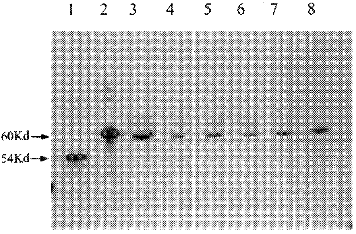
SDS-PAGE and immunoblotting were carried out as described previously. CYP1B1 was detected using the anti-CYP1 polyclonal antibody referred to above. The results are shown in FIG. 2; lane 1 human liver, lane 2 expressed CYP1B1, lanes 3-8 breast tumors. As can be seen, a single protein band of molecular weight 60 kDa corresponding to the molecular weight of the expressed CYP1B1 protein was identified. As previously, CYP1B1 was not detectable i...

example 3

Immunohistochemistry was used to demonstrate the presence of CYP1B1 specifically in a variety of normal and tumour tissues. The results are shown in Table 1 and in FIG. 3.

Immunohistological localisation of CYP1B1 was investigated in tumours and normal tissues from invasive ductal carcinoma of the breast, endometrial adenocarcinoma, transitional cell carcinoma of the bladder, diffuse high grade malignant lymphoma, high grade astrocytoma of the brain, soft tissue sarcoma (malignant fibrous histocytoma), normal liver, normal kidney, normal small intestine. The antibody used was the 218A anti-CYP1B1 polyclonal antibody described above.

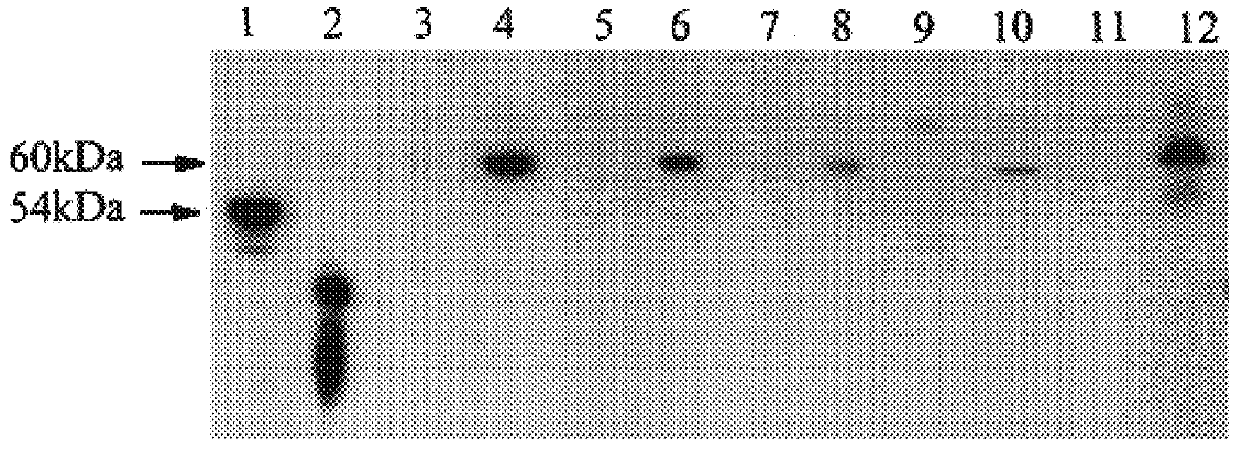
FIG. 3 shows an immunoblot of CYP1B1 in different types of tumours and normal tissues. Lane 1 normal colon, lane 2 colon adenocarcinoma, lane 3 normal kidney, lane 4 carcinoma of kidney, lane 5 normal breast, lane 6 breast cancer, lane 7 normal jejunum, lane 8 normal stomach, lane 9 normal liver, lane 10 malignant mixed Mullerian tumour, lane 11 endometria...

example 4

Experiments were conducted to defect CYP1B1 RNA in various tumour and normal tissues.

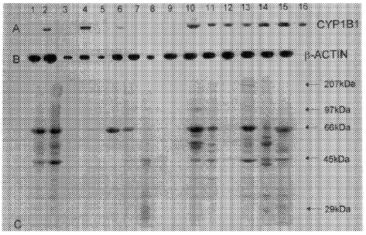
Reverse transcription polymerase chain reaction (RT-PCR) experiments to detect CYP1B1 mRNA were carried out as described in McKay et al (23). RNA was extracted from tissue samples and cDNA was synthesised from the isolated RNA using oligo (dT). The CYP1B1 primers had the following sequences: Forward 5'-AAC TCT CCA TCA GGT GAG GT-3' (nt 2104-2123); Reverse 5'-TAA GGA AGT ATA CCA GAA GGC-3' (nt 2573-3593) giving a PCR product of 489 bp. .beta.-actin was used as a positive control to confirm the presence and integrity of mRNA in each sample and the .beta.-actin primers which were brought from Stratagene (Cambridge, UK) had the following sequences: Forward 5'-TGA CGG GGT CAC CCA CAC TGT GCC CAT CTA-3' (nt 1067-1105); Reverse 5'-CTA GAA GCA TTT GCG GTG GAC GAT GGA GGG-3' (nt 1876-1905). PCR with 35 cycles of amplification for both CYP1B1 and .beta.-actin was performed as described (23). The positive cont...


PUM


Abstract

The discovery that Cyp1B1 protein is detectable in a wide range of human cancers of different histogenetic types, but is not detectable in non-cancerous tissues, gives rise to diagnostic methods for detecting tumors based on this protein as a marker, and to the possibility of tumor therapies involving the protein. A diagnostic method may include the steps of: (a) obtaining from a patient a tissue sample to be tested for the presence of cancer cells; (b) producing a prepared sample in a sample preparation process; (c) contacting the prepared sample with an antibody that reacts with human Cyp1B1 protein; and (d) detecting binding of the antibody to CYP1B1 protein in the prepared sample.

Description

This invention relates to tumour diagnosis and therapy, and to materials and methods for use therein.More particularly, the invention is based on the identification of a cytochrome P450 form, specifically CYP1B1, in a wide range of tumours, with a high frequency of expression in each type, and proposes the use of this enzyme as a tumour marker, and as the basis of a selective therapeutic approach involving the design of drugs, eg which are activated to a cytotoxic form by the action of CYP1B1.BACKGROUND TO THE INVENTIONThe major goal of cancer chemotherapy is the development of anti-cancer drugs that are effective in a wide range of cancers and produce no toxic effects in normal tissues. The target of such drugs should be expressed only in tumour cells and not normal cells. However, to date, no such tumour specific target, general to all types of cancers, has been identified.The cytochromes P450 are a multi-gene family of constitutive and inducible enzymes, which have a central role...

Claims


Application Information

Patent Timeline
IPC IPC(8): C12Q1/26C12N9/02G01N33/574A61K38/00A61K31/00A61K38/44A61K45/00A61P35/00C07K14/80G01N33/573
CPCA61K38/44C12N9/0077C12Q1/26G01N33/57492C12Y114/14001G01N2333/90245G01N2500/00A61P35/00A61P37/02
Inventor MELVIN, WILLIAM THOMASMURRAY, GRAEME IANBURKE, MICHAEL DANNYGREENLEE, WILLIAM FRANK
Owner ABERDEEN UNIV OF
Why Eureka
Try Eureka