Labelling of polymers and sequencing of nucleic acids
- Summary
- Abstract
- Description
- Claims
- Application Information
AI Technical Summary
Benefits of technology
Problems solved by technology
Method used
Image
Examples
example 2
Photochemical Labelling of Single-Stranded DNA
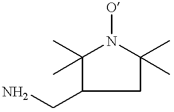
To a quartz cell were introduced 20 .mu.L of 1.02% single-stranded DNA (as used in Example 1), 0.5 mL of aqueous 36 mM amino-TEMPO, and 0.5 mL of aqueous 10 mM anthraquinone-2,6-disulfonate, disodium salt. The mixture was degassed with nitrogen for 15 minutes and then irradiated in a Rayonet RPR-100 photoreactor equipped with sixteen 300 nm low pressure mercury lamps. These lamps provide a broad band of light from 270 to 350 nm. Samples (300 .mu.L) were removed after 30, 60 and 100 minutes of irradiation. The three samples were purified, reacted with fluorescamine, and further purified as described in Example 1. At the end of purification each sample was diluted to 500 .mu.L with water.
Analysis was conducted by GPC as described in Example 1 and the three chromatograms are shown in FIG. 2. FIG. 2A shows the fluorescence curve 22 and the RI curve 24 from the 30 minutes irradiated sample. FIG. 2B shows the fluorescence curve 26 and the RI c...
example 3
Radiochemical Labelling of Single-Stranded DNA
A solution containing 0.2% single-stranded DNA and 61.9 mM amino-TEMPO was prepared and then 0.5 mL Was transferred to each of 4 vials. Each sample was degassed by bubbling with nitrogen for 5 minutes and then sealed with a rubber septum. The samples were then exposed to .gamma.-rays, produced by a .sup.60 Co source such that they received 0, 0.5, 1.0, or 2.0 Mrad doses of .gamma.-rays. The samples were purified as described in Example 1. To the retentate (ca. 20 .mu.L) was added 75 .mu.L of 0.17M sodium bicarbonate, and then 10 .mu.L of 11.8 mM fluorescein isothiocyanate (Isomer 1, Aldrich) in anhydrous dimethylformamide. The solutions were mixed on a vortex mixer, left to stand for 50 minutes at room temperature, and then purified as described in Example 1.
Analysis was conducted by GPC as described in Example 1. Each chromatogram displays an RI peak at 5.2 min but only samples exposed to .gamma.-rays display a fluorescence peak. The pr...
example 4
DNA (50 base-pair molecular weight ladder, 1 .mu.g / .mu.L in TE buffer, Pharmacia Biotech, catalog #27-4005-01) was used in these experiments. The DNA consists of nucleic acid polymer chains of 50, 100, 150, etc. bases in length, with the 250 base chains in roughly double the concentration of chains of any other length. As received this sample contains no fluorescent labels. It was labelled by the following procedure.
The TE buffer was removed from the sample so as not to interfere with labelling. 10 .mu.L of the DNA solution (10 .mu.g of DNA) was purified by centrifugal ultrafiltration (30,000 molecular weight cut-off) as outlined in Example 1. A 57.5 .mu.L solution containing the purified DNA (ca. 10 .mu.g), hydrogen peroxide (153.5 mM) and AT (40.8 mM) was deaerated, and then left for 90 min at room temperature. The DNA was then purified by centrifugal ultrafiltration. The purified DNA solution (ca. 20 .mu.L) was then mixed with ca. 100 .mu.L of 0.185 M NaHCO.sub.3 and 10 .mu.L of ...
PUM
| Property | Measurement | Unit |
|---|---|---|
| Time | aaaaa | aaaaa |
| Length | aaaaa | aaaaa |
| Temperature | aaaaa | aaaaa |
Abstract
Description
Claims
Application Information
 - R&D
- Intellectual Property
- Life Sciences
- Materials
- Tech Scout
- Unparalleled Data Quality
- Higher Quality Content
- 60% Fewer Hallucinations
Browse by: Latest US Patents, China's latest patents, Technical Efficacy Thesaurus, Application Domain, Technology Topic, Popular Technical Reports.
© 2025 PatSnap. All rights reserved.Legal|Privacy policy|Modern Slavery Act Transparency Statement|Sitemap|About US| Contact US: help@patsnap.com


