
Monoclonal antibody hybridoma immunoassay method and diagnosis kit

a hybridoma and antibody technology, applied in the field of monoclonal antibody hybridoma immunoassay method and diagnosis kit, can solve the problems of high cost, inconvenient diagnosis, and inability to judge the effect of bacterial eradication by antibody test, etc., and achieve excellent specificity, low cost, and free

Inactive Publication Date: 2005-02-01
WAKAMOTO PHARMA
View PDF16 Cites 17 Cited by
  • Summary
  • Abstract
  • Description
  • Claims
  • Application Information

AI Technical Summary

Benefits of technology

"The present invention provides a method for diagnosing Helicobacter pylori infection using a single monoclonal antibody that recognizes Helicobacter pylori catalase as an antigen. This antibody can be used in an immunological assay method that is cost-effective, painless, and highly specific, without causing cross-reactivity or variation between lots. The monoclonal antibody can be used in a diagnosis kit for making a judgment about Helicobacter pylori infection, which is also cost-effective and easy to use. The invention also provides a hybridoma that produces the monoclonal antibody."

Problems solved by technology

However, these test methods for Helicobacter pylori infection have the following problems.
The invasive test method inflicts a great deal of pain on subjects on the occasions of gastroscope insertion and biopsying.
Further, in carrying out the urea breath test, such apparatus as a mass spectrometer, an infrared spectrophotometer, etc. are required, hence the test can be carried out only in special institutions, and the resulting high cost is also a drawback.
The antibody test is not suited for the judgment about bacterial eradication since the antibody titer in blood remains at high levels for a long term after eradication of the bacteria.
As far as Helicobacter pylori is concerned, however, in spite of a large number of trials, there are few reports about the isolation, by culture, from feces.
However, polyclonal antibodies generally have cross reactivity and are inferior in specificity and, in addition, are disadvantageous in that the antibody titer and specificity may vary depending on the lot of antiserum.
Therefore, there is a problem that the production of diagnostic tests in which polyclonal antibodies are used is essentially difficult in terms of the quality control.

Method used


Image

  • Monoclonal antibody hybridoma immunoassay method and diagnosis kit
  • Monoclonal antibody hybridoma immunoassay method and diagnosis kit
  • Monoclonal antibody hybridoma immunoassay method and diagnosis kit

Examples


example 1

[0113][Monoclonal Antibody Production]

[0114](1) Preparation of a Helicobacter pylori Coccoid Cell Suspension (Immunogen)

[0115]Brain heart infusion agar medium (product of Difco) supplemented with 5% equine defibrinated blood was streaked with Helicobacter pylori (ATCC 43504) in series. The plate was incubated at 37° C. for 3 to 4 days in a microaerobic environment and then further incubated at 37° C. for 7 days in an anaerobic environment to cause the cells to become coccoid. Each colony obtained was scraped off with a platinum loop or the like and suspended in phosphate-buffered physiological saline (PBS). Helicobacter pylori cells were collected by 10 minutes of centrifugation at 10,000×g at 4° C. and suspended in 0.5 % formalin, and the suspension was allowed to stand at 4° C. for 4 days for inactivation. Thereafter, the cells were washed by three repetitions of the procedure comprising suspending in PBS followed by 10 minutes of centrifugation at 10,000×g at 4° C. and then suspe...

example 2

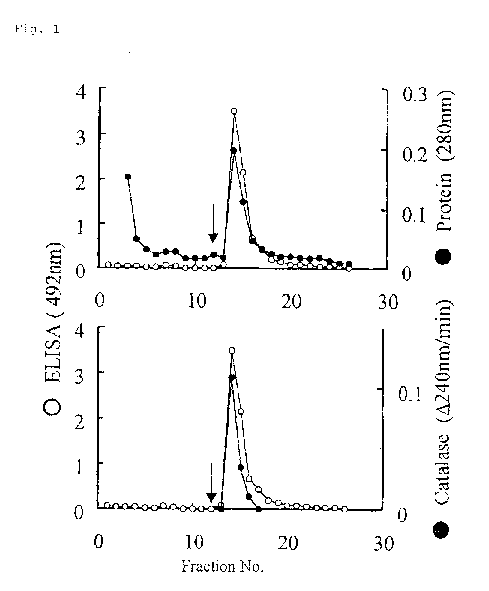
[0123][Selection of Monoclonal Antibodies Specifically Recognizing Helicobacter pylori in Digestive Tract Excreta by Sandwich ELISA]

[0124](1) Preparation of Monoclonal Antibody-Immobilized Plates

[0125]Each 1-mL portion of the ascetic fluids of the 32 clones was diluted two-fold with PBS, 2 mL of saturated ammonium sulfate was added dropwise, and the mixture was allowed to stand at 4° C. for 4 hours. Then, the mixture was centrifuged at 3000 rpm for 20 minutes, and the sediment was suspended in 2 mL of PBS and dialyzed. These monoclonal antibodies were immobilized on 96-well ELISA plates in the following manner. Thus, each monoclonal antibody was diluted to 5 μg / mL and 0.2 mL of the dilution was added to each well of 96-well ELISA plates. After overnight standing at 4° C., the plates were washed with PBS, then 0.25 mL of 1% skim milk-PBS was added to each well and the plates were allowed to stand at 4° C. for 1 hour for blocking. Thereafter, the plates were washed with the above wash...

example 3

[0131][Reactivity Comparison Among Strains and Reactivity with Other Bacterial Species]

[0132](1) Preparation of Helicobacter pylori Cell Suspensions

[0133]Brain heart infusion agar medium supplemented with 5% equine defibrinated blood was streaked with each of Helicobacter pylori strains (ATCC 43504; Tokai University Hospital clinical isolates No. 130 and No. 112; Hyogo Medical College clinical isolates No. 526, No. 4484, No. 5017, No. 5025, No. 5049, No. 5142, No. 5287, No. 5308, No. 5314 and No. 5330) in series. The plates were incubated at 37° C. under microaerobic conditions for 3 to 4 days to give colonies of helical cells, or further allowed to stand a: 37° C. for 7 days in an anaerobic environment to give colonies of coccoid cells. Each colony was scraped off with a platinum loop or the like and suspended in PBS. Cells were collected by 10 minutes of centrifugation at 10,000×g at 4° C. and suspended in 0.5% formalin, and the suspension was allowed to stand at 4° C. for 4 days ...


PUM

PropertyMeasurementUnit
molecular weightaaaaaaaaaa
concentrationaaaaaaaaaa

Abstract

The object of the present invention is to provide a diagnostic method by which Helicobacter pylori infection can be diagnosed at low cost without causing pain on subjects and without requiring particular equipment and which is free of cross reactivity and excellent in specificity without variation among lots as a result of the use of a single antibody facilitating the quality control and which shows good sensitivity even when a single monoclonal antibody is used.The present invention provides a monoclonal antibody which recognizes Helicobacter pylori catalase as an antigen.

Description

[0003]This is a National Stage Entry under 35 U.S.C. § 371 of PCT Application No. PCT / JP00 / 07554, filed Oct. 27, 2000.TECHNICAL FIELD[0004]The present invention relates to a monoclonal antibody recognizing Helicobacter pylori catalase as an antigen, a hybridoma producing said monoclonal antibody, an immunological assay method and a diagnosis kit.BACKGROUND ART[0005]Helicobacter pylori is a bacterial species found in the human gastric mucosa. The rate of Helicobacter pylori infection is closely related to social and economic factors, tending to be high in developing countries and low in advanced countries. However, the rate of infection among the Japanese is remarkably high among the advanced countries and it is even reported that 80% of people in and after the fourth decade of life have been infected. In recent years, it has been revealed that Helicobacter pylori may cause various gastric and duodenal diseases such as gastric ulcer, duodenal ulcer, chronic gastritis and, further, ga...

Claims


Application Information

Patent Timeline
Patent Type & Authority Patents(United States)
IPC IPC(8): C07K16/12G01N33/569C12P21/08
CPCC07K16/121G01N33/56922G01N2333/908C07K16/40
Inventor WAKASUGI, MASAHIKOMOCHIDA, RITSUKOHIRATA, HARUHISA
Owner WAKAMOTO PHARMA
Learn More