Multi-photon imaging installation

a multi-photon imaging and installation technology, applied in the field of biological material imaging, can solve the problems of insufficient lateral and depth resolution, time-consuming use of extrinsic luminescent contrast agents, and insufficient labelling procedures,

Inactive Publication Date: 2005-04-12
INSTITUT CURIE +1
View PDF2 Cites 26 Cited by
  • Summary
  • Abstract
  • Description
  • Claims
  • Application Information

AI Technical Summary

Benefits of technology

An object of the invention is to provide a solution to at least part of the drawbacks described above, and more particularly to make it possible i) to get images showing the geometric structure of a biological sample with resolution ranging from the sub-micron scale (cellular and sub-cellular scales) to the scale of tissular and organ structures, from a non-linear intrinsic imaging (microscopy, macroscopy or endoscopy), and ii) to combine such non-linear intrinsic imaging with spectroscopy for tissue exploration and diagnosis (or more generally detection of differences between normal and abnormal tissues).

Problems solved by technology

The use of extrinsic luminescent contrast agents is time-consuming and requires constraining labelling procedures.
Classically these methods offer lateral and depth resolutions which are insufficient for revealing the intimate tissue structures at cellular and subcellular scales.
It results from this paradox that i) linear imaging microscopy technique can hardly perform deep tissue imaging and ii) endoscopic fluorescence imaging cannot resolve signals coming from the surface from those produced by deep chromophores.
But, these so-called “spectral pathology” methods have been implemented with a linear excitation, thus leading to three major disadvantages:i) since excitation is not confined at a focus, the linear spectroscopy is not spatially resolved and intrinsically bears strong limitations in terms of lateral and depth resolutions, as well as deep imaging.ii) “Single-photon” fluorescence excitation spectra most often display a single and narrow peak, and one laser wavelength will only excite few molecular components.
This puts stringent conditions on the choice of excitation wavelengths, and often leads to complicated spectroscopic criteria for resolving pathologic situations from healthy ones.
Nevertheless, these local contributions are never dominant features in the final spectrum and therefore are difficult to extract, because they are spatially averaged out.
To summarize, every spectral diagnostic method implemented with linear excitation results in a very poor spatial resolution, a frequent need for multiple sources, a limited sensitivity, and an impossibility to get information at the cellular scale.

Method used


Image

  • Multi-photon imaging installation
  • Multi-photon imaging installation
  • Multi-photon imaging installation

Examples


Embodiment Construction

In the description below, reference is made to an installation and a method for multi-photon imaging of a biological material. More precisely, reference is made, purely by way of example, to a multi-photon imaging microscope.

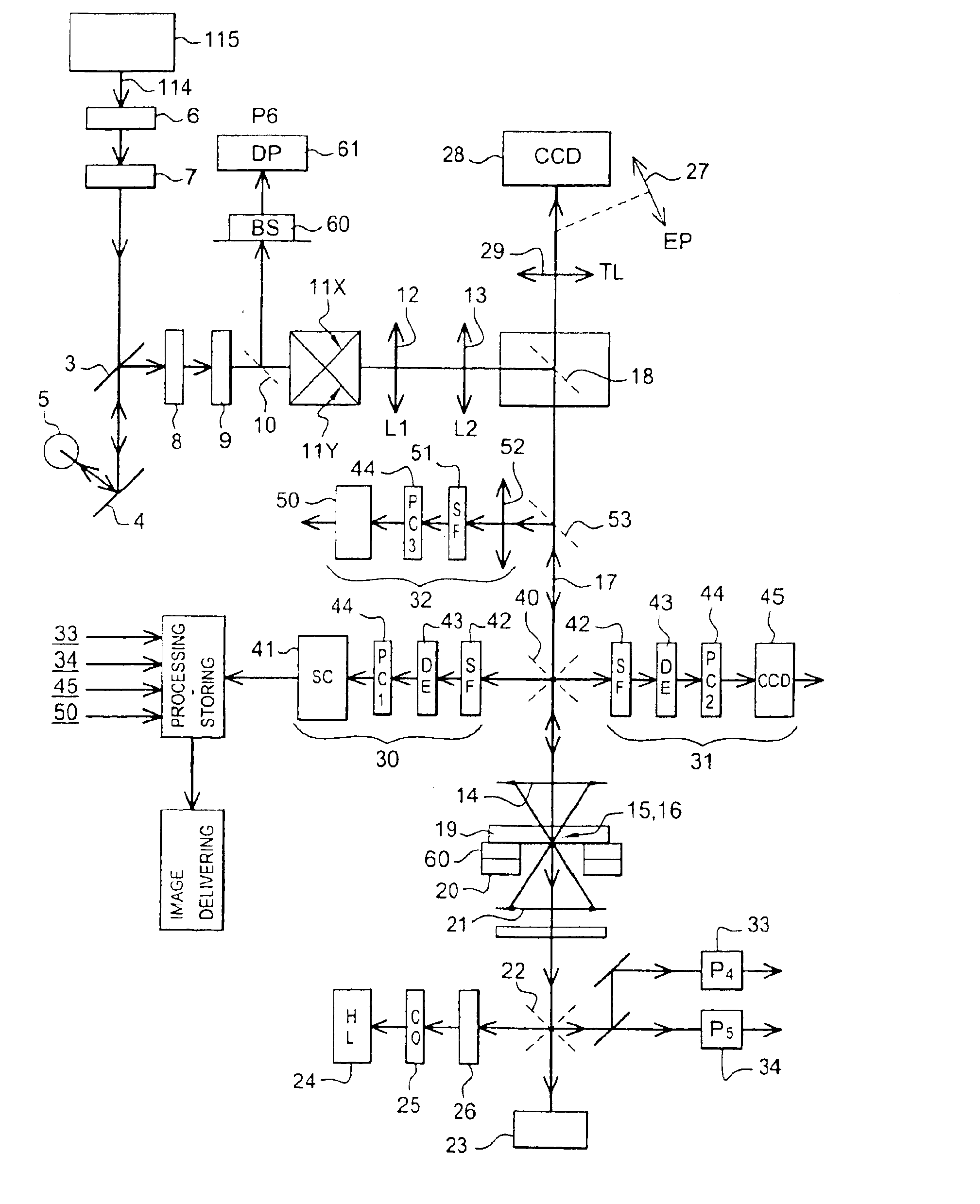
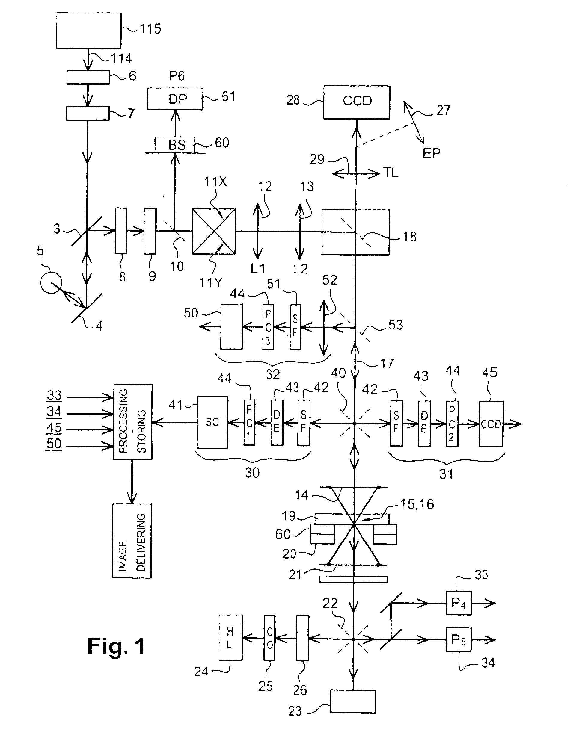
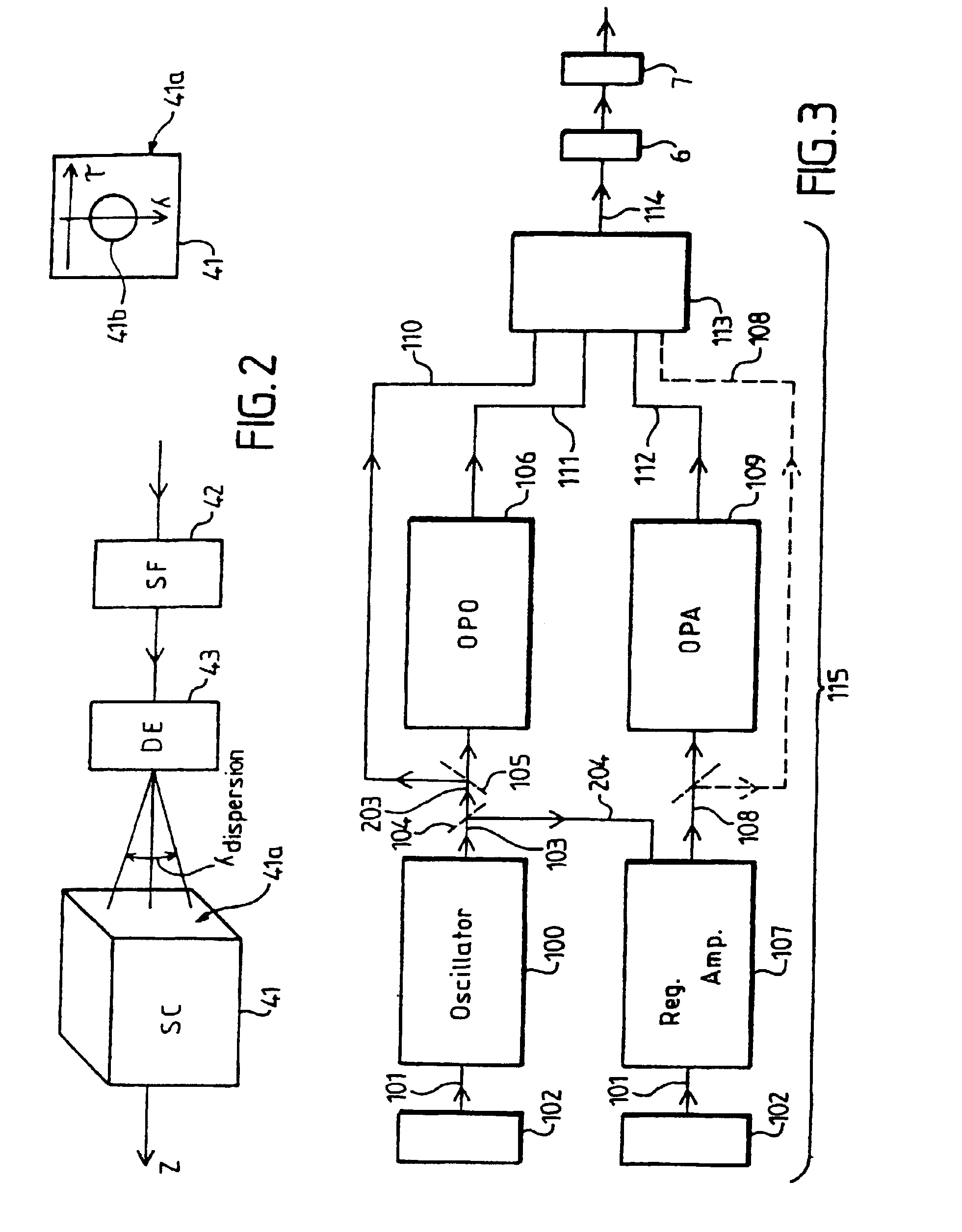
As mentioned above, the invention refers to an installation (or instrument) that permits laser scanning imaging of a biological sample chosen area, in an appreciably continuous way or at selected discrete points, in two (2D) or three (3D) dimensions, by moving either a laser beam or the sample holding stage, or both.

At each scanning point, with a dwell time between two successive points typically ranging from 100 microseconds up to seconds, a light emitted by a pulsed laser source is focused in a “focal volume” within or at the surface of the sample. The intrinsic chromophores comprised in the sample absorb groups of at least two synchronized photons to produce non-linear response photons which can be collected and integrated on several kinds of detectors: (1) a...


PUM

PropertyMeasurementUnit
wavelengthsaaaaaaaaaa

Abstract

An installation is provided for multi-dimensional non-linear imaging of a material comprising intrinsic chromophores, using laser scanning. This installation comprises i) at least one source (115) of time stamp pulses of synchronized photons, ii) means (12-14) for locally focusing the pulses on a material to cause its intrinsic chromophores to absorb groups of at least two synchronized photons to produce response photons, iii) means (22, 40, 53) for directing the response photons to at leat one collecting zone, iv) means (30-34) for collecting the response photons in the collecting zone(s) whatever their energy, v) processing means for converting the collected photons into data at least representative of their number and storing them in correspondence with at least the time stamp pulses that cause the material to produce the response photons, vi) means for scanning (11, 20) the pulses through a chosen area of the material, and vii) means for delivering from said data stored an image representative of said material chosen area, with a sub-millimeter resolution, and in function of the respective time stamp pulses of data.

Description

FIELD OF THE INVENTIONThe invention relates to the field of biological material imaging with the help of time stamp pulses of synchronized photons.This invention more particularly relates to installations and methods for imaging the intrinsic non-linear optical response of biological samples, in vivo or in vitro, for example, for the purpose of biological research, medical investigations (for screening normal from pathologic tissues, for example), forensic or biotechnology applications.The expression “non-linear optical response” designates the photons, hereafter called “response photons”, which are emitted by chromophores, naturally contained in a sample, when they are excited by several (at least two) synchronized photons. More precisely, the expression “non-linear” means that the efficiency for the excitation of molecules varies in a non linear way with the electromagnetic field amplitude. In fact, the efficiency of the excitation by n synchronized photons is proportional to the ...

Claims


Application Information

Patent Timeline
Patent Type & Authority Patents(United States)
IPC IPC(8): G01N21/64G02B21/00A61B1/00A61B3/10A61B3/12A61N5/06G01N21/65G02B23/24
CPCG01N21/6458G01N21/6486G02B21/0084G02B21/0076G02B21/0024
Inventor AMBLARD, FRAN.CEDILLA.OISSASTRE-GARAU, XAVIERROBINE, SYLVIE
Owner INSTITUT CURIE
Why Eureka
Try Eureka