
Multi-photon laser microscopy

a laser microscopy and multi-photon technology, applied in the field of multi-photon laser microscopy, can solve the problems of inability to achieve technologically feasible laser wavelengths and limited laser technology applications, and achieve the effect of extending the useful range of laser systems and less photodamag

Inactive Publication Date: 2002-02-05
WEBB WATT W +1
View PDF31 Cites 135 Cited by
  • Summary
  • Abstract
  • Description
  • Claims
  • Application Information

AI Technical Summary

Benefits of technology

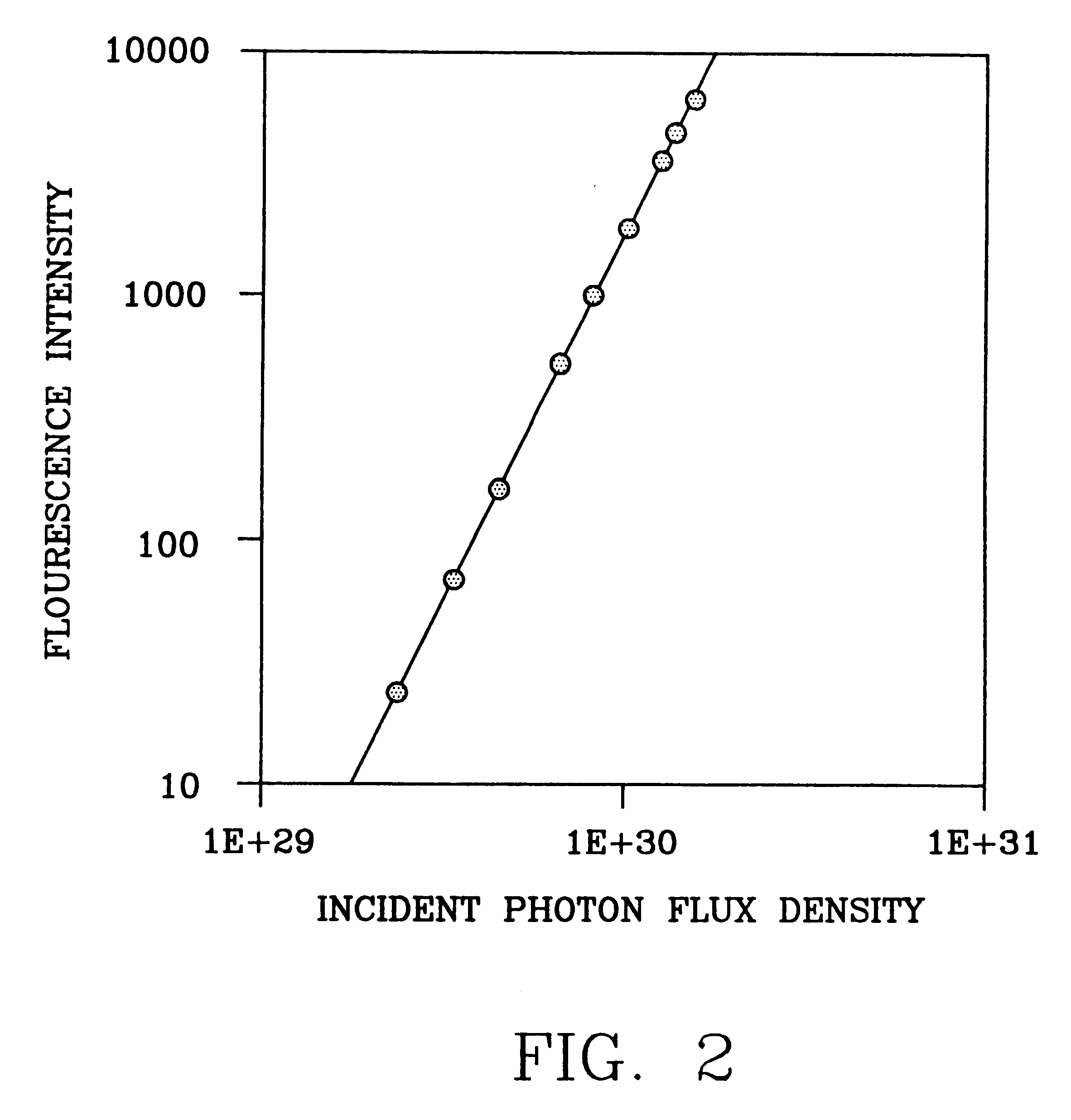
Because three-photon induced fluorescence obeys a cubic dependence on excitation intensity and four photon excitation obeys a quartic dependence, both provide intrinsic three-dimensional resolution in laser scanning microscopy. Although such 3-d resolution has already been achieved by the nonlinear microscopy technique based on two-photon excitation disclosed in the '613 patent, three-photon excitation provides a unique opportunity to excite molecules normally excitable in the UV range (230-350 nm) with near IR light (700-1100 nm). Interesting biomolecules, such as the amino-acids tryptophan and tyrosine, the neurotransmitter serotonin and nucleic acids, have one-photon absorption peaks at approximately 260-280 nm, and fluorescence can be excited in these biomolecules by three and four photon excitation. The advantages of using long wavelength, near IR light are possibly less photodamage to living cells and conveniently available solid state femtosecond laser sources for deep UV absorbers. In practice, the configuration of three-photon laser scanning microscopy can be identical to the existing two-photon systems. However, because three-photon and two-photon absorption spectra are in general quite different, the combination of two-and three-photon excited fluorescence microscopy extends the useful range of the laser systems currently employed in two-photon microscopy.
A particularly advantageous application of three or more photon excitation is the replacement of excimer lasers for certain applications which require absorption of wavelengths around 200 nm. Three and four photon excitation by lasers generating much longer wavelengths (say 550-900 nm) should provide similar energy absorption and provide 3-d spatial resolution as well. Because excimer lasers are extremely expensive and user unfriendly, several photon excitation could be highly desirable.
Alternatively, for microscopic photochemical activation, photoablation and optical surgery, the photo excitation can be advantageously accomplished by multi-photon excitation of intrinsic chromophores or even added chromophores that have very short wavelength absorption such as amino acids and nucleic acids. Multi-photon excitation allows the selection of more available lasers providing subpicosecond pulses at long wavelengths and long wavelength light transmission to the microscopic focal volume where photo excitation is desired.

Problems solved by technology

A drawback to the two-photon laser microscopy technique disclosed in the '613 patent is that its applications are limited by the available laser technology.
Unfortunately, some laser microscopy applications would require use of a laser having a wavelength which is not technologically feasible at the present time.

Method used


Image

  • Multi-photon laser microscopy
  • Multi-photon laser microscopy
  • Multi-photon laser microscopy

Examples


Embodiment Construction

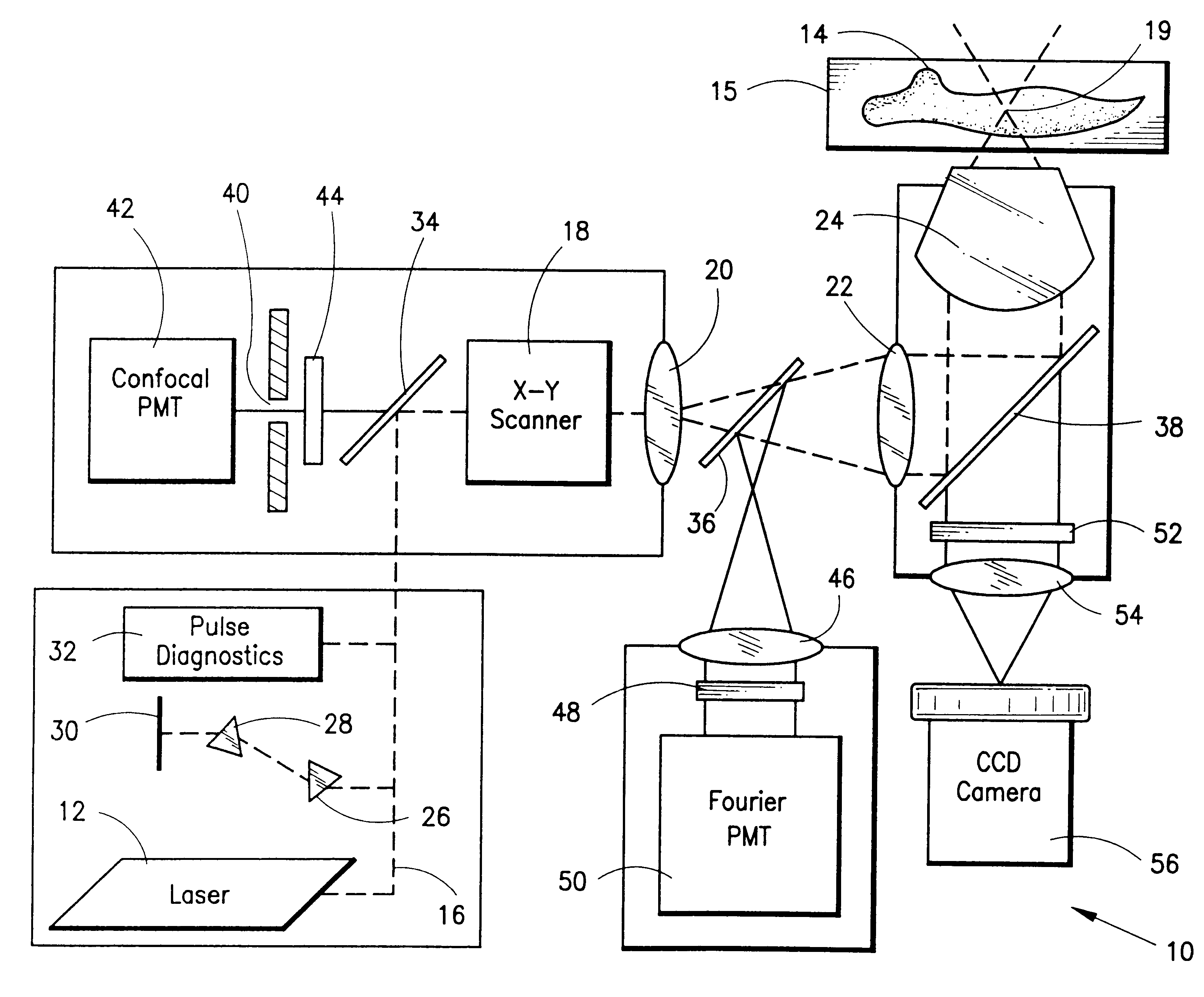
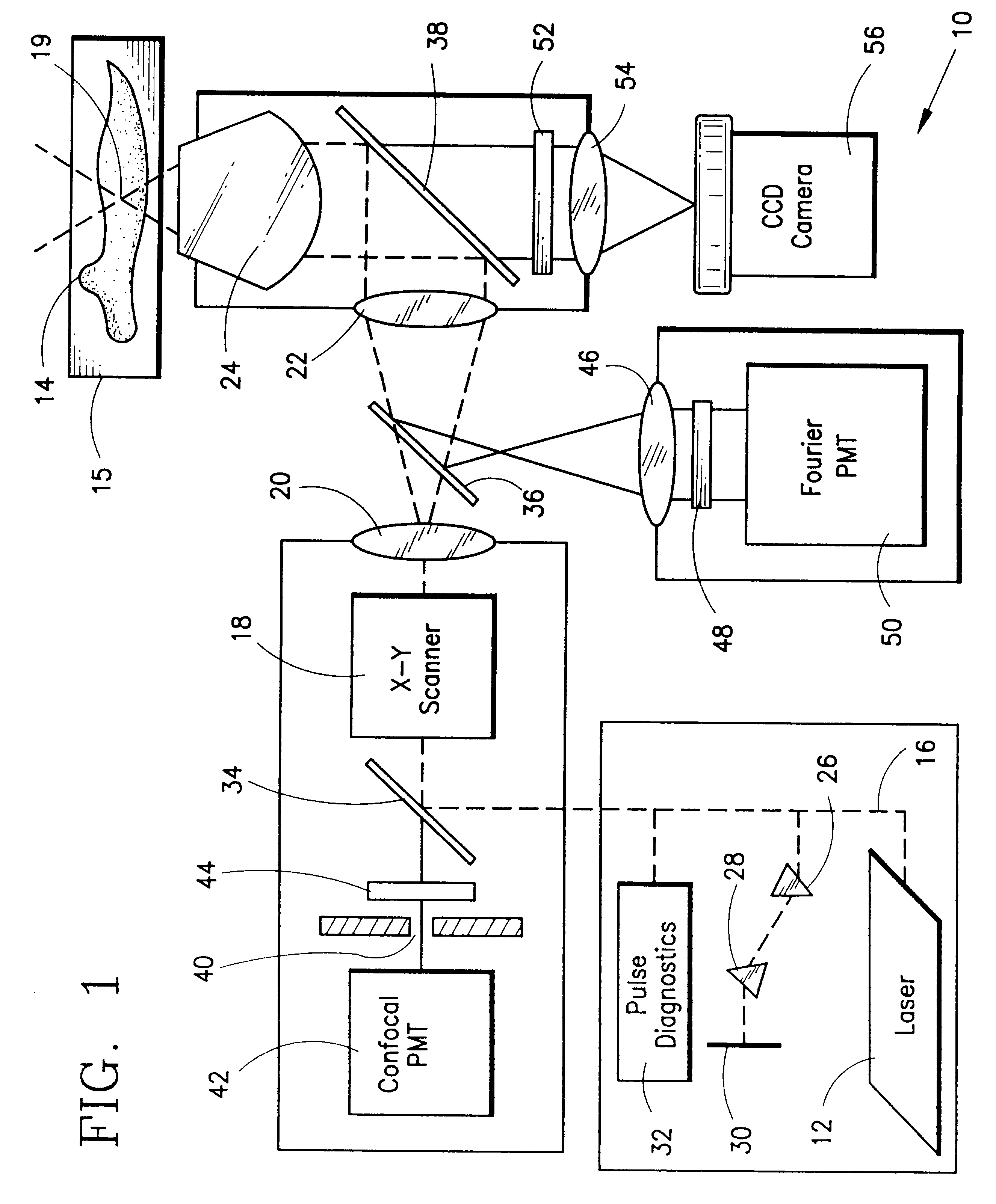
Turning now to a detailed description of a preferred embodiment of the present invention, FIG. 1 illustrates in diagrammatic form a conventional laser scanning microscope 10 which includes three detection alternatives. A subpicosecond pulsed laser source 12 provides the necessary excitation of a specimen or target material 14 which is positioned on a movable stage or other suitable support 15. The laser 12 may be, for example, a colliding pulse, mode-locked dye laser or a solid state laser which can generate pulses of light having a wavelength in the red region of the spectrum, for example about 630 nm, with the pulses having less than 100 fsec duration at about 80 MHz repetition rate. Other bright pulsed lasers may also be used to produce light at different relatively long wavelengths in the infrared or visible red region of the spectrum, for example, to generate the necessary excitation photon energies whose sum will equal the appropriate absorption energy band required by the flu...


PUM

PropertyMeasurementUnit
wavelength absorptionaaaaaaaaaa
wavelengthsaaaaaaaaaa

Abstract

A laser scanning microscope produces molecular excitation in a target material by simultaneous absorption of three or more photons to thereby provide intrinsic three-dimensional resolution. Fluorophores having single photon absorption in the short (ultraviolet or visible) wavelength range are excited by a beam of strongly focused subpicosecond pulses of laser light of relatively long (red or infrared) wavelength range. The fluorophores absorb at about one third, one fourth or even smaller fraction of the laser wavelength to produce fluorescent images of living cells and other microscopic objects. The fluorescent emission from the fluorophores increases cubicly, quarticly or even higher power law with the excitation intensity so that by focusing the laser light, fluorescence as well as photobleaching are confined to the vicinity of the focal plane. This feature provides depth of field resolution comparable to that produced by confocal laser scanning microscopes, and in addition reduces photobleaching and phototoxicity. Scanning of the laser beam by a laser scanning microscope, allows construction of images by collecting multi-photon excited fluorescence from each point in the scanned object while still satisfying the requirement for very high excitation intensity obtained by focusing the laser beam and by pulse time compressing the beam. The focused pulses also provide three-dimensional spatially resolved photochemistry which is particularly useful in photolytic release of caged effector molecules, marking a recording medium or in laser ablation or microsurgery. This invention refers explicitly to extensions of two-photon excitation where more than two photons are absorbed per excitation in this nonlinear microscopy.

Description

The present invention relates to a laser microscopy technique which produces molecular excitation in a target material by simultaneous absorption of three or more photons. The invention is an improvement over the two-photon laser microscopy technique disclosed in U.S. Pat. No. 5,034,613 to Denk et al. (hereinafter, the '613 patent), and this patent is hereby incorporated by reference.The '613 patent discloses a laser scanning microscope which produces molecular excitation in a target material by simultaneous absorption of two photons to provide intrinsic three-dimensional resolution. Fluorophores having single photon absorption in the short (ultraviolet or visible) wavelength range are excited by a stream of strongly focused subpicosecond pulses of laser light of relatively long (red or infrared) wavelength range. The fluorophores absorb at about one half the laser wavelength to produce fluorescent images of living cells and other microscopic objects. The fluorescent emission from t...

Claims


Application Information

Patent Timeline
Patent Type & Authority Patents(United States)
IPC IPC(8): G02B21/00G01N21/27G01N21/64G01N33/483H01S3/098
CPCG02B21/0084G02B21/0076G02B21/002G01N21/6458
Inventor WEBB, WATT W.XU, CHRIS
Owner WEBB WATT W
Learn More