Frozen tissue microarray technology for analysis RNA, DNA, and proteins

a tissue array and frozen technology, applied in the field of frozen tissue array technology, can solve the problems of insufficient array analysis of some proteins or rna using in situ hybridization, inability to prevent rna degradation, and inability to embed ethanol-fixed tissue paraffinly,

Inactive Publication Date: 2005-05-17
RGT UNIV OF CALIFORNIA
View PDF4 Cites 70 Cited by
  • Summary
  • Abstract
  • Description
  • Claims
  • Application Information

AI Technical Summary

Benefits of technology

[0006]The invention disclosed herein improves upon existing tissue microarray technology by arraying the specimens into a recipient block comprising tissue embedding compound. Tissue is not fixed prio

Problems solved by technology

A variety of technical problems exist with the current methodology, however, relating to the fact that the arrayed samples have to be pre-fixed and embedded in paraffin.
The quality of the studies performed on sections from tissue array technology may be limited by the fixation methods used on the original sample.
Nat Med. 1998 July; 4(7):844-7), but this approach is not optimal for array analysis of some proteins or for RNA using in situ hybridization.
Par

Method used


Image

  • Frozen tissue microarray technology for analysis RNA, DNA, and proteins
  • Frozen tissue microarray technology for analysis RNA, DNA, and proteins
  • Frozen tissue microarray technology for analysis RNA, DNA, and proteins

Examples


example 1

Frozen Array Construction

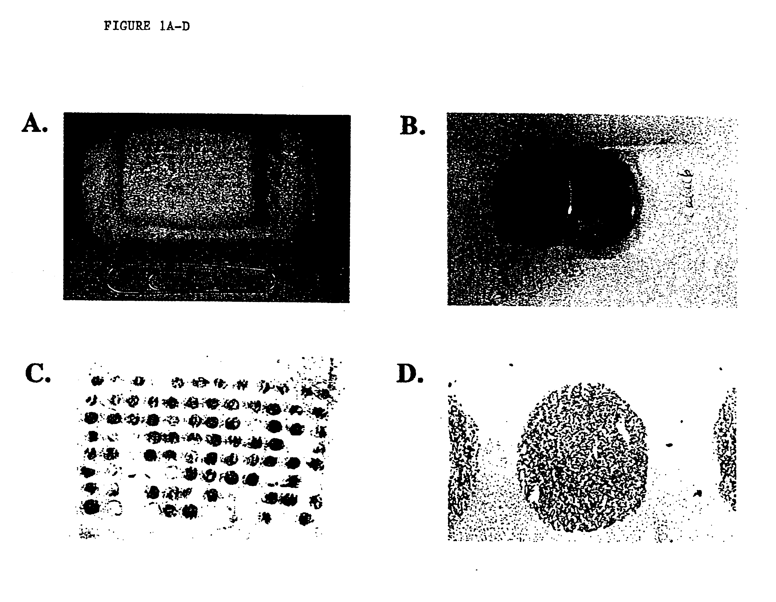
[0042]A human lung cancer cell line CALU-6 grown as a mouse xenograft and human breast cancer cells MDA-MB-231 grown in vitro and pelleted were frozen and embedded in O.C.T. compound embedding medium (Miles Inc. Diagnostic Division, Elkhart, Ind.) to test whether arrays could be cored and collected in this medium. Methodology was as published previously (1), except that tissue biopsies (diameter 0.6 and 1.0 mm; height 3-4 mm) were punched from tumors in O.C.T. and placed directly into an O.C.T. array block using a tissue microarrayer (Beecher Instruments, Silver Spring, Md., USA).

[0043]The recipient O.C.T. array block was made by filling a Tissue-Tek standard cryomold (Miles Inc., Elkhart Ind.) with O.C.T. and mounting the O.C.T. filled mold to the base of a plastic biopsy cassette (Simport Histosette II Biopsy Cassette from Fisher Scientific with lid removed), see FIG. 1A. The recipient O.C.T. block has the same size base as the paraffin recipient block tha...

example 2

Non radioactive RNA In Situ Hybridization Methods Employing Frozen Arrays

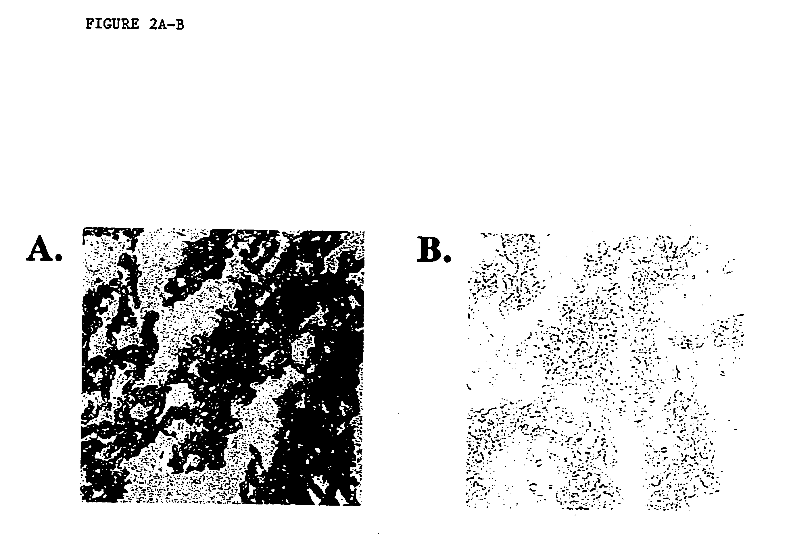
[0046]Nonradioactive RNA in situ hybridization was performed as published previously for frozen sections (see, e.g. Hogan et al. Manipulating the Mouse Embryo: A Laboratory Manual. New York: Cold Spring Harbor Laboratory Press (1994). Briefly, tissue array sections used for RNA in situ hybridization were fixed in 4% paraformaldehyde in phosphate buffered saline (PBS) for either 10 minutes, 2 hours, or overnight at 4 C. Slides were rinsed 3 times in PBS for 5 minutes each and drained. Sections were covered with a prehybridization buffer (50% deionized formamide, 5×SSC, 5× Denhardt's, 750 ug / ml torula RNA) and placed in a humid chamber at room temperature for 2 hours. Hybridization was performed by adding 0.5 micrograms of digoxigenenin-labeled actin RNA probe (Boehringer Mannheim, Germany) to 10 ml of prehybridization solution in a 5-slide mailer. Tissue nucleic acid was denatured at 85 C for 10 minutes and cool...

example 3

Fluorescent In Situ Hybridization Methods Employing Frozen Arrays

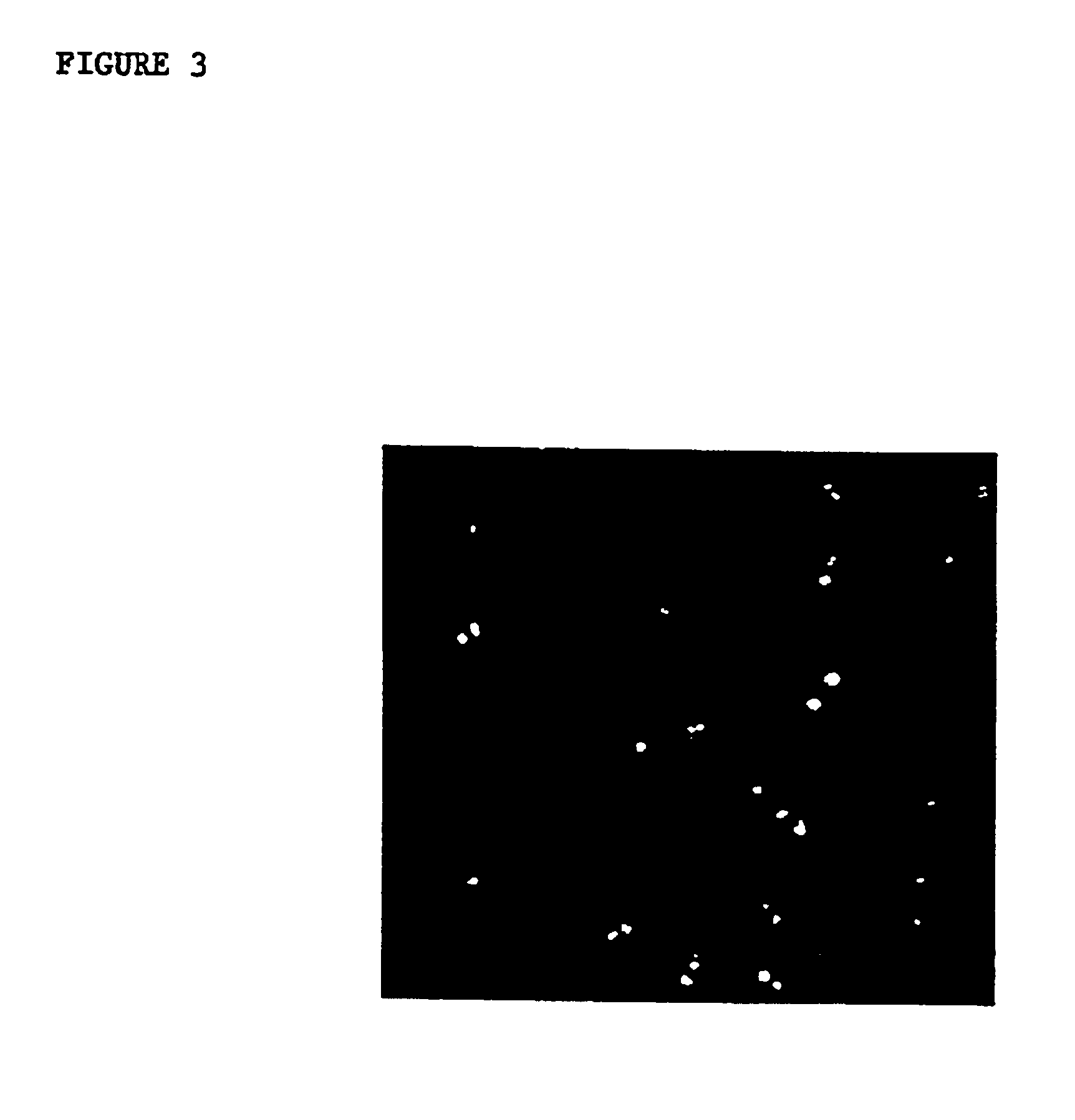
[0048]Frozen tissue microarray sections were fixed in Carnoy's fixative or 95% ethanol for 10 minutes. Slides were pretreated in 2×SSC at 37 C for 30 minutes, dehydrated, denatured in 70% formamide / 2×SSC for 5 minutes at 72 C, and dehydrated again. Slides were then treated either with or without 0.4 ug / ml proteinase K (Sigma, St. Louis, Mo., USA) at 37 C for 30 minutes. A spectrum orange chromosome 8 probe (Vysis Inc., Downer's Grove, Ill.) was prepared according to the manufacturer's instructions, denatured for 7 minutes at 72 C, and hybridized to the array slides overnight at 37 C in a humid chamber. Slides were washed (50% formamide / 2×SSC 44 C, 15 minutes; 2×SSC, 8 minutes) and counterstained with DAPI (Vysis, Downersgrove, Ill., USA). Slides were visualized using standard fluorescent microscopy and photographed with Ektachrome 400 ASA slide film (Eastman Kodak, Rochester, N.Y., USA).

[0049]Typical results of such an...


PUM


Abstract

The invention disclosed herein improves upon existing tissue microarray technology by using frozen tissues embedded in tissue embedding compound as donor samples and arraying the specimens into a recipient block comprising tissue embedding compound. Tissue is not fixed prior to embedding, and sections from the array are evaluated without fixation or post-fixed according to the appropriate methodology used to analyze a specific gene at the DNA, RNA, and/or protein levels. Unlike paraffin tissue arrays which can be problematic for immunohistochemistry and for RNA in situ hybridization analyses, the disclosed methods allow optimal evaluation by each technique and uniform fixation across the array panel. The disclosed arrays work well for DNA, RNA, and protein analyses, and have significant qualitative and quantitative advantages over existing methods.

Description

[0001]This Application is a Divisional application of U.S. patent application 09 / 935,507, filed Aug. 23, 2001, now U.S. Pat. No. 6,696,271, the contents of which are incorporated herein by reference.FIELD OF THE INVENTION[0002]This invention relates generally to tissue microarray technology. Microarray technology allows the rapid analysis of hundreds to thousands of genes, mRNAs, proteins, and tissue samples in expedited experimental approaches and is used to identify and characterize genes and markers involved in a variety of human pathologies.BACKGROUND OF THE INVENTION[0003]Recently developed high density tissue microarray technology involves arraying up to thousands of cylindrical tissue cores from individual tumors on a tissue microarray (see, e.g. Kononen et al. Nat Med. 1998 July; 4(7):844-7). More than two hundred serial sections can then be made from an individual microarray block and used for analysis of DNA, RNA, and / or proteins on a single glass slide. The technology is ...

Claims


Application Information

Patent Timeline
IPC IPC(8): G01N1/42G01N1/04G01N1/36G01N1/08
CPCG01N1/42G01N1/08G01N2001/368G01N1/36
Inventor SLAMON, DENNIS J.FEJZO, MARLENA SCHOENBERG
Owner RGT UNIV OF CALIFORNIA
Why Eureka
Try Eureka