Antegrade cardioplegia catheter and method

a cardioplegia and catheter technology, applied in the field of medical procedures, can solve the problems of long recovery time, high degree of pain and trauma, and the inability to meet the needs of patients, and achieve the effect of facilitating endovascular occlusion

Inactive Publication Date: 2005-08-23
EDWARDS LIFESCIENCES LLC
View PDF117 Cites 96 Cited by
  • Summary
  • Abstract
  • Description
  • Claims
  • Application Information

AI Technical Summary

Benefits of technology

[0010]The invention provides catheter systems and methods for inducing cardioplegic arrest that overcome many of the disadvantages of known techniques. The systems and methods of the invention facilitate endovascular occlusion of the ascending aorta, delivery of cardioplegic fluid, venting the aortic root, monitoring of aortic root pressure, and circulation of oxygenated blood through the patient's arterial system without the need for median sternotomy, aortic cross-clamping, or direct access to or puncture of the aorta. If peripheral vascular disease makes endovascular access through peripheral vessels difficult, the systems and methods of the invention may be utilized without such access. Optionally, the systems and methods of the invention further allow aortic occlusion to be discontinued and occlusion devices to be removed from the aorta without weaning the patient from cardiopulmonary bypass.
[0021]A method of inducing cardioplegic arrest according to the invention comprises the steps of: forming a penetration in a wall of a left chamber of the heart; positioning a cardioplegia catheter through the penetration into the left chamber of the heart; forming a seal between the wall of the heart and the cardioplegia catheter to inhibit leakage of blood through the penetration; advancing a distal end of the cardioplegia catheter in the direction of blood flow from the left chamber of the heart, through the aortic valve and into the ascending aorta; expanding an occlusion member on the cardioplegia catheter so as to occlude the ascending aorta downstream of the coronary ostia and upstream of the brachiocephalic artery; delivering cardioplegic fluid through the coronary vasculature to the myocardium so as to arrest cardiac function; and circulating oxygenated blood in the patient's arterial system downstream of the occlusion member.

Problems solved by technology

While cardiopulmonary bypass with cardioplegic arrest is currently the gold standard for performing most cardiac surgical procedures, conventional techniques suffer from several drawbacks.
Important among these are the high degree of pain and trauma, risk of complications and long recovery time which result from median sternotomy and other types of gross thoracotomy.
Furthermore, the use of a cross-clamp on a calcified or otherwise diseased aorta can produce serious complications such as embolization of plaque, potentially leading to stroke and other neurological problems.
While the endovascular technique proposed in the Peters patent is useful in many cardiac surgeries to avoid the need for median sternotomy and aortic cross-clamping, the technique can have disadvantages in some situations.
For example, in patients with severe peripheral vascular disease, the balloon occlusion catheter may be difficult to introduce into the femoral arteries and to advance transluminally into the ascending aorta.
First, because the catheter is introduced through the ascending aortic wall, the technique requires dissection and retraction of the various tissues surrounding the ascending aorta to expose the vessel.
This is particularly difficult if the procedure is to be performed minimally-invasively through trocars or small incisions, without a median sternotomy or other gross thoracotomy.
In such patients, it is undesirable to puncture the aortic wall with a catheter as taught by Grinfeld, as this could embolize plaque, initiate or aggravate an aortic dissection, or create other problems.
Finally, in Grinfeld's technique, because oxygenated blood is returned to the arterial system through a lumen within the aortic occlusion catheter itself, should the surgeon desire to remove the occlusion balloon from the aorta due to a balloon puncture or other problem, the patient must first be re-warmed and weaned from extracorporeal cardiopulmonary bypass before the balloon can be removed, lengthening the procedure considerably.

Method used


Image

  • Antegrade cardioplegia catheter and method
  • Antegrade cardioplegia catheter and method
  • Antegrade cardioplegia catheter and method

Examples


first embodiment

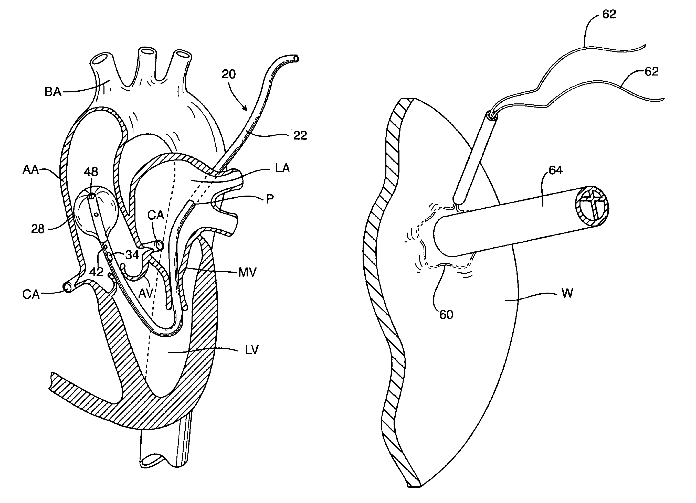
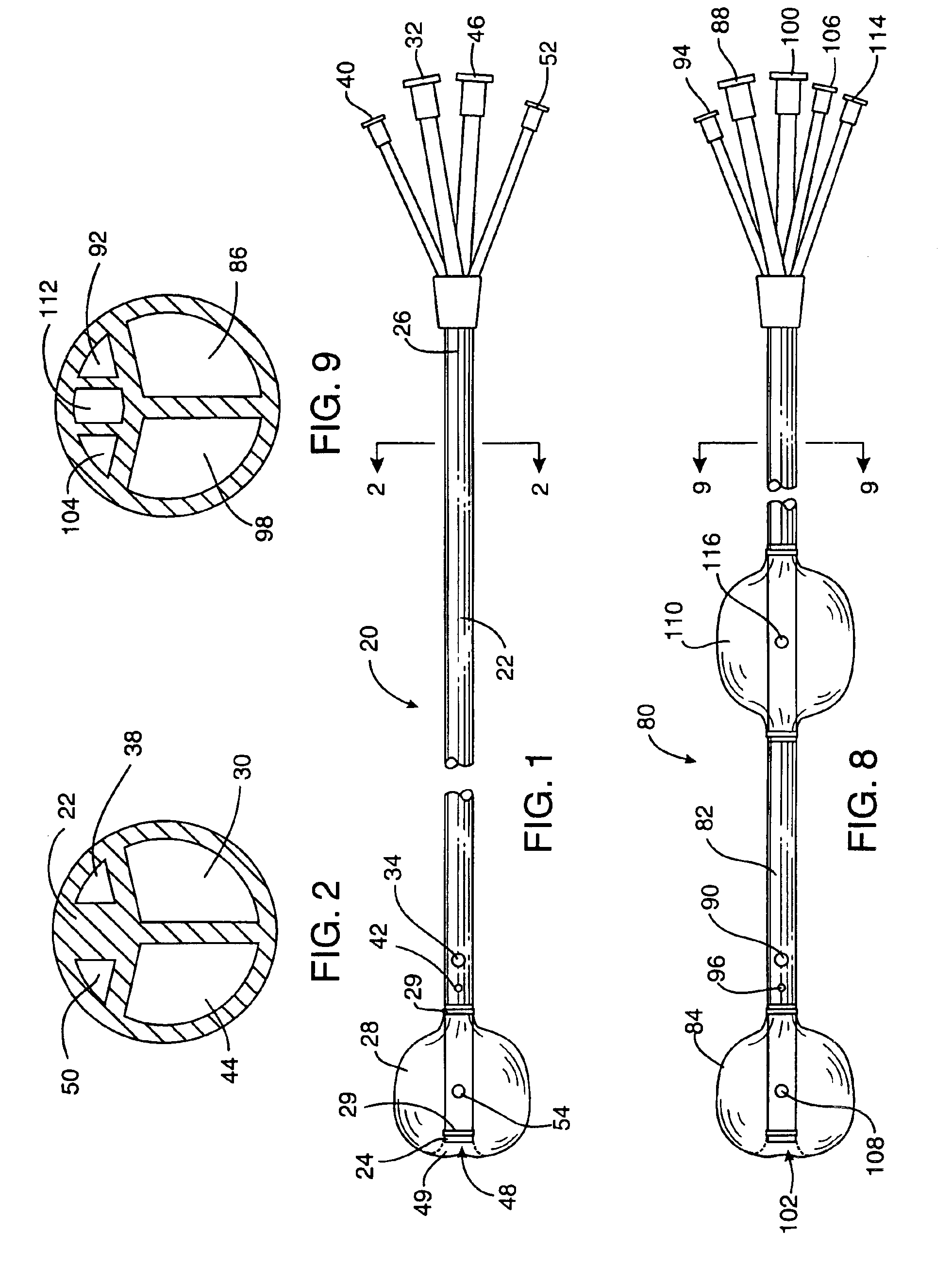
[0040]Referring to FIG. 1, a cardioplegia catheter according to the invention will be described. Cardioplegia catheter 20 has a shaft 22 having a distal end 24 and a proximal end 26. An occlusion balloon 28 is attached to shaft 22 near distal end 24 by adhesive bonding or other known technique. One or more radiopaque bands or markers 29 are provided on shaft 22 adjacent occlusion balloon 28 to permit fluoroscopic imaging of the catheter within the aorta.

[0041]Shaft 22 has a plurality of lumens, as shown in FIG. 2. A first lumen 30 extends from a first port 32 at proximal end 26 to a first opening 34 proximal to occlusion balloon 28. A second lumen 38 extends from a second port 40 at proximal end 26 to a second opening 42 proximal to occlusion balloon 28. The positions of first and second openings 34, 42 are selected such that the openings will be disposed within the ascending aorta downstream of the aortic valve when occlusion balloon 28 is disposed between the brachiocephalic arter...

second embodiment

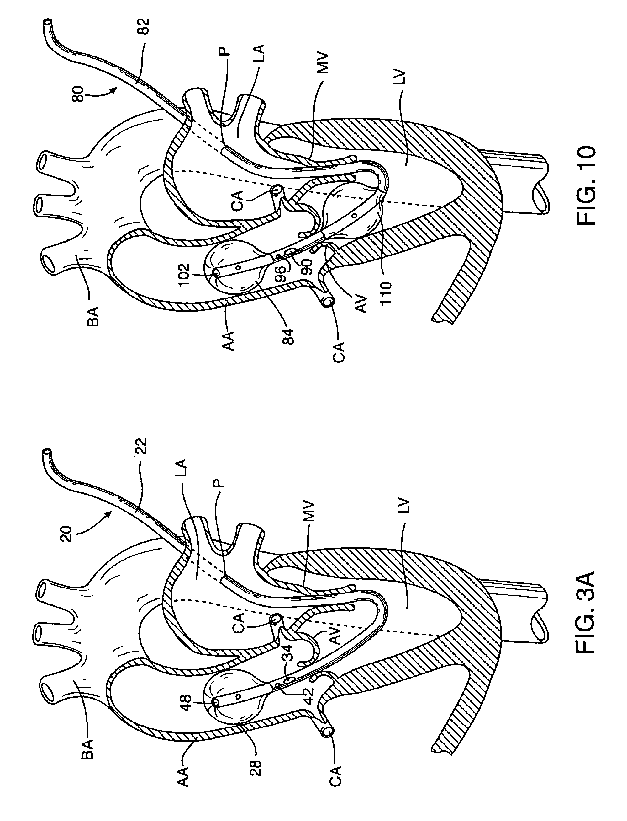
[0059]a cardioplegia catheter 80 according to the invention is illustrated in FIGS. 8-10. Cardioplegia catheter 80 is in many respects similar to cardioplegia catheter 20 of FIGS. 1-7, having a flexible shaft 82, an occlusion member 84 at the distal end of shaft 82, a first lumen 86 extending from a first port 88 to a first opening 90 proximal to occlusion member 94, a second lumen 92 extending from a second port 94 to second opening 96 proximal to occlusion member 84, and a third lumen 98 extending from a third port 100 to a third opening 102 distal to occlusion member 84. Occlusion member 84 is preferably an occlusion balloon, inflated by means of inflation fluid delivered through an inflation lumen 104 extending from an inflation port 106 to an inflation opening 108 within the occlusion balloon.

[0060]Unlike the previous embodiment, cardioplegia catheter 80 further includes a ventricular balloon 110 spaced proximally from occlusion member 84 and first and second openings 90,94. Th...


PUM


Abstract

A cardioplegia catheter is configured to extend into the ascending aorta with a proximal portion of the shaft extending into a left chamber of the heart through a aortic valve and out of the heart through a penetration in a wall thereof. The cardioplegia catheter has an occlusion member configured to occlude the ascending aorta between the brachiocephalic artery and the coronary ostia. An arterial return cannula delivers oxygenated blood to the arterial system downstream of the occlusion member, while cardioplegic fluid is delivered through a lumen in the cardioplegia catheter upstream of the occlusion member to induce cardioplegic arrest.

Description

CROSS-REFERENCE TO RELATED APPLICATION[0001]This application is continuation of U.S. patent application Ser. No. 08 / 839,189, filed Apr. 23, 1997, now issued as U.S. Pat. No. 6,090,086, the complete disclosure of which is incorporated herein by reference.FIELD OF THE INVENTION[0002]This invention relates generally to catheters for use in medical procedures, and more specifically to a catheter system for inducing cardioplegic arrest and maintaining circulation of blood.BACKGROUND OF THE INVENTION[0003]Cardiac surgery is conventionally performed with the patient's heart stopped and with circulation of blood maintained by extracorporeal cardiopulmonary bypass, a state known as cardioplegic arrest. Cardioplegic arrest conventionally requires the creation of a large incision in the chest, known as a sternotomy or thoracotomy, to expose the heart and great vessels of the thorax. Through this large incision, a steel clamp, called a cross-clamp, is placed around the aorta between the coronar...

Claims


Application Information

Patent Timeline
Patent Type & Authority Patents(United States)
IPC IPC(8): A61M25/10A61M25/00A61M29/02A61F2/958
CPCA61M25/10A61M25/1011A61M2025/1097A61M2210/125A61M2210/127A61M25/0032A61M2025/0003A61M2025/0031A61M2025/0036A61M2025/1052A61M2025/1095
Inventor ST. GOAR, FREDERICK G.STEVENS, JOHN H.GIFFORD, III, HANSON S.GRIFFITH, BARTLEY P.
Owner EDWARDS LIFESCIENCES LLC
Why Eureka
Try Eureka